
Improving early diagnosis of congenital malformations using detailed assessment of fetal anatomy
Piskulina A.A., Kostyukov K.V.
Relevance. Modern approaches to prenatal diagnosis demonstrate transition from the standard fetal anatomy screening in the second trimester to early detection of congenital anomalies at 11–14 weeks. In 2023, the updated guidelines of International Society of Ultrasound in Obstetrics & Gynecology (ISUOG) provided recommendations to follow the basic examination with a more detailed examination of the fetal anatomy. However, in Russia detailed examination of fetal anatomy in the first trimester is not regulated by the Order No.1130n (2020) of the Ministry of Health of Russia.
Objective. To compare the effectiveness of basic and detailed assessment of fetal anatomy in the first trimester of pregnancy based on the detection rate and congenital structural anomalies.
Materials and methods. Screening tests in the first trimester of pregnancy were analyzed in two groups. The group with standard protocol for assessment of fetal anatomy included 10,044 women who underwent screening tests in the first trimester of pregnancy in 2018–2023. The group with extended protocol for assessment of fetal anatomy included 2,741 patients who underwent screening tests in 2023–2025. The patients in both groups had singleton pregnancies, and underwent combined screening in the first trimester at 11+0–13+6 weeks (fetal crown-rump length (CRL) 45–84 mm)) at V.I. Kulakov National Medical Research Center for Obstetrics, Gynecology and Perinatology of the Ministry of Health of Russia. Ultrasound screening was performed in accordance with the Order No. 1130n of the Ministry of Health of Russia and ISUOG’s recommendations. The parametric and non-parametric statistical tests were used for data analysis depending on the type of variables. Statistical significance of differences was set at p<0.05. All calculations were made using software programs GraphPad Prism V. 9.5.1 and Microsoft Excel.
Results. In the group with basic assessment of fetal anatomy, the detection rate of CA was 4.3% (429/10,044). In the group with detailed assessment, the detection rate of CA significantly increased up to 8.2% (226/2741). Comparative analysis of congenital structural anomalies showed statistically significant increase in the detection rate almost in all anatomical structure groups using detailed assessment of fetal anatomy (p<0.005). In the studied period, the percentage of frequently detected anomalies in the first trimester increased from 42.7% (183/429) to 48.7% (110/226) (p<0.05). The percentage of anomalies with average detection increased from 50.6% (217/429) to 55.3% (125/226) (p<0.05). Also, the percentage of extremely rare detected anomalies proved to be statistically significant and increased from 6.7% (29/429) to 11.06% (25/226) (p=0.045).
Conclusion. Ultrasound examination in the first trimester allows to detect different congenital anomalies. The use of protocol for detailed assessment of fetal anatomy enabled to increase the detection rate of CA almost by 2 times – from 4.3% to 8.1%. Stratification of patients according to the underlying risk of chromosomal abnormalities is of key importance. The use of protocol for detailed assessment in the high-risk group increased detection rate of CA from 8.1 to 15.7%. This justifies the prioritized implementation of this approach specifically for this category of pregnant women for early diagnosis of anomalies and optimization of pregnancy management.
Authors' contributions. Piskulina A.A. – review of publications on the topic of the article, obtaining data for analysis, statistical data processing, manuscript writing; Kostyukov K.V. – the study design; manuscript editing.
Conflicts of interest. The authors confirm that they have no conflict of interest to declare.
Funding. The study was carried out without any sponsorship.
Ethical Approval. This study does not require Ethics Committee’s Approval, since this is a retrospective analysis of data obtained during standard screening tests at V.I. Kulakov National Medical Research Center for Obstetrics, Gynecology and Perinatology, Ministry of Health of Russia.
Generative Artificial Intelligence. No generative AI was used in preparing this article.
Patient Consent for Publication. The patients have signed informed consent for publication of their data.
Authors' Data Sharing Statement. The data supporting the findings of this study are available on request from the corresponding author after approval from the principal investigator.
For citation: Piskulina A.A., Kostyukov K.V. Improving early diagnosis of
congenital malformations using detailed assessment of fetal anatomy.
Akusherstvo i Ginekologiya/Obstetrics and Gynecology. 2026; (7): 72-79 (in Russian)
https://dx.doi.org/10.18565/aig.2026.34
Keywords
screening
ultrasound diagnosis
congenital malformations
prenatal diagnosis
Contemporary approaches to prenatal diagnostics demonstrate the shift from the traditional second-trimester scan of fetal anatomy to early detection of congenital anomalies (CA) at 11–14 weeks. The evolution of diagnostic ultrasonography and accumulation of knowledge and experience by the specialists of prenatal diagnosis have enabled to move beyond the basic assessment of sonography markers of fetal chromosomal abnormalities to comprehensive examination of fetal anatomy, that helps to identify up to 60% of structural anomalies in the first trimester of pregnancy [1]. The development of screening methods shows a gradual shift from nuchal translucency (NT) assessment that was used in the 1990s as a marker of trisomy 21 [2]. Over the following decade, a combined screening model was developed incorporating NT measurements and concentrations of biochemical markers – PAPP-A and free β-subunit of human chorionic gonadotropin (hCG) in maternal serum. [3]. This approach focused on searching for genetic abnormalities. In the 2010s, large-scale clinical studies showed that detailed assessment of fetal organs and systems at 11–14 weeks has a high diagnostic potential for detection of a wide range of congenital anomalies [4, 5]. This led to the revision of clinical protocols and the development of a concept for comprehensive prenatal screening that combines both risk assessment of chromosomal pathology and examination of fetal anatomy.
Large systematic reviews, for example, such as by Karim J.N. et al. [4], showed that about 40–60% of all major structural anomalies can be detected during detailed examination of fetal organs at 11–14 weeks of pregnancy. The International Society of Ultrasound in Obstetrics and Gynecology (ISUOG) provided recommendations (2013) for basic assessment of fetal anatomy including the main fetal structures: head and brain, face profile, anterior abdominal wall, stomach, urinary bladder, umbilical cord insertion site, and limbs [5]. This protocol included the minimum requirements for assessment of fetal anatomical structures and was an important step in shifting from screening aimed at assessment of the markers of chromosomal abnormalities to diagnosis of structural anomalies. It focused on confirmation of normal development of the main anatomical structures without detailed assessment, and laid the ground for the development of the current first-trimester screening protocol.
The updated ISUOG guidelines (2023) provided recommendations for systematic evaluation of fetal anatomical structures in the first trimester using two approaches – the basic protocol (the minimum requirements) and the detailed (expanded) protocol. In contrast to standard first-trimester screening, new approaches provide sequential “top-down” examination of all fetal anatomical structures [6]. The key difference between the two protocols is that the expanded protocol includes the detailed list of fetal structures that should be examined. Implementation of this protocol required to revise the organizational approaches to screening, including the extended length of ultrasound examination time (25–40 minutes), the use of expert-class, high-resolution ultrasound systems, and specialized training of ultrasound physicians. In the Russian Federation, first-trimester screening is officially regulated by the Order No. 1130n of the Ministry of Health of Russia “On approval of the procedure for providing medical care in the field of obstetrics and gynecology” (2020) [7]. However, the document stipulates only the basic examination, which is focused on assessment of the markers of chromosomal pathology and is limited to minimal requirements for evaluation of fetal anatomy.
According to a number of authors, the current conditional classification divides congenital anomalies into three groups based on the frequency of detection in the first trimester [8]. The first group includes anomalies that can be frequently detected (90–100%) in early pregnancy, such as acrania/anencephaly, holoprosencephaly, ectopia cordis, omphalocele, gastroschisis, megacystis, and body stalk anomaly. The second group includes anomalies, detection of which (10%–90%) depends on the doctor's experience, the quality of the equipment, and compliance with the ultrasound protocol: facial clefts, heart defects (ventricular and atrial septal defects, hypoplastic left heart syndrome), spina bifida, hydrocephalus, and limb reduction defects. The third group includes anomalies that are detected extremely rarely (<10%) due to late manifestation (intestinal obstruction, urinary tract obstruction and respiratory tract obstruction), or the impossibility of early diagnosis (small ventricular septal defects, agenesis of the corpus callosum, and others). This classification reflects implementation of first-trimester ultrasound screening, where detection effectiveness depends not only on the pathology of congenital anomalies, but also on the diagnostic protocol used for ultrasound assessment. This necessitates a differentiated approach to choosing between the basic and expanded screening protocols.
Despite the proposed recommendations, currently there is a lack of data indicating, in which cases ultrasound examination of fetal anatomy according to the basic protocol is sufficient, and in which cases it should be performed according to the expanded protocol. Our study is a comparative analysis of the effectiveness of the basic and expanded protocol for assessment of fetal anatomy at V.I. Kulakov National Medical Research Center for Obstetrics, Gynecology and Perinatology of the Ministry of Health of Russia to justify the feasibility of their implementation into clinical practice.
The objective of the study was to compare the effectiveness of the basic and detailed assessment of fetal anatomy in the first trimester of pregnancy based on the detection rate and congenital structural anomalies.
Materials and methods
The study included 12,785 patients with singleton pregnancy, who underwent first-trimester screening at 11+0–13+6 weeks (the fetal crown rump length (CRL) (КТР) of 45–84 mm) at V.I. Kulakov National Medical Research Center for Obstetrics, Gynecology and Perinatology, Ministry of Health of Russia (Kulakov Center). The women were followed-up through pregnancy until delivery or termination of pregnancy in the period from 2018 to August, 2025.
The combined screening was performed in accordance with the Order No. 1130n of the Ministry of Health of Russia [7], including ultrasound examination, assessment of biochemical markers and patient medical history.
Ultrasound examination was performed by ultrasound physicians, the experts of ultrasound and functional diagnostics using expert-class equipment Voluson E8 and Voluson E10 (General Electric, США), and convex transducer (2–9 MHz), 4–10 MHz linear transducer, and microconvex transvaginal transducers (5–9 MHz and 6–12 MHz). Ultrasound examination included evaluation of the following: confirmation of intrauterine pregnancy, fetal viability, and determination of the gestational age according to the CRL. The risk of chromosomal abnormalities was assessed using the following ultrasound biomarkers: NT, nasal bones, blood flow through the ductus venosus and the tricuspid valve, and heart rate. Ultrasound scan took 20 –40 minutes. When necessary, both transabdominal and transvaginal scans were used. In addition, placental parameters, the number of umbilical cord vessels, and conditions of the uterus, cervix, and uterine appendages were assessed.
Also, biochemical serum markers (PAPP-A and the free β-subunit of hCG) were evaluated, anamnestic data were collected, anthropometric measurements (height and weight) and blood pressure measurements were performed. Based on analysis of the anamnestic data the patients were divided into groups at low and high risk of fetal genetic pathology according to the woman’s age (over 35 years) and the presence of fetal chromosomal abnormalities in the previous pregnancy. The patients with diagnosed missed abortion were excluded from the study. The data on pregnancy outcomes were obtained from electronic medical records in Medialog system.
All patients were divided into two groups based on using the basic and expanded protocols for assessment of fetal anatomy. Group 1 included 10,044 patients who underwent combined first-trimester screening in the period from 2018 to 2023. Ultrasound examination was performed using the basic protocol for assessment of fetal anatomy (Table 1) [5].
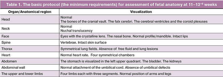
The basic protocol for assessment of fetal anatomy includes examination of the head and brain, scull bones (absence of bone defects), presence of the falx cerebri, and symmetrical appearance of the choroid plexus (the "butterfly" sign). Assessment of the fetal face includes the profile, nasal bone, eyes, the integrity of the lip and palate, measurement of the NT, the four-chamber view of the heart, as well cardiac axis and orientation within the thorax, the presence of the stomach and the integrity of the anterior abdominal wall, the spine (vertebrae and skin), the presence of the kidneys and bladder, and visualization of four limbs each with three segments.
Group 2 included 2,741 patients who underwent combined first-trimester screening in the period from 2023 to August, 2025. Ultrasound examination was performed in accordance with the updated ISUOG recommendations (2023) [6] using the expanded protocol for assessment of fetal anatomy (Table 2).
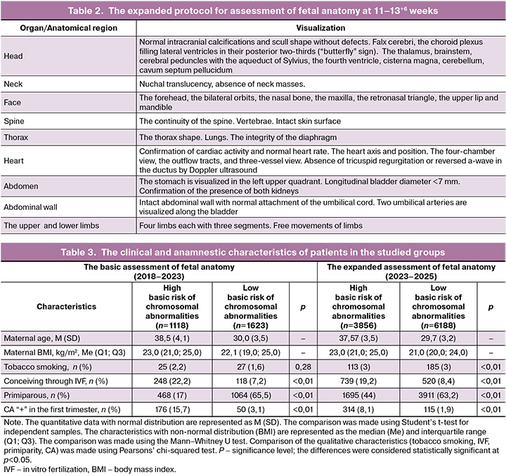
Expanded assessment of fetal anatomy included sequential examination of the brain structures (skull shape, the choroid plexuses, cerebellum, cavum septum pellucidum), facial structures (profile, nasal bone, eyes with the crystalline lens, the upper lip and palate), the neck (nuchal translucency measurement), the thorax (the thorax shape, the lungs, the diaphragm), heart (the four-chamber view, the heart axis, the outflow tracts), abdomen (integrity of the anterior abdominal wall, location of the stomach and the bladder), the spine (integrity of the spine in the sagittal and transverse planes), and the limbs (presence of all segments of the limbs, free movements of the limbs).
Each group was divided into subgroups based on high or low basic risk. The incidence of congenital anomalies and their structures detected by ultrasound examination were assessed in both groups. The diagnostic criterion for CA was visualization of the structures that deviated from normal anatomy. The cases with CA in the first trimester were discussed at case conference by ultrasound experts and multidisciplinary specialists. In most cases, invasive prenatal diagnostic testing was recommended.
Verification of the diagnosis was performed based on pathological examination when pregnancy was terminated, subsequent ultrasound examinations in the second and third trimesters, and after birth.
Statistical analysis
Statistical data processing was performed using software programs GraphPad Prism version 9.5.1 and Microsoft Excel. The normality of distribution of the quantitative characteristics was tested by the Shapiro–Wilk test. The parametric and non-parametric tests were used depending on the type of distribution.
The quantitative data with normal distribution are represented as the arithmetic mean and standard deviation – M (SD). Comparison of the quantitative data between the two independent groups was made using Student’s t-test for independent samples.
The quantitative data with non-normal distribution are represented as the median and interquartile range – Me (Q1; Q3). In this case, comparison of independent groups was conducted using the Mann–Whitney U test.
Comparison of the qualitative (categorical) variables represented as the absolute (n) and relative (%) frequencies was made using Pearson’s chi-squared test. Discrete variables are represented as frequencies (%). The quantitative data with normal distribution were compared using the Student's t-test for independent samples. The quantitative data with non-normal distribution were compared using the Mann–Whitney U test. The qualitative data were compared using Pearson’ chi-squared test.
For testing the null hypotheses, significance level was considered to be p<0.05.
Results
In the group with the basic assessment of fetal anatomy, 10.044 cases of first-trimester screening were analyzed. The subgroup of low-risk patients comprised 61.6% of women, and the subgroup of high-risk patients comprised 38,3% (Table 3). The median age of high-risk patients was 37.57 (3.5) years, 29.7 (3.2) years in the subgroup of low-risk patients. The differences were statistically significant according to the Student’s t-test for independent samples (p<0.001). High-risk patients became pregnant through assisted reproductive technologies more often – 19.2% compared with the subgroup of low-risk patients – 8.4% (p<0.01). In the subgroup of low risk, most women were primiparous – 63.2% compared with high-risk patients – 44%. In general, the frequency of fetal CA in the studied cohort was 4.3% and varied from 1.9% in the low-risk subgroup to 8.1% among high-risk pregnant women (p<0.01) [9].
The group with the expanded assessment of fetal anatomy (2023–2025) included 2,741 patients (Table 3). The subgroup of low risk comprised 59.2% (1,623/2,741) of patients, and 40.8% (1118/2741) in the high-risk subgroup. The median age of high-risk patients was 38.5 (4.1) years and 30.0 (3.5) years in the subgroup of low risk. The median body mass index in the group of high risk was 23.0 (21.0; 25.0) kg/m2 and 22.1 (19.0; 25.0) kg/m2 in the group of low risk. There was no statistically significant difference between the comparison groups in the frequency of tobacco smoking. High-risk patients conceived through IVF more often – 22.2% compared with the subgroup of low risk – 7.2% (p<0.01). The number of primiparous women was higher in the subgroup of low risk – 65.5% compared with the subgroup of high risk – 15.7%.
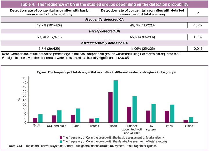
Comparative analysis of the clinical and anamnestic data of patients found similar distribution of high risk and low risk profiles in both cohorts. In the group with basic assessment of fetal anatomy (2018–2023), the percentage of high-risk patients was 38.4%, while in the cohort with expanded assessment of fetal anatomy (2023–2025) it increased up 40.8%. The incidence rate of CA in the group with basic assessment of fetal anatomy was 4.3% (429/10044), and in the group with detailed assessment of fetal anatomy it increased up to 8.2% (226/2741). According to the risk stratification of chromosomal abnormalities, analysis showed that in the group with basic assessment of fetal anatomy, the frequency of congenital anomalies in the low-risk subgroup was 1.9% (115/6188), while in the high-risk subgroup it reached 8.1% (314/3856). In the group with detailed assessment of fetal anatomy, the frequency of congenital anomalies in the low-risk subgroup was 3.1% (50/1623), and reached 15.7% (176/1118) in the high-risk group. The differences between the groups were statistically significant (p<0.01).
Note. CNS – the central nervous system; GI tract –the gastrointestinal tract; UG system – the urogenital system.
Analysis of CA distribution in the group with detailed assessment of fetal anatomy (Fig.) showed heart defects – 47.3% (107/226), anterior abdominal wall defects and gastrointestinal anomalies – 29.6% (67/226), urinary tract anomalies – 21.2% (48/226), limb anomalies – 20.3% (46/226), facial anomalies – 15.9% (36/226), abnormalities of the central nervous system and brain – 13.7% (31/226), spinal deformities – 6.2% (14/226), thoracic anomalies – 5.3% (12/226), anomalies of the skull – 4.0% (9/226). Comparative analysis of fetal congenital anomalies that were detected using the expanded protocol for ultrasound assessment showed statistically significant increase in the frequency of detecting malformations in all anatomical structures (p<0.005), except for thoracic anomalies (p<0.005).
Congenital anomalies that are frequently detected include acrania/anencephaly, holoprosencephaly, ectopia cordis, omphalocele, gastroschisis, megacystis, and body stalk anomaly. Congenital anomalies that are rarely detected include facial clefts, heart defects (ventricular septal defects and atrial septal defects, hypoplasia of the left heart structures), spina bifida, hydrocephalus, and limb reduction defects. Congenital anomalies that can be extremely rarely detected by ultrasound include intestinal atresia, urinary tract obstruction or airway obstruction, or impossibility of early diagnosis of small ventricular septal defects, agenesis of the corpus callosum, and other.
Comparative analysis showed statistically significant differences between the groups in the frequency of CA with different degree of detection during prenatal ultrasound screening (Table 4). During the research, the percentage of frequently detected anomalies increased from 42.7% (183/429) to 48.7% (110/226) (p<0.05). Rarely detected CA increased from 50.6% (217/429) to 55.3% (125/226) (p<0.05). The percentage of congenital anomalies that can be extremely rarely detected during the first-trimester screening increased from 6.7% (29/429) to 11.06% (25/226) (p=0.045).
Discussion
In recent years, the frequency of diagnosing CA in the first trimester of pregnancy has been increasing, and according to various studies on average reaches 30–60% [1, 4, 6, 10]. This progress is associated with the shift from the traditional second-trimester scan of fetal anatomy to early comprehensive ultrasound examination at 11–14 weeks. Through the development of ultrasound technologies, accumulation of clinical experience, and introduction of advanced diagnostic protocols, it has become possible not only to evaluate the markers of chromosomal abnormalities but also to examine fetal anatomy in detail, that significantly increases the detection of structural anomalies in early pregnancy.
Our study demonstrates that the use of the expanded protocol for first-trimester ultrasound screening increases the effectiveness of diagnosing congenital anomalies. The obtained results showed statistically significant increase in the detection rate of CA from 4.3% to 8.2% (p<0.01), that indicates improvement in the detection of CA in early pregnancy [9].
It is crucial to determine the indications for expanded examination of fetal anatomy, since performance of the routine detailed fetal ultrasound scan is not always justified (it has limitations, such as relation of the time the physician spends for ultrasound examination and workload to the probability of detecting congenital anomalies). Our study demonstrates an approach to determine the indications for expanded ultrasound examination through identification of the risk group based on high basic risk.
Special attention should be paid to differentiated analysis of the effectiveness of screening in the groups with different level of risk. In the high-risk group, the detection rate of CA increased from 8.1% to 15.7%, while in the low-risk group it increased from 1.9% to 3.1%. These data support the feasibility of using the expanded protocol for high-risk patients. Significant increase in the frequency of detection of CA in the high-risk group can be due to a combination of factors: objectively higher prevalence of structural anomalies in this group of patients and detailed examination of a broader range of anatomical structures.
Analysis of the structure of detected congenital anomalies showed that their distribution was consistent with the data in the international studies. Heart defects were diagnosed most frequently (47.3%), that was consistent with the results of the meta-analysis by Karim J.N. et al. [1], confirming the possibility of effective diagnosis of cardiovascular anomalies in the first trimester.
The percentage of detection of anterior abdominal wall defects and gastrointestinal anomalies is rather high (29.6%) and also corresponds to global data, given that CA, such as omphalocele and gastroschisis are detected in 90–100% of cases in the first trimester [10].
It is important to note that detection rate of CA that are detected extremely rarely in the first trimester also increased from 6.7 to 11.06% (p=0.045). This can be explained not only by detailed assessment of fetal anatomy, but also by changes in the clinical profile of patients, who were hospitalized at Kulakov Center, since these cases are more often referred to tertiary facilities, including rare and combined anomalies.
The obtained data confirm validation of the differentiated approach to prenatal diagnostics, namely, stratification of patients into risk groups. According to this approach, it is reasonable to perform first-trimester screening using the expanded protocol for high-risk patients in a specialized center, while screening using the basic protocol for low-risk patients can be performed in primary care facilities.
High effectiveness of screening using the expanded protocol in the federal center justifies its use for examination of patients in risk groups of different level, while the basic protocol for assessment of fetal anatomy in the primary health care centers ensures broad population coverage. This strategy allows for resource allocation. The results of using the expanded protocol at V.I. Kulakov National Medical Research Center for Obstetrics, Gynecology and Perinatology of the Ministry of Health of Russia show its diagnostic value and provide a basis for its implementation in routine clinical practice. The proposed screening model is consistent with the international experience in organizing a two-tier prenatal diagnostic system and patient referral to the specialized centers for expanded examinations, including those reported in the TRIDENT-2 study [11].
The obtained results have important practical implication to improve the prenatal diagnostic system in the Russian Federation. A 1.9-fold increase in the detection rate of congenital anomalies using the expanded protocol suggests the need to revise the existing regulations about first-trimester ultrasound screening. Implementation of a two-tier screening system with the use of the basic protocol in primary health care facilities and the expanded protocol in the expert centers can be an optimal solution.
Limitations of the study. The study has some limitations, since it was carried out at a single federal center, that makes it harder to directly apply the obtained data in clinical practice at primary health care facilities. [7]. Direct dependence of screening effectiveness on the type of ultrasound diagnostic system and the operator's qualification determines the importance of the programs for additional training of specialists and implementation of standardized uniform protocols.
Conclusion
Increased detection rate of CA from 4.3% to 8.2% (p<0.01) indicates that implementation of the expanded protocol for ultrasound examination makes it possible to increase the detection of fetal anomalies in the first trimester. Detection rate was higher in the high-risk group, and increased from 8.1% to 15.7%, that implies the importance of using the expanded protocol in this cohort.
The obtained data indicate the need to update the existing regulatory documents, namely implementation of the protocol for first-trimester ultrasound examination of fetal anatomy, that will significantly improve the effectiveness of prenatal diagnostics. Implementation of the expanded protocol for first-trimester ultrasound screening into clinical practice will promote early detection of congenital anomalies, optimization of patient routing, and reduction in perinatal morbidity and mortality.
References
- Karim J.N., Di Mascio D., Roberts N., Papageorghiou A.T. Detection of non‐cardiac fetal abnormalities on ultrasound at 11–14 weeks: systematic review and meta‐analysis. Ultrasound Obstet. Gynecol. 2024; 64(1): 15-27. https://dx.doi.org/10.1002/uog.27649
- Fantasia I., Catagini S., Zamagni G., Greco P., Bianchini I., Bussolaro S. et al. The clinical impact of the first‐trimester nuchal translucency between the 95th-99th percentiles. Prenat. Diagn. 2023; 43(7): 929-36. https://dx.doi.org/10.1002/pd.6390
- Bardi F., Kagan K.O., Bilardo C.M. First‐trimester screening strategies: a balance between costs, efficiency and diagnostic yield. Prenat. Diagn. 2023; 43(7): 865-72. https://dx.doi.org/10.1002/pd.6393
- Karim J.N., Roberts N.W., Salomon L.J., Papageorghiou A.T. Systematic review of first-trimester ultrasound screening for detection of fetal structural anomalies and factors that affect screening performance. Ultrasound Obstet. Gynecol. 2017; 50(4): 429-41. https://dx.doi.org/10.1002/uog.17246
- ISUOG Practice Guidelines: performance of first‐trimester fetal ultrasound scan. Ultrasound Obstet. Gynecol. 2013; 41(1): 102-13. https://dx.doi.org/10.1002/uog.12342
- Bilardo C.M., Chaoui R., Hyett J.A., Kagan K.O., Karim J.N., Papageorghiou A.T. et al. ISUOG Practice Guidelines (updated): performance of 11–14‐week ultrasound scan. Ultrasound Obstet. Gynecol. 2023; 61(1): 127-43. https://dx.doi.org/10.1002/uog.26106
- Приказ Министерства здравоохранения РФ от 20.10.2020 № 1130н «Об утверждении Порядка оказания медицинской помощи по профилю "акушерство и гинекология"». Доступно по: https://base.garant.ru/74840123/ [Order of the Ministry of Health of the Russian Federation No. 1130n dated 20.10.2020 «On approval of the Procedure for providing medical care in obstetrics and gynecology». Available at: https://base.garant.ru/74840123/ (in Russian)].
- Minnella G.P., Crupano F.M., Syngelaki A., Zidere V., Akolekar R., Nicolaides K.H. Diagnosis of major heart defects by routine first‐trimester ultrasound examination: association with increased nuchal translucency, tricuspid regurgitation and abnormal flow in ductus venosus. Ultrasound Obstet. Gynecol. 2020; 55(5): 637-44. https://dx.doi.org/10.1002/uog.21956
- Пискулина А.А., Костюков К.В. Диагностика врожденных пороков развития плода в первом триместре беременности. Акушерство и гинекология. 2024; 11: 90-7. https://dx.doi.org/10.18565/aig.2024.182 [Piskulina A.A., Kostyukov K.V. Diagnosis of fetal congenital anomalies in the first trimester of pregnancy. Obstetrics and Gynecology. 2024; (11): 90-7 (in Russian). https://dx.doi.org/10.18565/aig.2024.182].
- Syngelaki A., Hammami A., Bower S., Zidere V., Akolekar R., Nicolaides K.H. Diagnosis of fetal non‐chromosomal abnormalities on routine ultrasound examination at 11–13 weeks’ gestation. Ultrasound Obstet. Gynecol. 2019; 54(4): 468-76. https://dx.doi.org/10.1002/uog.20844
- van der Meij K.R.M., Sistermans E.A., Macville M.V.E., Stevens S.J.C., Bax C.J., Bekker M.N. et al. TRIDENT-2: National implementation of genome-widenon-invasive prenatal testing as a first-tier screening test in the Netherlands. Am. J. Hum. Genet. 2019; 105(6): 1091-101. https://dx.doi.org/10.1016/j.ajhg.2019.10.005
Received 03.02.2026
Accepted 24.06.2026
About the Authors
Alexandra A. Piskulina, PhD student, Academician V.I. Kulakov National Medical Research Center for Obstetrics, Gynecology and Perinatology, Ministry of Health of Russia, 117997, Russia, Moscow, Ac. Oparin str., 4, +7(904)888-36-22, piskulinaalexandra@yandex.ru, https://orcid.org/0009-0005-7845-690XKirill V. Kostyukov, Dr. Med. Sci., Head of the Department of Functional and Ultrasonic Diagnostics, Department of Visual Diagnostics, Academician V.I. Kulakov National Medical Research Center for Obstetrics, Gynecology and Perinatology, Ministry of Health of Russia, 117997, Russia, Moscow, Ac. Oparin str., 4, +7(926)214-97-84, kostyukov_k@yahoo.com, https://orcid.org/0000-0003-3094-4013
Similar Articles

