
The role of mitochondrial dysfunction in the pathogenesis of preterm birth
Medzhidova M.K., Tyutyunnik V.L., Kan N.E., Manukhova L.A., Marey M.V., Ivanova V.A., Vysokikh M.Yu.
Objective. To evaluate the role of placental and fetal membrane mitochondrial dysfunction in the pathogenesis of preterm birth (PTB).
Materials and methods. This study included 228 women with spontaneous preterm birth at 22.0–37.0 weeks of gestation. The participants were divided into four groups: Group 1 comprised 52 women with extremely preterm birth (22.0–27.6 weeks); Group 2 included 56 women with early preterm birth (28.0–31.6 weeks); Group 3 consisted of 60 women with preterm birth (32.0–33.6 weeks); and Group 4 included 60 women with late preterm birth (34.0–36.6 weeks). Given the different etiological pathways of PTB, each group was further subdivided into two subgroups: women with preterm prelabor rupture of membranes (PPROM) followed by spontaneous onset of labor (n=125) and women with spontaneous preterm birth (SPB) without PPROM (n=103). The control group comprised 72 women with uncomplicated pregnancies who delivered at term (37.0–41.0 weeks).
Results. Statistically significant differences in placental OPA1 protein expression were identified between the spontaneous preterm birth and term birth groups, with significantly higher OPA1 levels in the spontaneous preterm birth group (p<0.01).
PINK1 expression was significantly decreased in the placenta of spontaneous preterm birth compared with term birth (p<0.01). Analysis of PARKIN protein expression, functionally associated with PINK1 activity, revealed statistically significant differences between all PTB groups and the term birth group (p<0.01).
Placental P62 protein levels showed statistically significant differences between extremely preterm and late preterm births (p<0.01) and between extremely preterm and term births (p<0.001). Elevated P62 levels in extremely preterm births indicate autophagy activation in the context of mitophagy imbalance, resulting in impaired clearance of damaged mitochondria.
Conclusion. Disruption of mitophagy and autophagy pathways (P62, PINK1, and PARKIN) in the placenta represents a common but differentially regulated mechanism underlying the pathogenesis of both spontaneous preterm birth and PPROM. These findings support the hypothesis that mitochondrial quality control plays a critical role in maintaining pregnancy. Altered expression of P62, PARKIN, and PINK1 in spontaneous preterm birth and PPROM suggests that defects in mitochondrial quality control are key components in the pathogenesis of preterm birth.
Authors' contributions. Medzhidova M.K., Tyutyunnik V.L., Kan N.E., Vysokikh M.Yu., Marey M.V. – conception and design of the study; Medzhidova M.K., Manukhova L.A., Marey M.V., Ivanova V.A. – data collection and analysis and review of the relevant literature; Medzhidova M.K. – drafting of the manuscript; Tyutyunnik V.L., Kan N.E., Vysokikh M.Yu. – editing of the manuscript.
Conflicts of interest. The authors have no conflicts of interest to declare.
Funding. There was no funding for this study.
Ethical Approval. The study was reviewed and approved by the Research Ethics Committee of the V.I. Kulakov NMRC for OG&P.
Generative Artificial Intelligence. No artificial intelligence tools were used in the preparation of this manuscript.
Patient Consent for Publication. All patients provided informed consent for the publication of their data.
Authors' Data Sharing Statement. The data supporting the findings of this study are available upon request from the corresponding author after approval from the principal investigator.
For citation: Medzhidova M.K., Tyutyunnik V.L., Kan N.E., Manukhova L.A., Marey M.V., Ivanova V.A.,
Vysokikh M.Yu. The role of mitochondrial dysfunction in the pathogenesis of preterm birth.
Akusherstvo i Ginekologiya/Obstetrics and Gynecology. 2026; (7): 86-93 (in Russian)
https://dx.doi.org/10.18565/aig.2026.82
Keywords
preterm birth
spontaneous preterm birth (SPB)
preterm premature rupture of membranes (PPROM)
mitochondrial dysfunction
mitochondrial biogenesis
Recent advances in clinical obstetric practice have reshaped our understanding of preterm birth (PTB). PTB is widely recognized as a heterogeneous syndrome with multiple etiologies, necessitating its classification into distinct clinical subtypes. Currently, PTB is categorized as spontaneous preterm birth (SPB), preterm prelabor rupture of membranes (PPROM), or medically indicated PTB. This classification is essential for developing targeted strategies for the early diagnosis, prevention, and treatment of obstetric complications [1–3].
Complications associated with preterm birth remain the leading cause of mortality among children aged <5 years [4,5]. Despite substantial research efforts, the global incidence of PTB has remained unchanged over the past decade. According to Ohuma E.O. et al., 13.4 million infants were born preterm (<37 weeks of gestation) in 2020, accounting for 9.9% of all live births, compared with 13.8 million (9.8%) in 2010 [6].
The pathogenesis of PTB is thought to involve several interrelated mechanisms, including an imbalance between pro- and anti-inflammatory cytokines, reproductive tract dysbiosis accompanied by a localized inflammatory response [7, 8], and progressively increased oxidative stress [9, 10]. Placental dysfunction is a key triggering factor. Considerable attention has been focused on the central role of impaired energy metabolism, mitochondrial damage, and inflammation driven by mitochondria-derived proinflammatory factors [11]. Throughout pregnancy, mitochondria play a pivotal role in sustaining placental energy metabolism, supporting placental development, eliminating senescent cells, and maintaining trophic and endocrine functions. The functional integrity of mitochondrial energy-transducing membranes is closely linked to the involvement of mitochondria in programmed cell death. The extent of stress-induced mitochondrial damage determines the structural integrity of the placenta and its capacity to transport nutrients to the developing fetus [12–14].
To ensure an effective cellular stress response and post-stress adaptation, cells possess a mitochondrial quality control program that continuously monitors their integrity. This process comprises mitochondrial fission, recognition and labeling of dysfunctional organelles, their selective removal through mitophagy, followed by lysosomal degradation via autophagy and subsequent mitochondrial fusion into a reticular network [15–17].
In our previous studies, we demonstrated the existence of a causal cascade linking mitochondrial damage, oxidative stress, and inflammation in the development of PTB. We found that in extremely early PTB, mitochondrial lipid peroxidation reaches a critical threshold that affects low- and very-low-density lipoproteins [12]. These modified lipoproteins acquire spatial conformations resembling bacterial lipopolysaccharides and exhibit similar effects on Toll-like receptors (TLRs), particularly TLR4. This positive feedback loop drives a self-amplifying inflammatory response that plays a decisive role in reaching the threshold levels of free radicals required to trigger PTB. Consequently, maintaining mitochondrial integrity is critical for preventing this pathological cascade. Impairment of the mitochondrial quality control system leads to the rapid accumulation of dysfunctional organelles and initiates a detrimental cascade within the placenta, even in the absence of a microbial inflammatory trigger, ultimately resulting in PTB. Collectively, these findings underscore the importance of investigating mitochondrial dysfunction in the pathogenesis of PTB.
This study aimed to evaluate the role of mitochondrial dysfunction in the placenta and fetal membranes in PTB pathogenesis.
Materials and methods
The study included 228 women with spontaneous PTB at 22.0–36.6 weeks’ gestation. Participants were stratified into four groups: group 1 comprised 52 women with extremely preterm birth (22.0–27.6 weeks of gestation); group 2 included 56 women with very preterm birth (28.0–31.6 weeks); group 3 consisted of 60 women with moderate preterm birth (32.0–33.6 weeks); and group 4 comprised 60 women with late preterm birth (34.0–36.6 weeks). Given the heterogeneous etiology of PTB, each group was further subdivided into women with PPROM (n=125) and those with SPB without PPROM (n=103). The control group comprised 72 women with uncomplicated singleton pregnancies who delivered at term (37.0–41.0 weeks’ gestation).
The inclusion criteria were singleton pregnancy, first stage of spontaneous preterm labor, and PPROM occurring between 22.0 and 36.6 weeks of gestation.
The exclusion criteria were multiple pregnancy; severe extragenital disease; severe obstetric complications requiring elective preterm delivery; placental abruption; fetal growth restriction; congenital fetal anomalies; sexually transmitted infections; and maternal HIV infection.
The study was reviewed and approved by the Research Ethics Committee of the NMRC for OG&P. All participants provided written informed consent prior to enrollment.
Collection of placental tissue and fetal membrane samples
Placental tissue and fetal membrane samples were collected within 5 min of delivery. In each case, a placental biopsy was obtained 1.5–2 cm from the umbilical cord insertion site. Fetal membrane specimens were collected simultaneously with the placental biopsy. Samples designated for subsequent protein expression analysis were placed in 1-mL tubes and snap-frozen in liquid nitrogen. The tissues were then pulverized under liquid nitrogen and homogenized in a glass homogenizer using RIPA lysis buffer for western blotting analysis, according to the manufacturer's instructions (Santa Cruz Biotechnology).
Protein electrophoresis and western blot analysis of placental tissue and fetal membranes
Proteins were separated by sodium dodecyl sulfate–polyacrylamide gel electrophoresis (SDS-PAGE) according to the method described by Laemmli [18], followed by electrotransfer onto nitrocellulose membranes (Millipore, USA). To prevent nonspecific antibody binding, the membranes were incubated for 1 h in 5% nonfat dry milk powder (NDMP) prepared in TBST buffer (Bio-Rad, USA). The membranes were subsequently incubated for 12 h at 4°C with continuous agitation using primary antibodies against SQSTM1/p62 (catalog no. ab56416), and OPA1 (catalog no. ab42364), PINK1 (catalog no. ab23707), and Parkin (catalog no. ab77924) and β-actin (catalog no. A5441; Sigma-Aldrich, USA). Following washing with TBST buffer to remove unbound primary antibodies, the membranes were incubated with horseradish peroxidase-conjugated secondary antibodies (Goat Anti-Mouse ab6789 and Goat Anti-Rabbit ab6721; Abcam, USA). Chemiluminescent detection was performed using the Novex ECL detection kit (Invitrogen, USA), and signals were acquired using a ChemiDoc imaging system (Bio-Rad, USA). The chemiluminescent signal intensities of the target proteins were quantified using the Image Lab software (Bio-Rad, USA). Relative protein expression levels were normalized to that of β-actin.
Statistical analysis
Statistical analysis was performed using GraphPad Prism version 6.0 (GraphPad Software Inc., USA). Comparisons between two independent groups were conducted using the Mann–Whitney U test for non-parametric data. Comparisons between independent groups were performed using the Kruskal–Wallis test. Statistical significance was set at p<0.05. Data are presented in histograms as median, interquartile range, and minimum and maximum values.
Results and discussion
All patients included in the study groups were comparable with respect to their clinical characteristics. The maternal age ranged from 18 to 41 years. The anthropometric parameters were consistent with those observed in the general population. No statistically significant differences were observed in the family history or prevalence of childhood infectious diseases.
Analysis of reproductive history demonstrated a higher rate of spontaneous pregnancy loss in group 2 (15/56, 26.8%), which was significantly greater than that observed in groups 3 (6/60, 10.0%) and 4 (6/60, 10.0%) (p=0.029 and p=0.029, respectively). Missed miscarriage occurred in 16/52 (30.8%) patients in group 1, 7/56 (12.5%) in group 2, 5/60 (5.8%) in group 3, and 6/60 (10.0%) in group 4, with a significantly higher prevalence in group 1 (p=0.033, p=0.003, and p=0.008, respectively). A history of PTB was reported in 14/52 (26.9%) patients in group 1, 16/56 (28.6%) in group 2, 8/60 (13.3%) in group 3, and 7/60 (11.7%) in group 4, respectively. The prevalence was also significantly higher in groups 1 and 2 than in group 4 (p=0.052 and p=0.035, respectively).
A higher prevalence of chronic cystitis was observed in groups 2 (16/56, 28.5%) and 3 (18/60, 30.0%) than in group 1 (6/52, 11.5%) (p=0.033 and p=0.021, respectively) and group 4 (8/60, 13.3%) (p=0.065 and p=0.045, respectively). Similarly, chronic pyelonephritis occurred more frequently in groups 1 (17/52, 32.7%) and 2 (19/56, 33.9%) than in group 3 (8/60, 13.3%) (p=0.022 and p=0.015, respectively) and group 4 (6/60, 10.0%) (p=0.004 and p=0.003, respectively). Since infection is recognized as one of the principal contributors to the development of PTB, the prevalence of infectious and inflammatory diseases of the reproductive tract and the spectrum of potential pathogens were further evaluated. Bacterial vaginosis was more common in group 2 (11/56, 19.6%) than in groups 3 and 4, where it occurred in 3/60 (5.0%) and 5/60 (8.3%) patients, respectively. Likewise, vulvovaginal candidiasis was more frequent in group 2 (18/56, 32.1%) than in groups 3 and 4, where it was identified in 7/60 (11.6%) and 6/60 (10.0%) patients, respectively (p<0.01).
Clinical and anamnestic analyses identified several factors associated with an increased risk of PTB. The observed correlations demonstrated a high degree of statistical reliability and should be considered for risk stratification. Significant predictors included chronic infectious and inflammatory diseases of the genitourinary tract and adverse obstetric and gynecological histories characterized by spontaneous pregnancy loss, missed miscarriage, PTB, and prior intrauterine interventions. These findings indicate that the combined presence of these factors substantially increases the likelihood of PTB and supports the classification of such pregnancies as high-risk, warranting close clinical surveillance. Nevertheless, the complexity of PTB prediction underscores the need for further studies to elucidate the underlying mechanisms of PTB and facilitate the development of effective preventive strategies.
Accordingly, we investigated mitochondrial dysfunction associated with impaired mitophagy in the placenta and fetal membranes under conditions of transient hypoxia and oxidative stress, which accompany placental disorders.
Comparative analysis of mitochondrial biogenesis and autophagy protein expression
OPA1 protein. Placental OPA1 expression progressively increased throughout gestation in cases of spontaneous preterm birth (SPB). Statistically significant differences were observed among patients with extremely early, early, late PTB, and term deliveries. Relative placental OPA1 protein levels were significantly higher in the term delivery group than in the SPB group (p<0.01) (Fig. 1A).
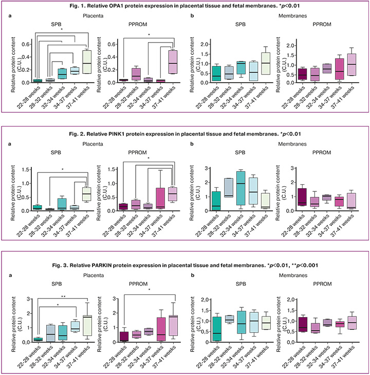
In PPROM, placental OPA1 protein levels were also significantly reduced compared with term delivery; however, no gestational age-dependent differences were observed (Figure 1C). No statistically significant differences in OPA1 expression were detected in the fetal membranes in either SPB or PPROM.
PINK1 kinase. Similar findings were obtained for the PINK1 kinase. Placental PINK1 expression was significantly reduced in SPB, irrespective of gestational age, compared to the peak expression observed at term delivery. In PPROM, significant differences in placental PINK1 expression were detected only in cases of extremely early PTB (Fig. 2A). No significant differences were observed in the fetal membranes (Fig. 2C).
PARKIN protein. Analysis of PARKIN, which functionally interacts with PINK1, demonstrated significant differences in protein expression between extremely early, early, and late PTB compared with term delivery. Similar expression patterns were observed in both the SPB and PPROM groups (Fig. 3A). In contrast, no significant differences in PARKIN expression were detected in the fetal membranes in either SPB or PPROM compared to term delivery (Fig. 3C).
P62 protein. Analysis of P62 protein expression (Fig. 4) in placental tissue revealed significant differences between extremely early and late SPB (p<0.01), as well as between extremely early SPB and term delivery (p<0.001) (Fig. 4A, SPB). In addition, significant differences (p<0.01) were observed among extremely early, early, and late PTB in the PPROM group and between late PTB and term deliveries (Fig. 4A, PPROM). Although no significant differences in P62 protein levels were detected in fetal membranes according to gestational age in either SPB or PPROM or relative to term delivery (Fig. 4C), correlation analysis demonstrated a positive correlation between placental and fetal membrane P62 expression in SPB (R=0.56, p=0.04) and a negative correlation in term delivery (R=-0.7, p=0.037). No such correlation was observed in the PPROM group.
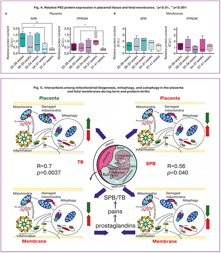
Disruption of mitochondrial fusion, regulated by OPA1, contributes to mitochondrial dysfunction and enhances oxidative stress. P62, also known as SQSTM1, functions as a selective autophagy receptor. Dysfunction of this protein is a common feature of numerous diseases because it mediates the degradation of polyubiquitinated proteins and participates in PINK1/PARKIN-dependent mitophagy. The increased P62 protein levels observed in women with extremely early PTB indicate the activation of autophagy in the setting of an imbalanced mitophagy cascade, resulting in the impaired clearance of damaged mitochondria (Fig. 5).
Dysregulation of mitophagy and autophagy (P62, PINK1, and PARKIN) in the placenta is a common but mechanistically divergent component of the pathogenesis of both SPB and PPROM, supporting the hypothesis that mitochondrial quality control plays a critical role in maintaining pregnancy. Altered expression of P62, PARKIN, and PINK1 in SPB and PPROM indicates that defects in mitochondrial quality control are an important pathogenic mechanism underlying both conditions. These findings are consistent with the current concepts of mitophagy as an integrated process linking cellular stress, inflammation, and apoptosis, which is particularly relevant in the context of sterile inflammation associated with PTB [19, 20].
Conclusion
The molecular mechanisms underlying SPB and PPROM appear to be fundamentally different. Mitochondrial damage in the placenta, rather than in fetal membranes, may serve as an initiating trigger. PPROM likely represents a state of secondary acute placental mitochondrial injury, characterized by the activation of the emergency mitochondrial clearance program (mitophagy) in response to mitochondria-derived proinflammatory factors entering the systemic circulation. These findings are consistent with the central role of infectious and inflammatory processes in the induction of PTB (Fig. 5). In contrast, SPB appears to result from chronic mitochondrial dysfunction, characterized by disruption of mitochondrial architecture and integrity (OPA1 deficiency), accompanied by less pronounced activation of the mitochondrial quality control pathway (reduced activity of PINK1 kinase and PARKIN ubiquitin ligase) in the presence of activated autophagy that is uncoupled from mitophagy (increased p62 levels).
These findings provide further insight into the mechanisms underlying PTB and support the identification of potential targets for future research aimed at improving the prediction, diagnosis, and pathogenetically based prevention of PTB. Such advances may enhance risk-stratification algorithms and enable preventive interventions to be focused on the most vulnerable gestational period, between 22 and 28 weeks.
References
- Министерство здравоохранения Российской Федерации. Клинические рекомендации. Преждевременные роды. М.; 2024. 40 с. [Ministry of Health of the Russian Federation. Clinical guidelines. Preterm birth. Moscow; 2024. 40 p. (in Russian)].
- Williams S.M., Rosenblatt K.P., Reznik S., Misra D.P., Siricilla S., Gomez-Lopez N. et al. Defining knowledge gaps in preterm birth research: can biomarkers fill the gaps? Front. Med. (Lausanne). 2025; 12: 1655833. https://dx.doi.org/10.3389/fmed.2025.1655833
- Ремнева О.В., Колядо О.В., Песоцкая А.В., Стародубцев Е.Г., Гуменюк И.С. Патоморфологические особенности последов у пациенток с различными клиническими фенотипами спонтанных преждевременных родов. Акушерство и гинекология. 2021; 8: 111-8. https://dx.doi.org/10.18565/aig.2021.8.111-118 [Remneva O.B., Kolyado O.V., Pesotskaya A.V., Starodubtsev E.G., Gumenyuk I.S. Pathomorphological features of placenta in patients with different clinical phenotypes of spontaneous preterm births. Obstetrics and Gynecology. 2021; (8): 111-8 (in Russian). https://dx.doi.org/10.18565/aig.2021.8.111-118].
- Perin J., Mulick A., Yeung D., Villavicencio F., Lopez G., Strong K.L. et al. Global, regional, and national causes of under-5 mortality in 2000-19: an updated systematic analysis with implications for the sustainable development goals. Lancet Child Adolesc. Health. 2022; 6(2): 106-15. https://dx.doi.org/10.1016/S2352-4642(21)00311-4
- Горина К.А., Ходжаева З.С., Белоусов Д.М., Баранов И.И., Гохберг Я.А., Пащенко А.А. Преждевременные роды: прошлые ограничения и новые возможности. Акушерство и гинекология. 2020; 1: 12-9. https://dx.doi.org/10.18565/aig.2020.1.12-119 [Gorina K.A., Khodzhaeva Z.S., Belousov D.M., Baranov I.I., Gokhberg Ya.A., Pashchenko A.A. Premature birth: past limitations and new opportunities. Obstetrics and Gynecology. 2020; (1): 12-9 (in Russian). https://dx.doi.org/10.18565/aig.2020.1.12-119].
- Ohuma E.O., Moller A.B., Bradley E., Chakwera S., Hussain-Alkhateeb L., Lewin A. et al. National, regional, and global estimates of preterm birth in 2020, with trends from 2010: a systematic analysis. Lancet. 2023; 402(10409): 1261-71. https://dx.doi.org/10.1016/S0140-6736(23)00878-4
- Супрун С.В., Кудерова Н.И., Супрун Е.Н., Морозова О.Н., Евсеева Г.П., Лебедько О.А. Комплексная оценка митохондриальных изменений иммунокомпетентных клеток крови у беременных женщин при срочных и преждевременных родах. Медицинская иммунология. 2021; 23(3): 557-68. https://dx.doi.org/10.15789/1563-0625-CEO-2118 [Suprun S.V., Kuderova N.I., Suprun E.N., Morozova O.N., Evseeva G.P., Lebedko O.A.Complex estimation of mitochondrial changes of immunocompetent blood cells in pregnant women with urgent and premature birth. Medical Immunology. 2021; 23(3): 557-68 (in Russian). https://dx.doi.org/10.15789/1563-0625-CEO-2118].
- Белоусова В.С., Стрижаков А.Н., Свитич О.А., Тимохина Е.В., Кукина П.И., Богомазова И.М., Пицхелаури Е.Г. Преждевременные роды: причины, патогенез, тактика. Акушерство и гинекология. 2020; 2: 82-7. https://dx.doi.org/10.18565/aig.2020.2.82-87 [Belousova V.S., Strizhakov A.N., Svitich O.A., Timokhina E.V., Kukina P.I., Bogomazova I.M., Pitskhelauri E.G. Preterm birth: causes, pathogenesis, and management. Obstetrics and Gynecology. 2020; (2): 82-7 (in Russian). https://dx.doi.org/10.18565/aig.2020.2.82-87].
- Lien Y.C., Zhang Z., Cheng Y., Polyak E., Sillers L., Falk M.J. et al. Human placental transcriptome reveals critical alterations in inflammation and energy metabolism with fetal sex differences in spontaneous preterm birth. Int. J. Mol. Sci. 2021; 22(15): 7899. https://dx.doi.org/10.3390/ijms22157899
- Кан Н.Е., Салпагарова З.Х., Тютюнник В.Л., Щипицына В.С., Красный А.М. Содержание про- и антиоксидантов в капиллярной крови при преждевременных родах. Акушерство и гинекология. 2022; 1: 56-61. https://dx.doi.org/10.18565/aig.2022.1.56-61 [Kan N.E., Salpagarova Z.Kh., Tyutyunnik V.L., Shchipitsyna V.S., Krasnyi A.M. Prooxidant and antioxidant content in capillary blood in preterm birth. Obstetrics and Gynecology. 2022; (1): 56-61 (in Russian) https://dx.doi.org/10.18565/aig.2022.1.56-61].
- Voros C., Stavros S., Sapantzoglou I., Mavrogianni D., Daskalaki M.A., Theodora M. et al. The role of placental mitochondrial dysfunction in adverse perinatal outcomes: a systematic review. J. Clin. Med. 2025; 14(11): 3838. https://dx.doi.org/10.3390/jcm14113838
- Меджидова М.К., Тютюнник В.Л., Кан Н.Е., Высоких М.Ю. Роль митохондриальной дисфункции в патогенезе преждевременных родов. Акушерство и гинекология. 2023; 5: 5-11. https://dx.doi.org/10.18565/aig.2023.31 [Medzhidova M.K., Tyutyunnik V.L., Kan N.E., Vysokikh M.Yu. The role of mitochondrial dysfunction in the pathogenesis of premature birth. Obstetrics and Gynecology. 2023; (5): 5-11 (in Russian). https://dx.doi.org/10.18565/aig.2023.31].
- Toledano J.M., Puche-Juarez M., Galvez-Navas J.M., Moreno-Fernandez J., Diaz-Castro J., Ochoa J.J. Pregnancy disorders: a potential role for mitochondrial altered homeostasis. Antioxidants (Basel). 2024; 13(8): 979. https://dx.doi.org/10.3390/antiox13080979
- Wu Y., Li M., Ying H., Gu Y., Zhu Y., Gu Y. et al. Mitochondrial quality control alterations and placenta-related disorders. Front. Physiol. 2024; 15: 1344951. https://dx.doi.org/10.3389/fphys.2024.1344951
- Berry B.J., Trewin A.J., Amitrano A.M., Kim M., Wojtovich A.P. Use the protonmotive force: mitochondrial uncoupling and reactive oxygen species. J. Mol. Biol. 2018; 430(21): 3873-91. https://dx.doi.org/10.1016/j.jmb.2018.03.025
- Ardalan A., Smith M.D., Jelokhani-Niaraki M. Uncoupling proteins and regulated proton leak in mitochondria. Int. J. Mol. Sci. 2022; 23(3): 1528. https://dx.doi.org/10.3390/ijms23031528
- Kumari S., Barton G.P., Goss K.N. Increased mitochondrial oxygen consumption in adult survivors of preterm birth. Pediatr. Res. 2021; 90(6): 1147-52. https://dx.doi.org/10.1038/s41390-021-01387-9
- Laemmli U.K. Cleavage of structural proteins during the assembly of the head of bacteriophage T4. Nature. 1970; 227(5259): 680-5. https://dx.doi.org/10.1038/227680a0
- Menon R., Moore J.J. Fetal membranes, not a mere appendage of the placenta, but a critical part of the fetal-maternal interface controlling parturition. Obstet. Gynecol. Clin. North Am. 2020; 47(1): 147-62. https://dx.doi.org/10.1016/j.ogc.2019.10.004
- Gustafsson Å.B., Dorn G.W. Evolving and expanding the roles of mitophagy as a homeostatic and pathogenic process. Physiol. Rev. 2018; 99(1): 853-92. https://dx.doi.org/10.1152/physrev.00005.2018
Received 05.03.2026
Accepted 15.06.2026
About the Authors
Marzhanat К. Medzhidova, PhD, Doctoral Student, Academician V.I. Kulakov National Medical Research Center for Obstetrics, Gynecology and Perinatology, Ministry of Health of Russia, 117997, Russia, Moscow, Ac. Oparina str., 4, +7(926)381-17-10, marzhana-m@yandex.ru, Researcher ID: GRR-7195-2022, SPIN-code: 5942-2320,Authors ID: 116298611:49, Scopus Author ID: 57191960453, https://orcid.org/0000-0001-6938-4207
Victor L. Tyutyunnik, Professor, Dr. Med. Sci., Leading Researcher at the Center for Scientific and Clinical Research, Academician V.I. Kulakov National Medical Research Center for Obstetrics, Gynecology and Perinatology, Ministry of Health of Russia, 117997, Russia, Moscow, Ac. Oparina str., 4, +7(903)969-50-41, tioutiounnik@mail.ru, Researcher ID: B-2364-2015, SPIN-code: 1963-1359, Authors ID: 213217, Scopus Author ID: 56190621500, https://orcid.org/0000-0002-5830-5099
Natalia E. Kan, Professor, Dr. Med. Sci., Deputy Director of Science, Academician V.I. Kulakov National Medical Research Center for Obstetrics, Gynecology and Perinatology, Ministry of Health of Russia, 117997, Russia, Moscow, Ac. Oparina str., 4, +7(926)220-86-55, kan-med@mail.ru, Researcher ID: B-2370-2015,
SPIN-code: 5378-8437, Authors ID: 624900, Scopus Author ID: 57008835600, https://orcid.org/0000-0001-5087-5946
Lyudmila A. Manukhova, Researcher at the Mitochondrial Medicine Research Group, Academician V.I. Kulakov National Medical Research Center for Obstetrics, Gynecology and Perinatology, Ministry of Health of the Russia, 117997, Russia, Moscow, Ac. Oparina str., 4.
Maria V. Marey, PhD (Bio), Senior Researcher at the Mitochondrial Medicine Research Group, Academician V.I. Kulakov National Medical Research Center for Obstetrics, Gynecology and Perinatology, Ministry of Health of the Russia, 117997, Russia, Moscow, Ac. Oparina str., 4, m_marey@oparina4.ru
Victorya A. Ivanova, Russian University of Medicine, Ministry of Health of the Russian Federation, 127006, Russia, Moscow, Dolgorukovskaya str., 4,
viktorya12311@gmail.com, https://orcid.org/0009-0008-4193-2548
Mikhail Yu. Vysokikh, PhD, Head of Mitochondrial Medicine Research Group, Academician V.I. Kulakov National Medical Research Center for Obstetrics, Gynecology and Perinatology, Ministry of Health of the Russia, 117997, Russia, Moscow, Ac. Oparina str., 4, +7(495)438-76-33 (ex. 1472), m_vysokikh@oparina4.ru,
Researcher ID: H-4744-2014, SPIN-код: 2742-0833, Authors ID: 6602218584, Scopus Author ID: 57008835600, https://orcid.org/0000-0002-4047-6201
Corresponding author: Marzhanat К. Medzhidova, marzhana-m@yandex.ru
Similar Articles

