

Matrix metalloproteinase-11 as a potential tumor marker of uterine leiomyoma
Kuznetsova M.V., Pozdnyakova N.V., Bykova D.V., Karyagina V.Ye., Tonoyan N.M., Trubnikova E.V.
Objective. To evaluate the specific activity of serum immunoglobulins against the matrix metalloproteinase-11 (MMP11) protein in the groups of mothers of many children and patients with uterine fibroids.
Materials and methods. A cDNA fragment containing the MMP11 gene from a cell line with increased expression of this protein was cloned. Recombinant human MMP11 protein produced in E. coli was purified and used to investigate the limit titer of antibodies in blood sera of patients using indirect ELISA. Donor blood serum samples (100 samples) were collected from the patients undergoing treatment at V.I. Kulakov National Medical Research Center for Obstetrics, Gynecology and Perinatology. Informed consent to participate in the study was obtained from the patients. To investigate the immunological activity of sera against the MMP11 protein, four groups of patients were formed. Group 1 consisted of women without diagnosed fibroids who had a history of five or more successful pregnancies. Group 2 consisted of women with fibroids, who also had five or more successful pregnancies. Group 3 included women with recurrent fibroids. Group 4 was the control group of men. An expression construct containing the MMP11 gene was inserted into the E. coli genome. As a result, preparative protein was obtained in sufficient amount to investigate its reaction with antibodies in the blood sera of different patient groups.
Results. Western blot analysis showed that the blood sera of women without fibroids, who had many children, contain antibodies to the MMP11 protein, that provided grounds for a broader study of the immunological reactivity of the blood sera in different groups of patients against this protein. In gentral, the highest protein reactivity was shown in the sera of women with many children. However, the average value for the group of women with fibroids, who had many childeren, was higher, but not statistically significant. In patients with fibroids the reactivity was lower than in group 1, and the lowest was in the control group of men. At the same time, statistical analysis showed significant differences between the groups using different dilutions, that is, there was a dose dependence of the immunological response. The obtained results confirm statistically significant differences in serum reactivity between the studied groups. At the same time, the identified dose-dependent dynamics corresponds to the expected immunological patterns.
Conclusion. Our study showed that the MMP11 protein reacts with antibodies from the sera of different patient groups. At the same time, higher immune response to this protein was found in the group of mothers with many children. The MMP11 protein may further be useful in the development and testing of new targeted preventive therapy for uterine fibroids.
Authors' contributions. Kuznetsova M.V. – literature review, the study concept and design; Pozdnyakova N.V. – protein cloning; Karyagina V.Ye. – protein identification, Western blot analysis; Tonoyan N.S. – biomaterial collection; Bykova D.V. – performance of indirect enzyme-linked immunosorbent assay; Trubnikova E.V. – statistical data processing; Kuznetsova M.V., Trubnikova E.V. – manuscript writing and editing.
Conflicts of interest. The authors confirm that they have no conflict of interest to declare.
Funding. Project No. 23-15-00069 of the Russian Science Foundation “The development of a prophylactic vaccine to prevent the development of uterine leiomyoma in patients planning pregnancy”.
Ethical Approval. The study was approved by the Biomedical Research Ethics Committee at V.I. Kulakov National Medical Research Center for Obstetrics, Gynecology and Perinatology, Ministry of Health of Russia (Protocol of May 23, 2024).
Generative Artificial Intelligence. No generative AI tools was used in preparing this article.
Patient Consent for Publication. All patients have signed informed consent for publication of the obtained data.
Authors' Data Sharing Statement. The data supporting the findings of this study are available on request from the corresponding author after approval from the principal investigator.
For citation: Kuznetsova M.V., Pozdnyakova N.V., Bykova D.V., Karyagina, V.Ye., Tonoyan N.M.,
Trubnikova E.V. Matrix metalloproteinase-11 as a potential tumor marker of uterine leiomyoma.
Akusherstvo i Ginekologiya/Obstetrics and Gynecology. 2026; (6): 125-134 (in Russian)
https://dx.doi.org/10.18565/aig.2025.322
Keywords
uterine fibroids
gene expression
MMP11
Uterine fibroids are the most common benign tumors of the female reproductive system. The prevalence of uterine fibroids in women aged 35–50 years is 30–35%. However, recently there is a trend in tumor occurrence in young adults of 25 years of age and younger. Furthermore, over the past 40 years the incidence of uterine fibroids in women under the age of 30 has increased from 2% to 12.5%, and the peak incidence of uterine fibroids is during reproductive years [1–3].
The studies on metatranscriptomic and metamethylomic analysis of leiomyomas [4, 5] report that a number of specific genes are expressed in leiomyomas, and are non-functional in any type of normal adult tissue, but are expressed during embryonic development. These genes include, in particular, the matrix metalloproteinase genes – MMP11 and MMP16.
The matrix metalloproteinases are extracellular enzymes that are responsible for degrading the extracellular matrix and also regulate the activity of cytokines and growth factors [6]. Matrix metalloproteinase-11 (MMP11), also known as stromelysin-3, is a member of the stromelysin subgroup of the metalloproteinases superfamily. MMP11 plays a role in tissue remodeling during embryogenesis, tissue involution, wound healing, and metamorphosis [7–9]. The MMP11 gene is not expressed in normal adult tissues, and MMP11 was first identified as a highly expressed protein in breast cancer – invasive carcinoma [10].
In addition, it is known that high expression of MMP11 is found in tumor tissues in different types of cancer, including bladder cancer [11], oral cavity cancer [12], desmoid fibroma [13], lung cancer [14], esophageal and pancreatic carcinoma [15, 16], aggressive meningioma [17], ovarian carcinoma [18], and colon cancer [19].
According to our data [20], human gene MMP11 is an important specific marker of MED12-dependent type leiomyomas, namely myomas that make up 50% or more of all fibroids [21].
It is believed that during pregnancy, some of fetal proteins penetrate the mother's body and can trigger immunization. In our previously published study we hypothesized that reduced risk of developing leiomyomas in women with a history of five or more successful pregnancies is due to the production of antibodies to fetal proteins during embryonic development, which can act as tumor markers for leiomyomas [22]. Given this, the hypothesis was put forward by us about the possibility of developing a prophylactic vaccine based on such tumor markers, that would prevent the risk of leiomyoma recurrence due to the ability of antibodies to identify and destroy myometrial cells that have undergone transformation and conversion to fibroid initiating cells.
The possibility of developing prophylactic anti-tumor vaccines using the MMP11 gene is confirmed by experimental data. In the study by Peruzzi D. et al. (2009) murine MMP11-based antitumor vaccine was used in a mouse model. This effectively protected the animals from developing colon cancer [23].
The objective of this study was to evaluate the specific activity of serum immunoglobulins against MMP11 protein in the groups of mothers who gave birth to many children, and patients with uterine fibroids.
Materials and methods
Clinical materials
The study was carried out from 2023 to 2025. Donor blood serum samples were collected from the patients who underwent treatment at V.I. Kulakov National Medical Research Center for Obstetrics, Gynecology and Perinatology (Kulakov Center) of the Ministry of Health of Russia, and volunteers. Inclusion criteria in the group of patients with leiomyomas were age from 18 to 60 years, the diagnosis of leiomyomas confirmed by laboratory tests and instrumental examinations. Exclusion criteria were age below 18 years, malignant diseases in active phase; absence of data confirming the diagnosis of leiomyomas; patient’s refusal to be included in the study. The study design was approved by the Ethics Committee at Kulakov Center of the Ministry of Health of Russia. All patients have signed informed consent. Due to this, the clinical anamneses of all donors were available. Blood samples were collected from 38 women, who had a history of five or more successful pregnancies. Of them, the subgroup of 14 women with small myomas was formed. Also, blood samples were obtained from 50 women with myomas, and from 12 men as the controls.
Venous blood from each donor (5–7 ml) was collected in vacutainer tubes, incubated at room temperature for 15 minutes, and centrifuged at 3000g for 10 minutes. Then, serum was obtained after allowing blood to clot and transferred into a clean tube, divided into aliquots (0.5 mL), and stored at -20°C until their use. The aliquots used in the study were stored at +4°C for no more than 7 days.
MCF7 cell line (ATCC HTB-22, https://www.atcc.org/products/htb-22) was used for cloning of human MMP11 transcripts.
Obtaining of genetically engineered protein construct
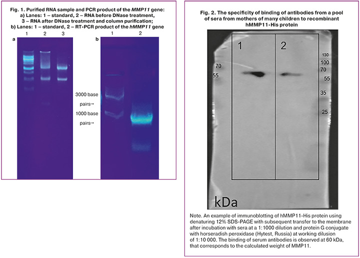
The cells of MCF7 cell line were cultured in RPMI medium (Sigma-Aldrich R8758). RNA was isolated from E. coli cells using the Direct-Zol miniprep RNA kit (R2052, Zymo Research, USA) according to the manufacturer's protocol. The quality and quantity of the purified RNA was verified by electrophoresis on a 1% agarose gel in 1×TAE buffer. The primers Eco‑hM11pro‑F (tcatcgaattcagcagcctcaggcctcc) and Hind‑hM11‑R (tctaagctttcagaggaaagtgttggcaggc), (Lyumiprob, Russia) were used in reverse transcription reaction. TransScript One-Step RT-PCR SuperMix (+dye) (AT411-02, Transgenbiotech) was used for reverse transcription (RT) according to the manufacturer’s recommendations. The expected theoretical PCR product size was 1252 bp.
The obtained RT_PCR product of the hMMP11 gene corresponded to the theoretical size and was purified from the agarose gel after electrophoresis using Zymo gel extraction kit (ZymoResearch, D4002) in accordance with the manufacturer’s protocol.
Preparation of competent cells, obtaining expression construct, fermentation and purification of MMP11 protein
The E. coli XL1 Blue strain (CC004M, Eurogen) was used to obtain gene constructs. Competent cells were prepared using Mix&Go E. coli Transformation Kit (T3001, ZymoResearch, USA). The recombinant protein production included cell lysis and protein extraction, protein purification using immobilized metal affinity chromatography (IMAC) with HisPur Ni-NTA resin, and subsequent purification from low molecular weight impurities using gel filtration chromatography.
Purified protein quality was confirmed by sodium dodecyl sulfate-polyacrylamide gel electrophoresis (SDS-PAGE). Protein concentration was measured using bicinchoninic acid kit (BCA Assay kit, Sigma) according to the manufacturer's recommendations.
Electrophoresis and Western blotting (immunoblotting)
Protein electrophoresis under denaturing conditions was performed on a 12% polyacrylamide SDS-PAGE gel prepared using a standard method. The samples of hMMP11-His protein were prepared at a concentration of 4 μg/well and pre-incubated at 95°C in Laemmli SDS sample buffer containing reducing agents, SDS and DTT.
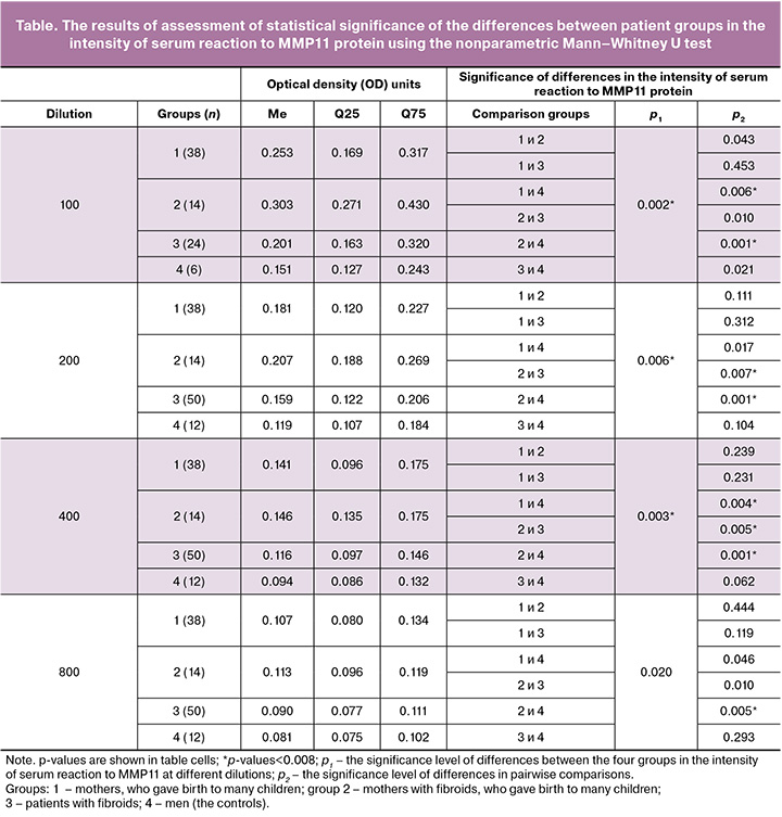
During Western blotting procedure proteins were transferred from the gel to the Immobilon PVDF membrane (Millipore, USA) by semi-dry electroblotting using a Mini-Protean 3 cell system (Bio-Rad, USA), followed by staining with 1% Ponceau S staining solution in 10% acetic acid. Then, the specificity of binding of antibodies from a pool of sera from mothers with many children to the resulting recombinant hMMP11-His protein was tested by incubation with sera at a 1:1000 dilution and protein G conjugate with horseradish peroxidase (Hytest, Russia).
Determination of antibody titers to MMP11 protein by indirect enzyme-linked immunosorbent assay (ELISA)
Purified recombinant human hMMP11-His protein was added to high-quality 96-well Maxibinding immunoplates (SPL Lifesciences, South Korea) at a final concentration of 5 μg/mL in carbonate-bicarbonate buffer, pH 9.5 (0.3 g Na2CO3 and 0.56 g NaHCO3 per 100 ml mQ) at 100 μL per well. The plates were sealed with plate film and incubated for 14–16 hours at temperature +4°C.
Non-specific binding of reagents to the plate surface was blocked with a 0.5% bovine serum albumin (BSA) solution, adding 100 µL of 1% BSA in PBS buffer prepared from tablets (PanEco, Russia) to each well with 100 µl of previously added antigen solution. The well plates were sealed with the plate sealing film and incubated for 1 hour on a shaker at room temperature.
After incubation, the solution was removed from the wells, and each well was washed four times in PBS-T (PBS with 0.05% Tween 20), 300 μL (two times with 150 μL) per well.
Two-fold serial dilutions (1:100, 1:200, 1:400, and 1:800) of the testing sera were performed in PBS-T buffer with 1% BSA on a separate empty plate, and 100 µl of each was transferred to the wells containing the adsorbed antigen. The wells of the last row were filled with 1% BSA in PBS-T as the control for nonspecific binding of the secondary antibodies. The well plates were sealed with the plate sealing film and incubated for 1 hour on a shaker at room temperature. After incubation, the wells were washed four times in PBS-T, 300 μL (two times with 150 μL) per well to remove unbound antibodies.
Protein G, HRP conjugate (Hytest, Russia) was added to all wells of the plates at working dilution of 1:8000 in PBS-T (100 µl per well). The plates were sealed with the plate sealing film and incubated for 45 minutes on a shaker at room temperature. After incubation, to remove unbound conjugate the wells were washed six times in PBS-T with 150 μL per well. The first three washes were done with two volumes of buffer solution.
For detection of the binding of antibodies to the antigen, 100 μL of TMB chromogenic substrate solution was added to the wells, incubated in the dark at room temperature on a shaker for 5 minutes until color changed to bright blue color at least in some wells. At the same time, the control wells without sera were not coloured. The reaction was stopped by adding 50 μL of stop reagent for TMB substrate based on 20% sulfuric acid. Optical density was measured in optical density units at 450 nm using Multiskan FC microplate photometer (ThermoFisher Scientific, USA).
Statistical analysis
Statistica 13.3 (StatSoft Inc., USA) software package was used for statistical data processing. The normality of distribution of quantitative data was assessed using the Shapiro–Wilk test, р≤0.05. The quantitative parameters were described as the median (Me) and interquartile range [Q25; Q75], as well as the arithmetic mean and standard deviation (M±SD). Absolute and relative frequencies (the percentage of the total number of observations) were calculated. The Kruskal–Wallis test and the Mann–Whitney U test were used for comparative analysis. The results were interpreted with the Bonferroni correction for multiple comparisons at р=0.008.
Results
The product of human MMP11 protein was obtained for the first time in Russia (Fig. 1), that made it possible to extract this protein in preparative quantities necessary for further study.
Step 1 in the study was immunoblotting, that confirmed the specificity of membrane binding of antibodies from a pool of sera from mothers with many children to MMP11 (Fig. 2). The result of RT-PCR analysis is shown in Fig. 2, indicating the presence of a clear signal on both lanes with the applied recombinant protein, that proves immunological activity related to this protein in the experimental group.
Step 2 was analysis of immunological activity of blood serum related to MMP11 protein by testing sera samples from 4 patient groups. Group 1 comprised 38 women without diagnosed fibroids, who had a history of five or more successful pregnancies. Group 2 comprised 14 women with small fibroids, who also had five or more successful pregnancies. Group 3 comprised women with fibroids. Group 4 was the control group of men.
According to ELISA data analysis, sera from all patient groups contained antibodies reactive with recombinant MMP11 protein. Mean optical density values (M±SD) showed the expected dependence on serum dilution, decreasing from 1:100 to 1:800. At the same time, significant intergroup differences were found in concentrations of MMP11 antibodies, especially in patients with fibroids in group 3 with minimum (0.092), and extremely high (1.664) concentrations at a 1:100 dilution. Significant difference between the groups in the arithmetic mean (M) and the median (Me) (Table 1), as well as large interquartile range (Q25; Q75) confirmed assymetric data distribution (Fig. 3). This justified the use of non-parametric methods for subsequent comparative analysis of the intensity of the immune response between the groups.
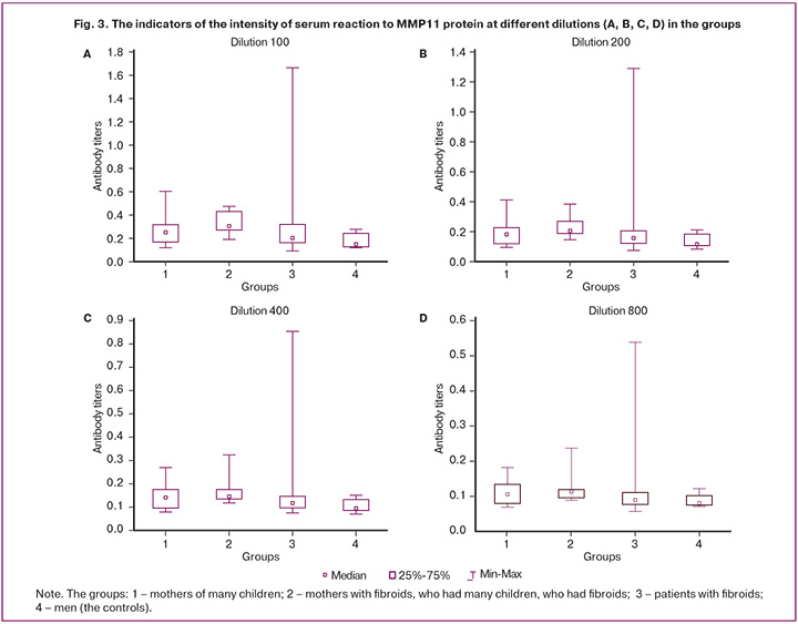
The Kruskal–Wallis test and the Mann–Whitney U test for subsequent pairwise comparisons with the Bonferroni correction were used to assess the significance of differences between the groups at the significance level of p*<0.008). The results are represented in the Table.
Statistical analysis found a clear hierarchy of serum immune reactivity to MMP11. With all tested dilutions (1:100–1:800), sera from mothers with fibroids, who gave birth to many children (group 2) exhibited the highest median of the reaction intensity, which was significantly higher than in the control group of men (p<0.001–0.005) and, with the exception of 1:800 dilution in group 1 of healthy mothers with many children (p=0.002–0.006).
In contrast, the control group 4 was characterized by the lowest median reactivity, that significantly differed not only from group 2, but also from group 1 (at dilutions of 1:100, 1:200, and 1:400), and group 3 (at a 1:100 dilution). It is logical that the level of anti-MMP11 antibodies in men was lower, given the absence of organ-specific (uterine) processes associated with remodeling of the intracellular matrix. This confirmed the relationship between the immune response and reproductive events.
Significant differences were found between group 3 (patients with fibroids) and group 2 with the maximum response at dilutions of 1:100, 1:200, and 1:400 (p=0.010–0.005). However, no differences were found between group 3 and group 1 (healthy mothers with many children) at any of the dilutions (p=0.119–0.453). A wide scatter of indicators in group 3, from very low to extremely high, can reflect different biological activity of tumors (for example, the differences in cell composition, tumor growth rate, MMP11 expression in fibrous stroma), as well as individual immunological characteristics of patients. Similar variability has been described for autoimmune responses in other tumor and proliferative diseases.
It is interesting that with increasing dilution, the number of statistically significant pairwise differences decreased, that indicates the decline in antibody concentration at high dilutions due to decreased antibody concentration at serial dilutions. However, the key differences in the indicators between mothers with fibroids, who gave birth to many children, and the controls (men) remained significant at all stages of titration.
The obtained data confirm the mechanism of humoral innate immune response (not vaccination-induced response) to MMP11 in the general population, that is consistent with the concept of autoantibodies as biomarkers reflecting the processes of physiological and pathological tissue remodeling.
The observed pattern of intergroup differences suggests the clinical and diagnostic significance of our study and confirms that the intensity of sera reaction to MMP11 protein can serve as a differential diagnostic marker for distinguishing the studied patient groups, especially at a 1:100 dilution, showing the maximum differences.
Dose-dependent trends were observed in all groups. Increase in the titer (dilution) from 100 to 800 led to subsequent reduction in the mean values of the indicators. For example, in group 1, the values reduced from 0.272 to 0.111, that demonstrated a 2.5-fold decrease and confirmed a dose-dependent effect.
Thus, the tests conducted by us showed significant, well-detectable titers of MMP11 antibodies in all sera samples. However, the differences found between the groups were that the data in the group of women who gave birth to many children demonstrated the highest titers to MMP11 protein, that significantly differed from the indicators in women with fibroids and the control group of men (Fig. 3). At the same time, the highest intensity of reactivity was in the group of women with many children, who were diagnosed with small fibroids. In patients with fibroids requiring surgical treatment, the intensity of reaction was significantly lower. However, it was higher compared with the controls.
Discussion
The obtained data confirm statistically significant differences in the reactivity of sera between the studied groups. At the same time, the observed dose-dependent dynamics corresponded to the expected immunological patterns.
According to modern concepts, MMP11 protein is not expressed in adult tissues, but is frequently expressed in different types of tumors, often in malignant tumors, as well as in fibroids. Furthermore, high MMP11 protein expression level is observed during embryonic development. Previous hypothesis [24] suggested, that fetal proteins can penetrate the mother's body and trigger immunization. It is probable that the immunogenicity of such proteins is low. However, via repeated boosting during subsequent pregnancies, the production of antibodies suppressing the growth of leiomyomas can be gradually increased. This can explain the findings in our study that serum reactivity was high in mothers with many children. Slightly higher reactivity in the group of mothers with fibroids who gave birth to many children can be explained by the cumulative effect of MMP11 boosting in embryos and growing fibroids. It should be noted that fibroids detected in these women were small and seldom seen by ultrasound. We can suggest that anti-MMP11 antibodies can activate some of immunological mechanisms, preventing rapid growth of fibroids, even if there is a predisposition to uterine fibroids.
The absence of immune tolerance against embryonic antigens and the possibility of creating preventive anti-tumor vaccines based on immunization with such proteins has already been confirmed by experimental data. For example, in 2009, the experiments in a mouse model using MMP11 as the vaccine antigen showed effective protection of animals from the development of chemically induced colon tumors [23]. The attention of researchers has been long focused on metalloproteinases in general and MMP11 in particular as promising agents for the development of new types of therapy. New molecular genetic technologies are being developed to either completely block the MMP11 gene in cells or to identify and block cell populations with increased expression of this gene, that will ultimately lead to the development of new antitumor agents [24, 25].
The role of high expression of the MMP11 gene in uterine fibroids requires further research. It's possible that increased expression of MMP11 in fibroids is promoted by fibroblasts, rather than only by fibroid cells, since they are abundant in fibroids with somatic mutations in the MED12 gene [26], and MMP11 expression is higher [20] in the same way as was found in populations of fibroblasts in cancer tumors that produce MMP11 protein leading to tumor progression [27].
The data obtained in our study show that most of the tested blood sera yield significant, easily detectable titers of anti-MMP11 antibodies. Statistically significant differences between the groups that prevailed in women with many children open up a series of experiments to explore the possibility of using MMP11 protein as the basis for the development of new targeted preventive therapy for uterine fibroids.
Conclusion
Human MMP11 protein was obtained for the first time in preparative quantities sufficient to analyze its reaction to antibodies from the sera of different patient groups. The tests showed that the majority of samples from all groups had significant titers of anti-MMP11 antibodies. Antibody activity varied widely in sera from men and women. Anti-MMP11 antibodies were detected in women, who gave birth to many children. However, the titers were slightly higher in the subgroup of patients with small leiomyomas, whereas the titers were lower in patients with fibroids compared with both groups of women, who gave birth to many children.
Moreover, it is unlikely that MMP11 immunization is associated with the risk of pathologies. The titers of anti-MMP11 antibodies in the general group of women who gave birth to many children were significantly higher than in patients with recurrent fibroids and in the control group of men. MMP11 protein appears to be a potential target for the development of new therapeutic approaches for treatment of uterine fibroids.
The results obtained in our study showed that analysis of MMP11 as a candidate protein for sera antibody responses in different patient groups found high immune response to this protein in the group of mothers with many children. MMP11 protein may further be useful in the development and testing of new targeted preventive therapy for uterine fibroids.
References
- Миома матки. Клинические рекомендации. Акушерство и гинекология. 2025; 8 (Приложение). https://dx.doi.org/10.18565/aig.2025.234 [Uterine fibroids. Clinical guidelines. Obstetrics and Gynecology. 2025; 8 (Suppl.) (in Russian). https://dx.doi.org/10.18565/aig.2025.234].
- Pavone D., Clemenza S., Sorbi F., Fambrini M., Petraglia F. Epidemiology and risk factors of uterine fibroids. Best Pract. Res. Clin. Obstet. Gynaecol. 2018; 46: 3-11. https://dx.doi.org/10.1016/j.bpobgyn.2017.09.004
- Уварова Е.В., Адамян Л.В., Стрижакова М.А. и др. Рецидивирующая миома матки у юной пациентки (клиническое наблюдение). Репродуктивное здоровье детей и подростков. 2005; 1: 53-7. [Uvarova E.V., Adamyan L.V., Strizhakova M.A. et al. Recurrent uterine fibroids in a young patient (clinical observation). Reproductive Health of Children and Adolescents. 2005; 1:53-7 (in Russian)].
- Maekawa R., Sato S., Tamehisa T., Sakai T., Kajimura T., Sueoka K., Sugino N. Different DNA methylome, transcriptome and histological features in uterine fibroids with and without MED12 mutations. Sci. Rep. 2022; 12(1): 8912. https://dx.doi.org/10.21203/rs.3.rs-1041773/v1
- Navarro A., Yin P., Monsivais D., Lin S.M., Du P., Wei J.J. et al. Genome-wide DNA methylation indicates silencing of tumor suppressor genes in uterine leiomyoma. PLoS One. 2012; 7(3): e33284. https://dx.doi.org/10.1371/journal.pone.0033284
- Andreini C., Bertini I., Cavallaro G., Holliday G.L., Thornton J.M. Metal ions in biological catalysis: from enzyme databases to general principles. JBIC J. Biol. Inorg. Chem. 2008; 13(8): 1205-18. https://dx.doi.org/10.1007/s00775-008-0404-5
- Lefebvre O., Régnier C., Chenard M.P., Wendling C., Chambon P., Basset P. et al. Developmental expression of mouse stromelysin-3 mRNA. Development. 1995; 121(4): 947-55. https://dx.doi.org/10.1242/dev.121.4.947
- Okada A., Saez S., Misumi Y., Basset P. Rat stromelysin 3: cDNA cloning from healing skin wound, activation by furin and expression in rat tissues. Gene. 1997; 185(2): 187-93. https://dx.doi.org/10.1016/S0378-1119(96)00615-4
- Wang Z., Brown D.D. Thyroid hormone-induced gene expression program for amphibian tail resorption. J. Biol. Chem. 1993; 268(22): 16270-8.
- Basset P., Bellocq J.P., Wolf C., Stoll I., Hutin P., Limacher J.M. et al. A novel metalloproteinase gene specifically expressed in stromal cells of breast carcinomas. Nature. 1990; 348(6303): 699-704. https://dx.doi.org/10.1038/348699a0
- Chen C., Liu X., Jiang J., Li S., Wang G., Ju L. et al. Matrix metalloproteinase 11 is a potential biomarker in bladder cancer diagnosis and prognosis. Onco Targets Ther. 2020; 13: 9059-69. https://dx.doi.org/10.2147/OTT.S243452
- Panara K., Hui T.L., Keshari D., Tong C.C.L., Palmer J.N., Adappa N.D. et al. Matrix metalloproteinase-11 regulates inverted papilloma epithelial cell migration and invasion. Int. Forum Allergy Rhinol. 2024; 14(11): 1714-23. https://dx.doi.org/10.1002/alr.23405
- Denys H., De Wever O., Nusgens B., Kong Y., Sciot R., Le A.T. et al. Invasion and MMP expression profile in desmoid tumours. Br. J. Cancer. 2004; 90(7): 1443-9. https://dx.doi.org/10.1038/sj.bjc.6601661
- Yoon M.L., Kim S.Y., Chun H., Park J., Seo W.J., Lee J.Y. et al. Diagnostic accuracy of serum biomarkers MMP11 and SPP1 in non-small cell lung cancer: an analysis of sensitivity, specificity, and area under the curve. Transl. Lung Cancer Res. 2025; 14(4): 1197-211. https://dx.doi.org/10.21037/tlcr-2024-1068
- Xu W., Liang T., Fang H., Fu L., Deng D., Tan X. et al. Single-cell RNA Sequencing identifies MMP11+cancer-associated fibroblasts as drivers of angiogenesis and bladder cancer progression. Adv. Sci. (Weinh). 2025; 12(32): e02774. https://dx.doi.org/10.1002/advs.202502774
- von Marschall Z., Riecken E.O., Rosewicz S. Stromelysin 3 is overexpressed in human pancreatic carcinoma and regulated by retinoic acid in pancreatic carcinoma cell lines. Gut. 1998; 43(5): 692-8. https://dx.doi.org/10.1136/gut.43.5.692
- Perret A.G., Duthel R., Fotso M.J., Brunon J., Mosnier J.F. Stromelysin-3 is expressed by aggressive meningiomas. Cancer. 2002; 94(3): 765-72. https://dx.doi.org/10.1002/cncr.10270
- Xu J., Fang Y., Chen K., Li S., Tang S., Ren Y. et al. Single-cell RNA sequencing reveals the tissue architecture in human high-grade serous ovarian cancer. Clin. Cancer Res. 2022; 28(16): 3590-602. https://dx.doi.org/10.1158/1078-0432.CCR-22-0296
- Chu J., Wang D., Guo F., Li D., Li Z., Ding H. et al. Expression and clinical significance of FOXQ1, MMP11, and CST1 in colorectal cancer. Clin. Lab. 2026; 72(2). https://dx.doi.org/10.7754/Clin.Lab.2025.250764
- Акуленко Л.В., Нерсесян Е.А., Кузнецова М.В., Адамян Л.В. Профилирование дифференциальной экспрессии генов в миоматозных узлах и миометрии при первичной и рецидивной миоме матки. Медицинская генетика. 2025; 24(8): 29-31. https://dx.doi.org/10.25557/2073-7998.2025.08.29-31 [Akulenko L.V., Nersesyan E.A., Kuznetsova M.V., Adamyan L.V. Profiling of differential gene expression in leomyoma and myometrium in primary and recurrent uterine fibroids. Medical Genetics. 2025; 24(8): 29-31 (in Russian) https://dx.doi.org/10.25557/2073-7998.2025.08.29-31].
- Кузнецова М.В., Трофимов Д.Ю., Тихончук Е.Ю., Согоян Н.С., Адамян Л.В., Сухих Г.Т. Молекулярные механизмы патогенеза миомы матки: анализ мутаций гена MED12 в российской популяции. Акушерство и гинекология. 2016; 10: 73-9. https://dx.doi.org/10.18565/aig.2016.10.73-9 [Kuznetsova M.V., Trofimov D.Yu., Tikhonchuk E.Yu., Sogoyan N.S., Adamyan L.V., Sukhikh G.T. Molecular mechanisms of uterine fibroids pathogenesis: analysis of MED12 gene mutations in the Russian population. Obstetrics and Gynecology. 2016; (10): 73-9 (in Russian). http://dx.doi.org/10.18565/aig.2016.10.73-9].
- Kuznetsova M.V., Tonoyan N.M., Trubnikova E.V., Zelensky D.V., Svirepova K.A., Adamyan L.V. et al. Novel approaches to possible targeted therapies and prophylaxis of uterine fibroids. Diseases. 2023; 11(4): 156. https://dx.doi.org/10.3390/diseases11040156
- Peruzzi D., Mori F., Conforti A., Lazzaro D., De Rinaldis E., Ciliberto G. et al. MMP11: a novel target antigen for cancer immunotherapy. Clin. Cancer Res. 2009; 15(12): 4104-13.
- Umezawa K., Lin Y. Inhibition of matrix metalloproteinase expression and cellular invasion by NF-κB inhibitors of microbial origin. Biochim. Biophys. Acta Proteins Proteom. 2020; 1868(6): 140412. https://dx.doi.org/10.1016/j.bbapap.2020.140412
- Kou Y.B., Zhang S.Y., Zhao B.L., Ding R., Liu H., Li S. Knockdown of MMP11 inhibits proliferation and invasion of gastric cancer cells. Int. J. Immunopathol. Pharmacol. 2013; 26(2): 361-70. https://dx.doi.org/10.1177/039463201302600209
- Goad J., Rajkovic A. Uterine fibroids at single-cell resolution: unveiling cellular heterogeneity to improve understanding of pathogenesis and guide future therapies. Am. J. Obstet. Gynecol. 2025; 232(4S): S124-34. https://dx.doi.org/10.1016/j.ajog.2024.08.037
- Wang Z., Guo X., Li X., Wang J., Zhang N., Amin B. et al. Cancer-associated fibroblast-derived MMP11 promotes tumor progression in pancreatic cancer. Cancer Sci. 2025; 116(3): 643-55. https://dx.doi.org/10.1111/cas.16418
Received 07.11.2025
Accepted 09.06.2026
About the Authors
Maria V. Kuznetsova, PhD, Senior Researcher at the Laboratory of Molecular Genetic Methods, Institute of Reproductive Genetics, V.I. Kulakov National Medical Research Center for Obstetrics, Gynecology and Perinatology, Ministry of Health of Russia, 4 Oparina str., Moscow, 117997, Russia, mkarja@mail.ru,https://orcid.org/0000-0003-3790-0427
Natalia V. Pozdniakova, PhD, Senior Researcher at the Laboratory of Radionuclide and Radiation Technologies in Experimental Oncology, Blokhin National Research Medical Center, Ministry of Health of Russia, 23 Kashirskoe Shosse, Moscow, 115230, Russia, +7(977)767-99-90, natpo2002@mail.ru, https://orcid.org/0000-0002-5765-3016
Daria V. Bykova, Researcher at the Central Pathoanatomical Laboratory, Research Institute of Human Morphology, 3 Tsyurupy str., Moscow, 117418, Russia,
+7(916)594-89-06, dashasam@mail.ru, https://orcid.org/0000-0001-5639-0835
Viktoria E. Karyagina, PhD, Senior Researcher at the Institute of Translation Medicine, V.I. Kulakov National Medical Research Center for Obstetrics, Gynecology and Perinatology, Ministry of Health of Russia, 4 Oparina str., Moscow, 117997, Russia, vypryazhkina.viktoriya@mail.ru
Narine M. Tonoyan, PhD, Doctor, V.I. Kulakov National Medical Research Center for Obstetrics, Gynecology and Perinatology, Ministry of Health of Russia,
4 Oparina str., Moscow, 117997, Russia, tonnar.13@bk.ru, SPIN: 8547-9399, Scopus Author ID: 57213609878, https://orcid.org/0000-0002-1631-1829
Elena V. Trubnikova, Dr. Bio. Sci., Associate Professor, Chief Researcher at the Research Institute of Genetics, Kursk State University, 33 Radishcheva str., Kursk, 305000, Russia; Leading Researcher, Vavilov Institute of General Genetics of the Russian Academy of Sciences, 3 Gubkina str., GSP-1, Moscow, 119991, Russia, +7(910)311-18-86, tr_e@list.ru, https://orcid.org/0000-0001-5025-9406
Corresponding author: Maria V. Kuznetsova, mkarja@mail.ru
Similar Articles

