
Genetic determinants of sex hormone-binding globulin and myoma nodule volume in patients with uterine myoma
Ponomarenko M.S., Reshetnikov E.A., Churnosova M.M., Churnosov M.I., Ponomarenko I.V.
Objective. To investigate the association between polymorphisms in genes linked to sex hormone-binding globulin (SHBG) levels and myoma nodule volume in patients with uterine myomas.
Materials and methods. The study group comprised 329 patients with uterine myomas. Molecular genetic analysis was performed for eight gene loci associated with SHBG levels according to genome-wide association studies (GWAS): (rs17496332 [A/G] (1хр.) PRMT6, rs780093 [C/T] (2хр.) GCKR, rs3779195 [T/A] (7хр.) BAIAP2L1, rs440837 [A/G] (8хр.) ZBTB10, rs7910927 [G/T] (10хр.) JMJD1C, rs4149056 [T/C] (12хр.) SLCO1B1, rs8023580 [T/C] (15хр.) NR2F2, SHBG rs12150660 [G/T] (17хр.)). Associations between these candidate SHBG-related polymorphic loci and myoma nodule volume were assessed using the gPLINK software (linear regression).
Results. Associations with myoma nodule volume were identified for two of the eight molecular genetic markers analyzed. The minor G alleles of rs440837 [A/G] ZBTB10 (dominant model: β=-0.142, pperm=0.050) and rs17496332 [A/G] PRMT6 (allelic model: β=-0.131, pperm=0.018; additive model: β=-0.135, pperm=0.014; recessive model: β=-0.328, pperm=0.004) were associated with smaller myoma nodule volumes. The rs17496332 [A/G] PRMT6 polymorphism and 14 SNPs in linkage disequilibrium with it are associated with DNA interactions in the PRMT6 gene region involving 59 transcription factors and influence PRMT6 expression in more than 20 organs/tissues implicated in the pathophysiology of uterine myoma. The rs440837 [A/G] ZBTB10 polymorphic locus and five loci in strong linkage with it are located within the ZBTB10/RP11-48B3.3/RP11-48B3.4 gene region and influence DNA interactions with 22 transcription factors.
Conclusion. The G allelic variants of rs17496332 [A/G] PRMT6 and rs440837 [A/G] ZBTB10 are associated with the development of smaller myoma nodules in patients with uterine myoma.
Authors' contributions. Ponomarenko M.S. – conception and design of the study, data synthesis, drafting of the manuscript; Reshetnikov E.A. – drafting of the manuscript; Churnosova M.M. – literature search and analysis; Churnosov M.I. – review, final editing; Ponomarenko I.V. – statistical analysis, editing of the manuscript.
Conflicts of interest. The authors have no conflicts of interest to declare.
Funding. The study was conducted with the support of Russian Science Foundation grant No. 25-25-00034,
https://rscf.ru/project/25-25-00034/.
Ethical Approval. The study was reviewed and approved by the Research Ethics Committee of the Belgorod State National Research University.
Generative Artificial Intelligence. No artificial intelligence tools were used in the preparation of this manuscript.
Patient Consent for Publication. All patients provided informed consent for the publication of their data.
Authors' Data Sharing Statement. The data supporting the findings of this study are available upon request from the corresponding author after approval from the principal investigator.
For citation: Ponomarenko M.S., Reshetnikov E.A., Churnosova M.M., Churnosov M.I., Ponomarenko I.V. Genetic determinants of sex hormone-binding globulin and myoma nodule volume in patients with uterine myoma.
Akusherstvo i Ginekologiya/Obstetrics and Gynecology. 2026; (7): 94-101 (in Russian)
https://dx.doi.org/10.18565/aig.2026.53
Keywords
sex hormone-binding globulin
polymorphism
myoma nodule volume
Uterine myoma is a common benign pelvic neoplasm in women, predominantly composed of myometrial smooth muscle cells and fibrous connective tissue [1, 2]. The prevalence of uterine myoma increases with age, peaking in women older than 40 years [3]. This condition affects women of all ethnic groups but is most frequently reported in Black women [4]. Uterine myoma substantially impairs quality of life, particularly through menstrual disturbances such as heavy, prolonged, or irregular bleeding, chronic pelvic pain, dyspareunia, and related symptoms [5].
Sex hormones play an important role in initiating myoma development [6–10]. The activity of sex hormones in the female body is largely mediated by sex hormone-binding globulin (SHBG) [11], which plays a key role in regulating the bioavailability of sex hormones in target tissues and cells [11]. Only a single study by Wang et al. examined the correlations between the genetic determinants of SHBG and uterine myoma, yielding highly inconsistent results [12]. Using Mendelian randomization, the authors found no association between SHBG and uterine myoma in a meta-analysis of GWAS data from two cohorts, FinnGen and FibroGENE, yet identified such an association (p=0.016) within the FibroGENE cohort alone [12]. No other experimental genetic studies have been published on this topic. This near-complete lack of data on the role of SHBG genetic determinants in myoma formation underscores the need for further research in this area.
This study aimed to investigate the association between polymorphisms in genes related to SHBG levels and myoma nodule volume.
Materials and methods
The study group comprised 329 patients with uterine myomas (continuous sampling). Patients were recruited by physicians from the Gynecology Department of the Perinatal Center at St. Ioasaph Belgorod Regional Clinical Hospital between 2008 and 2013. The inclusion criteria were as follows: 1) a confirmed diagnosis of uterine myoma (by ultrasound examination or following hysterectomy); 2) Russian ethnicity; 3) birthplace and residence in the Central Chernozem region of Russia; 4) absence of close biological relatedness among participants; and 5) voluntary informed consent to participate. The exclusion criteria were as follows: 1) presence of oncological disease; 2) severe somatic or autoimmune disease; 3) non-Russian ethnicity; 4) birthplace or residence outside the Central Chernozem region of Russia; and 5) refusal to participate.
The myoma (uterine) nodule volume was calculated using the formula: V (cm³)= (A + B + C) × 0.4186, where A, B, and C represent the longitudinal, anteroposterior, and transverse radii of the myoma nodule (uterus), respectively (cm), and the coefficient 0.4186 corresponds to π4/3, used to calculate the volume of ellipsoidal structures [13]. The total combined nodule volume was calculated for patients with multiple nodules.
Eight polymorphic gene loci associated with SHBG levels according to GWAS data were selected for analysis [14–17] (coordinates given for GRCh38): rs17496332 [NC_000001.11:g.107003753A>G] PRMT6, rs780093 [NC_000002.12:g.27519736T>C] GCKR, rs3779195 [NC_000007.14:g.98364050T>A] BAIAP2L1, rs440837 [NC_000008.11:g.80549739A>G] ZBTB10, rs7910927 [NC_000010.11:g.63379150T>G] JMJD1C, rs4149056 [NC_000012.12:g.21178615T>C] SLCO1B1, rs8023580 [NC_000015.10:g.96165062T>C] NR2F2, and rs12150660 [NC_000017.11:g.7618597G>T] SHBG. Genotyping of the study loci (DNA samples at concentrations of 10–20 ng/μL) was performed using a CFX-96 thermocycler, applying the TaqMan probe method with genotyping kits developed specifically for this purpose by TestGen LLC [18, 19]. Quality control of the resulting genotype data was performed by assessing the Hardy–Weinberg equilibrium (concordance between observed and expected genotype distributions) [20].
Statistical analysis
Statistical analyses were performed using STATISTICA software. The distribution of the continuous variable (myoma nodule volume) was assessed for normality using the Shapiro–Wilk test [21]. As the distribution of this parameter deviated from normal, it was described using the median (Me) and interquartile range (Q1 and Q3). Associations between the single-nucleotide polymorphisms (SNPs) under study and myoma nodule volume were analyzed using transformed values of this parameter (owing to its non-normal distribution), with adjustment for covariates, including age, body mass index, coexistence of uterine myoma with other hyperplastic conditions (endometrial hyperplasia, endometriosis), and myoma nodule localization. Calculations were performed using gPLINK (linear regression method) under four genetic models: allelic, additive, dominant, and recessive [22]. A permutation test was used to correct for multiple comparisons, with pperm<0.05 considered statistically significant [23].
For the molecular genetic markers rs17496332 [A/G] PRMT6 (chromosome 1) and rs440837 [A/G] ZBTB10 (chromosome 8), which were associated with myoma nodule volume, and for loci in strong linkage disequilibrium with them (threshold r² ≥ 0.80), functional significance was evaluated by in silico analysis [24] using HaploReg (accessed January 09, 2026) [25] and GTEx Portal (accessed 26.09.2026) [26].
Results and discussion
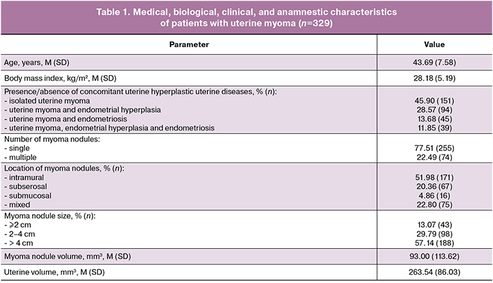
The medical, biological, clinical, and anamnestic characteristics of the study patients with uterine myoma are presented in Table 1. The mean age of patients diagnosed with uterine myoma was 43.69 (7.58) years (range, 24–74 years), and the mean body mass index was 28.18 (5.19) kg/m² (range, 17–49 kg/m²). Among the women included in the analysis, 45.90% had isolated uterine myoma, whereas the remaining 54.10% had concomitant uterine pathology. Most patients (77.51%) had a single myoma nodule. In more than half of the women, the myoma nodules were intramural (51.98%) and larger than 4 cm in diameter (57.14%).
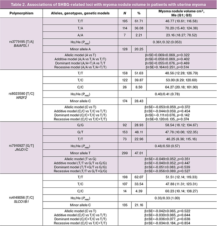
Among patients with uterine myoma, the genotype distributions of all eight analyzed single-nucleotide polymorphisms (SNPs) were in Hardy–Weinberg equilibrium (p>0.05) (Table 2). Associations between two of the eight SHBG candidate gene polymorphic loci and myoma nodule volume were identified (Table 2). The minor G allele of the rs17496332 [A/G] polymorphism in PRMT6 (1p) was associated with a smaller myoma nodule volume under the allelic (β=-0.131, p=0.019, pperm =0.018), additive (β=-0.135, p=0.015, pperm =0.014), and recessive (β=-0.328, p=0.003, pperm =0.004) genetic models (Table 2). In women with uterine myoma carrying the rs17496332 PRMT6 GG genotype, the myoma nodule volume was 20.82 (4.02; 63.48) cm³, which was 2.7–3.1 times lower than in individuals with the AG and AA genotypes (55.79 [16.31; 139.22] and 65.30 [10.13; 138.27] cm³, respectively) (Table 2).
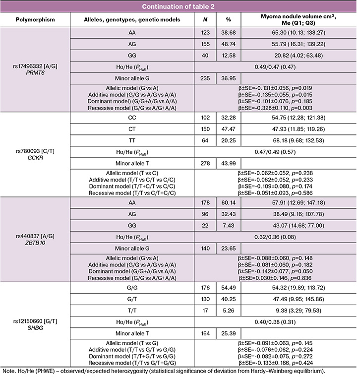
The rs440837 [A/G] polymorphism in ZBTB10 (8p) was also associated with myoma nodule volume under the dominant model (for the G allele: β=-0.142, p=0.050, pperm=0.050) (Table 2). Among women with uterine myoma carrying the rs440837 ZBTB10 GG and AG genotypes, the median myoma nodule volumes were 43.07 (14.68; 77.00) cm³ and 38.49 (9.16; 107.78) cm³, respectively, representing the smallest volumes compared with patients carrying the AA genotype, for whom the corresponding value was 57.91 (12.69; 147.18) cm³ (Table 2).
According to the in-silico analysis performed using HaploReg and GTEx Portal, the rs17496332 [A/G] variant in PRMT6 is functionally important. Polymorphic loci in linkage disequilibrium with this variant (14 SNPs) also demonstrated substantial functional relevance. Specifically, nearly all of these SNPs (93.33%) determine interactions between the genomic region encompassing PRMT6 (29–60 kb upstream of PRMT6) and 59 transcription factors (TFs). Notably, two loci, rs111232683 and rs4914939, among the 13 SNPs in linkage disequilibrium with rs17496332 [A/G] PRMT6, were involved in regulating DNA interactions with the greatest numbers of TFs (21 and 15 TFs, respectively). More than half of the evaluated SNPs (60.00%) influenced PRMT6 expression across more than 20 tissues and organs, including those involved in both SHBG production (liver) and the pathophysiology of uterine myoma (thyroid gland, adrenal glands, brain, adipose tissue, blood, etc.). Importantly, according to our in silico data, the G allele of rs17496332 was associated with reduced PRMT6 expression in virtually all examined tissues and organs: liver (NES=-0.49, p=1.7×10⁻³⁷), thyroid gland (NES=-0.29, p=2.0×10⁻²⁶), adrenal glands (NES=-0.35, p=1.3×10⁻¹¹), brain (hypothalamus; NES=-0.21, p=0.000016), visceral adipose tissue (NES=-0.28, p=2.7×10⁻¹⁴), subcutaneous adipose tissue (NES=-0.32, p=1.3×10⁻¹⁹), and blood (NES=-0.31, p=4.7×10⁻³⁰). Genome-wide association study (GWAS) findings reported by Coviello A.D. et al. demonstrated an association between the rs17496332 [A/G] polymorphism in PRMT6 and circulating SHBG levels: the common A allele was associated with lower SHBG levels (β=-0.028, p=1×10⁻¹¹), whereas the minor G allele was associated with higher SHBG concentrations [14]. Thus, the G allele of rs17496332 is associated with higher SHBG levels (according to the GWAS data of Coviello A.D. et al. [14]) and a smaller myoma nodule volume in patients with uterine myoma (our data: β=-0.135 to -0.328, pperm=0.004–0.018).
The rs440837 [A/G] polymorphism in ZBTB10 and five loci in strong linkage disequilibrium with it are located within the ZBTB10/RP11-48B3.3/RP11-48B3.4 genomic region and influence DNA interactions with 22 transcription factors (TFs). In the liver, the primary site of sex hormone-binding globulin (SHBG) synthesis, the rs440837 [A/G] variant in ZBTB10 and the linked rs7013042 variant in RP11-48B3.4 are located within DNA regions containing putative promoters/enhancers (H3K4me3/H3K4me1) as well as active promoters/enhancers (H3K9ac/H3K27ac). The protein encoded by ZBTB10 is a key regulator of gene transcription through its modulatory effects on the binding of RNA polymerase II to genomic DNA [27], which may be relevant to the regulation of SHBG synthesis in the liver. According to GWAS data, the rs440837 [A/G] polymorphism in ZBTB10 is associated with circulating SHBG levels: the common A allele is associated with lower SHBG concentrations (β=-0.030, p=3×10⁻⁹), whereas the minor G allele is associated with higher SHBG concentrations [14]. Thus, the G allele of rs440837 [A/G] ZBTB10 is associated with both higher SHBG concentrations (GWAS data from Coviello A.D. et al. [14]) and smaller myoma nodule volume in patients with uterine myoma (our data: β=-0.142).
Notably, SNPs in linkage disequilibrium with rs440837 [A/G] ZBTB10 are also associated with SHBG concentrations at the genome-wide significance level: rs72688090 (D'=0.85; r²=0.33) [28], rs388922 (D'=0.96; r²=0.53), rs575452 (D'=0.71; r²=0.28), and rs117921873 (D'=1.00; r²=0.26) [15]. These findings underscore the important role of the genomic region encompassing rs440837 [A/G] ZBTB10 in the regulation of circulating SHBG concentrations and suggest its potential involvement in the pathophysiology of uterine myoma.
One possible mechanism underlying the association between SHBG-associated gene polymorphisms and myoma nodule volume is the role of SHBG in testosterone transport and sequestration [6]. Current evidence indicates that approximately 80% of circulating testosterone is bound to SHBG, whereas only 1% circulates as the biologically active free fraction [6]. Consequently, by directly regulating the balance between SHBG-bound and free testosterone, SHBG is likely to substantially influence the biological effects of testosterone in uterine myoma. The close relationship between SHBG and testosterone is further supported by the strong negative genetic correlation between SHBG and circulating free testosterone, which reaches -0.75 in women [17, 28]. Wong J.Y. et al. demonstrated that higher bioavailable testosterone levels are associated with an increased risk of uterine myoma (OR=1.33) [7]. In addition, several studies have shown that testosterone is converted to estradiol by aromatase within myoma cells, thereby promoting local hyperestrogenism in myoma tissue and stimulating myoma growth [6].
SHBG also plays an important role in estrogen transport. Approximately 38% of circulating estrogens are bound to SHBG, whereas only 2% circulate as the biologically active free fraction [29]. Through activation of estrogen receptors (ERα and ERβ), estrogens promote myoma cell proliferation [12, 30] by increasing the expression of growth factors involved in the pathogenesis of this disease, including insulin-like growth factor 1 (IGF-1), epidermal growth factor (EGF), and platelet-derived growth factor (PDGF) [10].
Taken together, our findings and the available literature suggest that SHBG-increasing allelic variants, which are associated with lower levels of biologically active testosterone and estrogens, are associated with smaller myoma nodule volume in patients with uterine myoma.
Conclusion
The SHBG candidate gene polymorphisms rs17496332 [A/G] in PRMT6 and rs440837 [A/G] in ZBTB10 are associated with myoma nodule volume in patients with uterine myoma. The G alleles of rs17496332 [A/G] PRMT6 and rs440837 [A/G] ZBTB10 are associated with the smallest myoma nodule volume.
References
- Munro M.G., Tchaikovski S.N., Murji A. The epidemiology and pathogenesis of uterine fibroids. Int. J. Gynaecol. Obstet. 2025; 171(3): 1029-45. https://dx.doi.org/10.1002/ijgo.70527
- Пономаренко М.С., Решетников Е.А., Пономаренко И.В., Чурносов М.И. Факторы риска развития миомы матки. Акушерство и гинекология. 2024; 3: 20-7. https://dx.doi.org/10.18565/aig.2023.275. [Ponomarenko M.S., Reshetnikov E.A., Ponomarenko I.V., Churnosov M.I. Risk factors for uterine fibroids. Obstetrics and Gynecology. 2024; (3): 20-7 (in Russian). https://dx.doi.org/10.18565/aig.2023.275].
- Wu C., Zhou L., Chen R., Li H., Li J., Guo F. et al. The epidemiology of uterine fibroids: global disease burden from 1990 to 2021 and future trend predictions. Front. Reprod. Health. 2025; 7: 1629834. https://dx.doi.org/10.3389/frph.2025.1629834
- Mitro S.D., Dyer W., Lee C., Bindra A., Wang L., Ritterman Weintraub M. et al. Uterine fibroid diagnosis by race and ethnicity in an integrated health care system. JAMA Netw. Open. 2025; 8(4): e255235. https://dx.doi.org/10.1001/jamanetworkopen.2025.5235
- Bulun S.E., Yin P., Wei J., Zuberi A., Iizuka T., Suzuki T. et al. Uterine fibroids. Physiol. Rev. 2025; 105(4): 1947-88. https://dx.doi.org/10.1152/physrev.00010.2024
- Lv M., Yu J., Huang Y., Ma J., Xiang J., Wang Y. et al. Androgen signaling in uterine diseases: new insights and new targets. Biomolecules. 2022; 12(11): 1624. https://dx.doi.org/10.3390/biom12111624
- Wong J.Y., Gold E.B., Johnson W.O., Lee J.S. Circulating sex hormones and risk of uterine fibroids: study of women's health across the nation (SWAN). J. Clin. Endocrinol. Metab. 2016; 101(1): 123-30. https://dx.doi.org/10.1210/jc.2015-2935
- Alsudairi H.N., Alrasheed A.T., Dvornyk V. Estrogens and uterine fibroids: an integrated view. Research Results in Biomedicine. 2021; 7(2): 156-63. https://dx.doi.org/10.18413/2658-6533-2021-7-2-0-6
- Пономаренко М.С., Решетников Е.А., Пономаренко И.В., Чурносов М.И. Этиопатогенетические механизмы развития миомы матки. Акушерство и гинекология. 2024; 1: 34-41. https://dx.doi.org/10.18565/aig.2023.241 [Ponomarenko M.S., Reshetnikov E.A., Ponomarenko I.V., Churnosov M.I. Etiopathogenetic mechanisms of uterine fibroids development. Obstetrics and Gynecology. 2024; (1): 34-41 (in Russian). https://dx.doi.org/10.18565/aig.2023.241].
- Пономаренко И.В., Чурносов М.И. Современные представления об этиопатогенезе и факторах риска лейомиомы матки. Акушерство и гинекология. 2018; 8: 27-32. https://dx.doi.org/10.18565/aig.2018.8.27-32 [Ponomarenko I.V., Churnosov M.I. Current views on the etiopathogenesis and risk factors of uterine leiomyoma. Obstetrics and Gynecology. 2018; (8): 27-32 (in Russian). https://dx.doi.org/10.18565/aig.2018.8.27-32].
- Hadad A., França V.L.B., Amaral J.L., Carvalho H.F., Freire V.N. Quantum biochemistry characterization of representative conformations of the sex hormone-binding globulin monomer bound to estradiol, dihydrotestosterone and testosterone. J. Steroid Biochem. Mol. Biol. 2026; 255: 106873. https://dx.doi.org/10.1016/j.jsbmb.2025.106873
- Wang H., Li C., Chen L., Zhang M., Ren T., Zhang S. Causal relationship between female reproductive factors, sex hormones and uterine leiomyoma: a Mendelian randomization study. Reprod. Biomed. Online. 2024; 48(2): 103584. https://dx.doi.org/10.1016/j.rbmo.2023.103584
- Леваков С.А., Буданов П.В., Зайратьянц О.В., Мовтаева Х.Р., Азадова Г.Я., Левакова С.Е. Персонифицированный подход к ведению пациенток с миомой матки. Акушерство и гинекология. 2019; 12: 174-82. https://dx.doi.org/10.18565/aig.2019.12.174-82 [Levakov S.A., Budanov P.V., Zairatyants O.V., Movtaeva Kh.R., Azadova G.Ya., Levakova S.E. A personalized approach to managing patients with uterine fibroids. Obstetrics and Gynecology. 2019; (12): 174-82 (in Russian). https://dx.doi.org/10.18565/aig.2019.12.174-182].
- Coviello A.D., Haring R., Wellons M., Vaidya D., Lehtimäki T., Keildson S. et al. A genome-wide association meta-analysis of circulating sex hormone-binding globulin reveals multiple Loci implicated in sex steroid hormone regulation. PLoS Genet. 2012; 8(7): e1002805. https://dx.doi.org/10.1371/journal.pgen.1002805
- Harrison S., Davies N.M., Howe L.D., Hughes A. Testosterone and socioeconomic position: Mendelian randomization in 306,248 men and women in UK Biobank. Sci. Adv. 2021; 7(31): eabf8257. https://doi.org/10.1126/sciadv.abf8257
- Haas C.B., Hsu L., Lampe J.W., Wernli K.J., Lindström S. Cross-ancestry genome-wide association studies of sex hormone concentrations in pre- and postmenopausal women. Endocrinology. 2022; 163(4): bqac020. https://dx.doi.org/10.1210/endocr/bqac020
- Ruth K.S., Day F.R., Tyrrell J., Thompson D.J., Wood A.R., Mahajan A. et al. Using human genetics to understand the disease impacts of testosterone in men and women. Nat. Med. 2020; 26(2): 252-8. https://dx.doi.org/10.1038/s41591-020-0751-5
- Решетникова Ю.Н. Оценка вклада межлокусных взаимодействий полиморфных локусов генов матриксных металлопротеиназ и генов фолатного цикла материнского организма в формирование веса новорожденного. Научные результаты биомедицинских исследований. 2026; 12(2): 183-201. https://dx.doi.org/10.18413/2658-6533-2026-12-2-0-2 [Reshetnikova Yu.N. Investigation of SNP-SNP interactions of maternal organism in matrix metalloproteinases genes and folate cycle genes and their influence on birth weight. Research Results in Biomedicine. 2026; 12(2): 183-201 (in Russian). https://dx.doi.org/10.18413/2658-6533-2026-12-2-0-2].
- Пономаренко И.В., Полоников А.В., Чурносов М.И. Полиморфные локусы гена LHCGR, ассоциированные с развитием миомы матки. Акушерство и гинекология. 2018; 10: 86-91. https://dx.doi.org/10.18565/aig.2018.10.86-91 [Ponomarenko I.V., Polonikov A.V., Churnosov M.I. Polymorphic LHCGR gene loci associated with the development of uterine fibroids. Obstetrics and Gynecology. 2018; (10): 86-91 (in Russian). https://dx.doi.org/10.18565/aig.2018.10.86-91].
- Пономаренко М.С. Связь генетических детерминант SHBG с гормональным профилем больных миомой матки. Научные результаты биомедицинских исследований. 2025; 11(4): 628-42. https://dx.doi.org/10.18413/2658-6533-2025-11-4-0-3 [Ponomarenko M.S. The relationship between the genetic determinants of SHBG and the hormonal profile of patients with uterine fibroids. Research Results in Biomedicine. 2025; 11(4): 628-42 (in Russian). https://dx.doi.org/10.18413/2658-6533-2025-11-4-0-3].
- Чурносов В.И. Ассоциации полиморфных локусов генов-кандидатов с уровнем половых гормонов у больных гиперплазией эндометрия. Научные результаты биомедицинских исследований. 2025; 11(2): 243-62. https://dx.doi.org/10.18413/2658-6533-2025-11-2-0-3 [Churnosov V.I. Associations of polymorphic loci of candidate genes with the level of sex hormones in patients with endometrial hyperplasia. Research Results in Biomedicine. 2025; 11(2): 243-62 (in Russian). https://dx.doi.org/10.18413/2658-6533-2025-11-2-0-3].
- Абрамова М.Ю. Генетические маркеры тяжелого течения преэклампсии. Научные результаты биомедицинских исследований. 2022; 8(3): 305-16. https://dx.doi.org/10.18413/2658-6533-2022-8-3-0-4 [Abramova M.Yu. Genetic markers of severe preeclampsia. Research Results in Biomedicine. 2022; 8(3): 305-16 (in Russian). https://dx.doi.org/10.18413/2658-6533-2022-8-3-0-4].
- Жабин С.Н., Лазаренко В.А., Азарова Ю.Э., Клёсова Е.Ю., Башкатов Д.А., Кононов С.И., Солодилова М.А., Полоников А.В. Миссенс-вариант rs11556924 в гене ZC3HC1 характеризуется протективным эффектом в отношении риска развития облитерирующего атеросклероза сосудов нижних конечностей у женщин. Научные результаты биомедицинских исследований. 2026; 12(1): 37-51. https://dx.doi.org/10.18413/2658-6533-2026-12-1-0-3 [Zhabin S.N., Lazarenko V.A., Azarova I.E.,Klyosova E.Yu., Bashkatov D.A., Kononov S.I., Solodilova M.A., Polonikov A.V. The missense variant rs11556924 in ZC3HC1 has a protective effect against the risk of peripheral artery disease in women. Research Results in Biomedicine. 2026; 12(1): 37-51 (in Russian). https://dx.doi.org/10.18413/2658-6533-2026-12-1-0-3].
- Полоников А.В., Клёсова Е.Ю., Азарова Ю.Э. Биоинформатические инструменты и интернет-ресурсы для оценки регуляторного потенциала полиморфных локусов, установленных полногеномными ассоциативными исследованиями мультифакториальных заболеваний (обзор). Научные результаты биомедицинских исследований. 2021; 7(1): 15-31. https://dx.doi.org/10.18413/2658-6533-2020-7-1-0-2 [Polonikov A.V.,Klyosova E.Yu., Azarova I.E. Bioinformatic tools and internet resources for functional annotation of polymorphic loci detected by genome wide association studies of multifactorial diseases (review). Research Results in Biomedicine. 2021; 7(1): 15-31 (in Russian). https://dx.doi.org/10.18413/2658-6533-2020-7-1-0-2].
- Ward L.D., Kellis M. HaploReg v4: systematic mining of putative causal variants, cell types, regulators and target genes for human complex traits and disease. Nucleic Acids Res. 2016; 44(D1): 877-81. https://dx.doi.org/10.1093/nar/gkv1340
- GTEx Consortium. The GTEx Consortium atlas of genetic regulatory effects across human tissues. Science. 2020; 369(6509): 1318-30. https://dx.doi.org/10.1126/science.aaz1776
- Giordano C., Accattatis F.M., Gelsomino L., Del Console P., Győrffy B., Giuliano M. et al. miRNAs in the box: potential diagnostic role for extracellular vesicle-packaged miRNA-27a and miRNA-128 in breast cancer. Int. J. Mol. Sci. 2023; 24(21): 15695. https://dx.doi.org/10.3390/ijms242115695
- Sinnott-Armstrong N., Tanigawa Y., Amar D., Mars N., Benner C., Aguirre M. et al. Genetics of 35 blood and urine biomarkers in the UK Biobank. Nat. Genet. 2021; 53(2): 185-94. https://dx.doi.org/10.1038/s41588-020-00757-z
- Balogh A., Karpati E., Schneider A.E., Hetey S., Szilagyi A., Juhasz K. et al. Sex hormone-binding globulin provides a novel entry pathway for estradiol and influences subsequent signaling in lymphocytes via membrane receptor. Sci. Rep. 2019; 9(1): 4. https://dx.doi.org/10.1038/s41598-018-36882-3
- Ponomarenko M.S., Reshetnikov E.A., Churnosova M.M., Reshetnikova Y.N., Churnosov V.I., Ponomarenko I.V. Comorbidity and syntropy of benign proliferative diseases of the female reproductive system: non-genetic, genetic, and epigenetic factors (review). Research Results in Biomedicine. 2023; 9(4): 544-56. https://dx.doi.org/10.18413/2658- 6533-2023-9-4-0-9
Received 10.02.2026
Accepted 26.06.2026
About the Authors
Marina S. Ponomarenko, PhD student at the Department of Biomedical Disciplines, Belgorod State National Research University, 308015, Russia, Belgorod, Pobedy str., 85, +7(4722)30-13-83, ponomarenkomc@yandex.ru, https://orcid.org/0009-0009-0312-0829Evgeny A. Reshetnikov, Dr. Bio. Sci., Professor at the Department of Medical and Biological Disciplines, Belgorod State National Research University, 308015, Russia, Belgorod, Pobedy str., 85, +7(4722)30-13-83, reshetnikov@bsuedu.ru, http://orcid.org/0000-0002-5429-6666
Maria M. Churnosova, student at the Medical Institute, Belgorod State National Research University, 308015, Russia, Belgorod, Pobedy str., 85, +7(4722)30-13-83, churnosovamary@gmail.com, https://orcid.org/0000-0001-6444-8806
Mikhail I. Churnosov, Dr. Med. Sci., Head of the Department of Medical and Biological Disciplines, Belgorod State National Research University, 308015, Russia, Belgorod, Pobedy str., 85, +7(4722)30-13-83, churnosov@bsuedu.ru, https://orcid.org/0000-0003-1254-6134
Irina V. Ponomarenko, Dr. Med. Sci., Professor at the Department of Medical and Biological Disciplines, Belgorod State National Research University,
308015, Russia, Belgorod, Pobedy str., 85, +7(4722)30-13-83, ponomarenko_i@bsuedu.ru, https://orcid.org/0000-0002-5652-0166
Corresponding author: Irina V. Ponomarenko, ponomarenko_i@bsuedu.ru
Similar Articles

