
Dysregulation of synaptogenesis in fetal growth restriction
Kan N.E., Gusar V.A., Zolotareva A.P., Tyutyunnik V.L., Leonova A.A., Chagovets V.V., Alieva L.E.
Objective. To evaluate the expression of synaptic proteins within fetal neuronal vesicles (FNVs) in early- and late-onset fetal growth restriction (FGR) and to determine their associations with neonatal complications.
Materials and methods. One hundred and twenty pregnant women were enrolled in the study. The study group comprised 60 patients with a postnatally confirmed diagnosis of FGR: 30 with early-onset FGR and 30 with late-onset FGR. The control group comprised 60 pregnant women matched by gestational age at delivery. The expression of the synaptic proteins synapsin 1 (SYN1), synaptotagmin 1 (SYT1), synaptophysin (SYP), synaptopodin (SYNPO), and postsynaptic density protein 95 (PSD95) within FNVs was assessed using western blotting.
Results. Early-onset FGR was associated with statistically significant reductions in SYN1 and SYT1 expression and an elevation in SYP expression. Late-onset FGR was associated with reduced SYN1 expression and elevated SYNPO and SYP levels. The most pronounced difference in the proteomic profiles between early- and late-onset FGR was SYP, with its expression significantly higher in late-onset FGR. Analysis of neonatal outcomes revealed that in early-onset FGR, altered synaptic protein expression was associated with central nervous system (CNS) depression syndrome and intraventricular hemorrhage (IVH), whereas in late-onset FGR, it was associated exclusively with IVH. Correlations were identified between synaptic protein expression levels and Doppler indices of uteroplacental and fetoplacental blood flows.
Conclusion. The assessment of synaptic protein expression within FNVs represents a promising approach for the non-invasive prenatal diagnosis of synaptogenesis impairment in FGR. The observed alterations may reflect the disruption and/or dysregulation of synaptic contact formation in the fetus and may serve as potential biomarkers for adverse neurological outcomes.
Authors’ contributions. Kan N.E., Gusar V.A., Zolotareva A.P., Tyutyunnik V.L., Leonova A.A., Chagovets V.V., Alieva L.E. – concept and design of the study, data acquisition, literature review, processing and analysis of material on the topic, drafting of the manuscript, editing of the manuscript.
Conflicts of interest. The authors have no conflicts of interest to declare.
Funding. The study was conducted within the framework of the independent research topic «Epigenetic criteria for diagnosing fetal growth restriction from the perspective of neurogenesis dysfunction» Research Work No. 19-I23 (dated 08.12.22) (Registration number in the Unified State Information System for Accounting of Research, Development, and Technological Works (state registration) – 123060500032-8).
Ethical Approval. The study was reviewed and approved by the Research Ethics Committee of the V.I. Kulakov NMRC for OG&P (Protocol No. 10 dated October 20, 2022).
Use of generative artificial intelligence. None.
Patient Consent for Publication. All patients provided informed consent for the publication of their data.
Authors' Data Sharing Statement. The data supporting the findings of this study are available upon request from the corresponding author after approval from the principal investigator.
For citation: Kan N.E., Gusar V.A., Zolotareva A.P., Tyutyunnik V.L., Leonova A.A.,
Chagovets V.V., Alieva L.E. Dysregulation of synaptogenesis in fetal growth restriction.
Akusherstvo i Ginekologiya/Obstetrics and Gynecology. 2026; (5): 78-88 (in Russian)
https://dx.doi.org/10.18565/aig.2026.132
Keywords
fetal growth restriction
fetal neuronal vesicles
extracellular vesicles
synaptogenesis
synapsin 1
synaptophysin
synaptotagmin 1
synaptopodin
postsynaptic density protein 95
SYN1
SYT1
SYP
SYNPO
PSD95
perinatal brain injury
Fetal growth restriction (FGR) remains one of the most clinically significant challenges in contemporary obstetrics, associated not only with a high burden of adverse perinatal outcomes [1–4] but also with the development of long-term neurological sequelae. [5–8]. Chronic placental insufficiency and sustained fetal hypoxia disrupt normal development of the fetal CNS, with the severity and character of these disturbances dependent on the gestational age at FGR onset [9–11].
In recent years, considerable attention has been directed toward extracellular vesicles of neuronal origin [12, 13], as they enable the non-invasive assessment of fetal brain status in a liquid biopsy format [14, 15]. Our previous work demonstrated that various forms of FGR are associated with altered expression of neurogenesis-related proteins within FNVs [16–18], and that these alterations correlate with early neonatal complications, potentially reflecting the emergence of fetal neurological injury.
The present study extends this line of investigation to the subsequent stage of neuroontogenesis, synaptogenesis [19, 20]. Synaptic formation and maturation are essential for the integration of neurons into functional networks, and disruptions at this stage may underlie subsequent cognitive, motor, and behavioral disorders [19, 21].
This study aimed to evaluate the expression of synaptic proteins within FNVs in early- and late-onset FGR and determine their associations with neonatal complications.
Materials and methods
This study was conducted at the V.I. Kulakov NMRC for OG&P of the Ministry of Health of the Russian Federation and was approved by the Institutional Research Ethics Committee. Written informed consent was obtained from all the participants. A total of 120 pregnant women were enrolled in the study. The study group comprised 60 patients with a postnatally confirmed diagnosis of FGR: 30 with early-onset FGR and 30 with late-onset FGR. The control group consisted of 60 pregnant women matched by gestational age at delivery. Obstetric management and delivery were conducted in accordance with the clinical guidelines on «Insufficient fetal growth requiring maternal care (fetal growth restriction)» (2025), approved by the Ministry of Health of the Russian Federation.
The inclusion criteria were as follows: singleton pregnancy at 22–41 weeks of gestation complicated by FGR (study group) or uncomplicated by FGR (control group); maternal age 18–45 years; and provision of written informed consent. The exclusion criteria were as follows: pre-eclampsia, multiple gestation, severe maternal comorbidities (including autoimmune and oncological conditions), sexually transmitted infections, and fetal chromosomal abnormalities or congenital malformations.
The assessment of synaptic protein expression involved the isolation of the FNV subpopulation by immunoprecipitation, followed by electrophoretic protein separation and detection using western blotting. Densitometric analysis was performed using the Bio-Rad ImageLab 6.0 software.
Neonatal anthropometric parameters, including birth weight and length, were assessed using established criteria, the INTERGROWTH-21st standards, and Fenton growth charts. Neonatal growth restriction was diagnosed according to the consensus-based definition of growth restriction in newborns. All neonates underwent cranial ultrasonography (neurosonography).
Statistical analysis
Statistical analysis was performed using StatTech v. 4.7.0 software (Stattech LLC, Russia). The distribution of continuous variables was assessed using the Shapiro–Wilk test. Normally distributed data are presented as mean (M) and standard deviation (SD), and non-normally distributed data are presented as median (Me) with interquartile range (Q1; Q3). Comparisons of three independent groups on non-normally distributed continuous variables were performed using the Kruskal–Wallis one-way analysis of variance. Categorical variables across the three groups were compared using Pearson's chi-squared test for contingency tables. Where statistically significant differences were identified (p<0.05), post-hoc analysis was conducted. Pairwise comparisons of proportions were performed using Bonferroni correction, applying an adjusted significance threshold of p<0.017. Correlation analysis was performed using Spearman's rank correlation coefficient. Correlation strength was classified as follows: 0–0.3, weak; 0.3–0.7, moderate; >0.7, strong. Statistical significance was set at p<0.05.
Results
The clinical and anamnestic characteristics of the pregnant women are shown in Table 1.
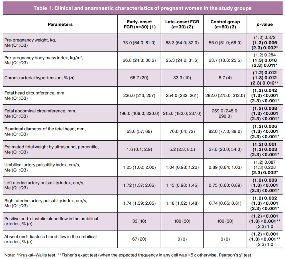
Patients with early-onset FGR had a significantly higher prevalence of chronic arterial hypertension (p=0.012) and higher pre-pregnancy body weight (p=0.006) than those in the control group. Ultrasound and Doppler assessments demonstrated statistically significant between-group differences in fetal abdominal circumference (p<0.001), estimated fetal weight (p=0.003), and uterine artery pulsatility index (p<0.001) measurements. Similar findings were observed for late-onset FGR (Table 2).
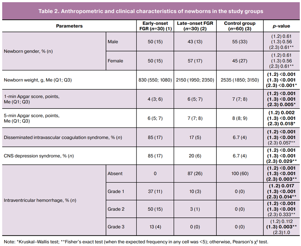
During the early neonatal period, newborns with early FGR demonstrated a significantly higher incidence of disseminated intravascular coagulation (DIC) syndrome (p<0.001), central nervous system (CNS) depression syndrome (p<0.001), and intraventricular hemorrhage (IVH) detected by neurosonography (p<0.001). Notably, 50% of these cases (15/30) had grade 2 IVH. In late-onset FGR, grade 1 IVH was diagnosed in 10% (3/30) of the cases and grade 2 IVH in 3% (1/30).
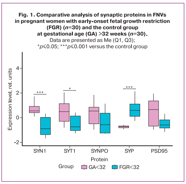
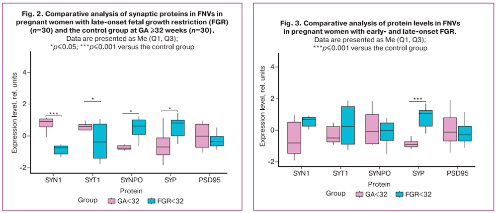
Analysis of the expression of synaptogenesis-related proteins within fetal neuronal vesicles (FNVs) in early-onset FGR demonstrated a statistically significant decrease in SYN1 (-0.89; 0.55; p≤0.001) and SYT1 (-0.74; 0.49; p≤0.04) levels compared with the control group (Fig. 1). SYP levels were also significantly elevated (0.59; -0.72; p≤0.001) in the early-onset FGR group. No significant differences were observed in SYNPO or PSD95 levels. In late-onset FGR, a distinct pattern of protein expression changes was identified (Fig. 2). This group demonstrated a significant reduction in SYN1 expression (-0.74; 0.91; p≤0.001), whereas SYNPO (0.62; -0.77; p≤0.03) and SYP (0.82; -0.70; p≤0.02) levels were significantly increased compared with those in the control group. Changes in SYT1 and PSD95 expression in the late-onset FGR group were not statistically significant.
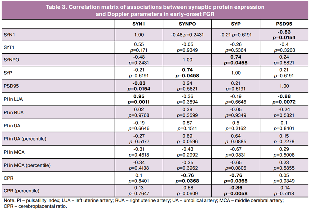
A comparison of the proteomic profiles of early- and late-onset FGR (Fig. 3) revealed the most pronounced differences in SYP expression and distinct involvement patterns of individual synaptic proteins. SYP levels were significantly higher in late-onset FGR, whereas reduced SYN1 expression was common to both forms. While decreased SYT1 expression was predominantly associated with early-onset FGR and increased SYNPO expression was more characteristic of late-onset FGR, these differences did not reach statistical significance.
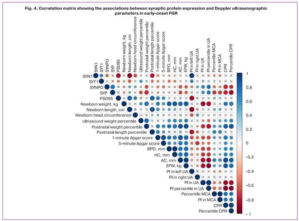
Particular interest was directed toward the relationship between the levels of the investigated proteins and the hemodynamic parameters. Correlation analysis in early-onset FGR demonstrated a strong positive correlation between SYN1 and the pulsatility index in the left uterine artery (r=0.95), and a strong negative correlation between PSD95 and the same parameter (r=-0.88). In addition, SYNPO and SYP levels were negatively correlated with the cerebroplacental ratio (CPR) (r=-0.76) (Fig. 4, Table 3).
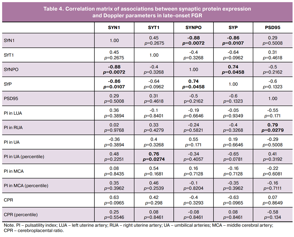
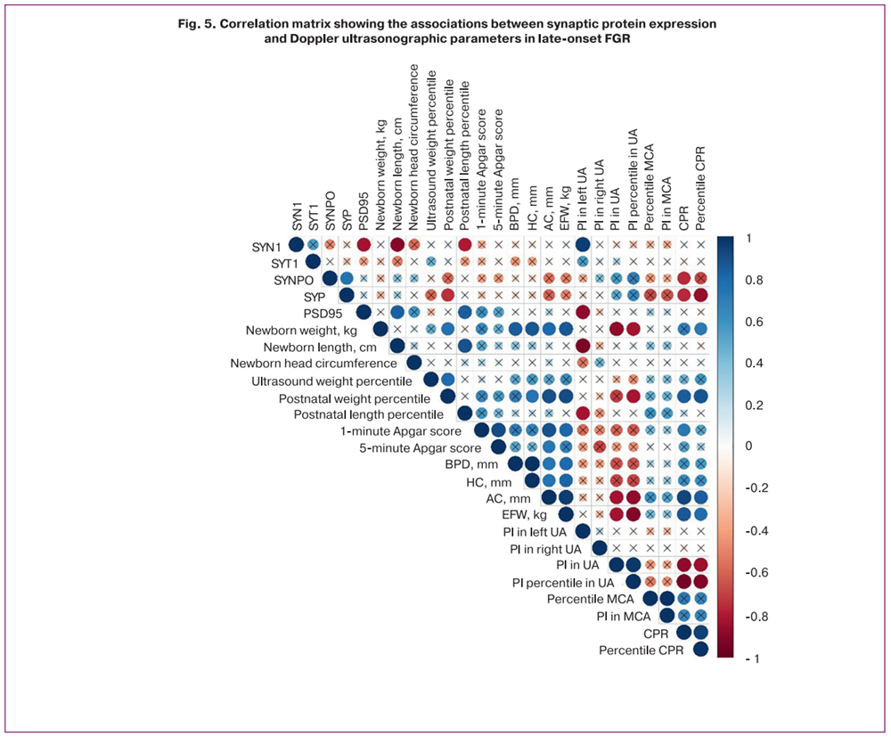
In late-onset FGR, PSD95 levels were negatively correlated with the pulsatility index in the right uterine artery (r=0.79), whereas SYT1 levels were positively correlated with the pulsatility index percentile in the umbilical arteries (r=0.76) (Fig. 5, Table 4).
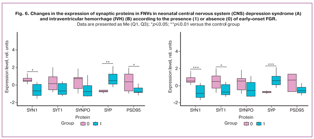
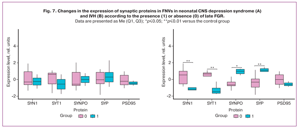
Among the neonatal complications identified in newborns with growth restriction, alterations in synaptic protein expression were associated with CNS depression syndrome and IVH. In early-onset FGR, newborns with CNS depression syndrome demonstrated reduced expression levels of SYN1 (-0.67; 0.52; p≤0.01) and PSD95 (-0.8; 0.37; p≤0.04), along with increased SYP expression (0.5; -0.7; p≤0.005) (Fig. 6A). In cases of IVH, a statistically significant reduction in SYN1 levels was observed (-0.89; 0.55; p≤0.01) (Fig. 6B). In late-onset FGR, statistically significant changes were identified only in association with IVH. This group demonstrated significantly reduced levels of SYN1 (-1.18; 0.54; p≤0.004) and SYT1 (-1.48; 0.66; p≤0.007), accompanied by increased SYNPO (0.98; -0.64; p≤0.02) and SYP (1.09; -0.32; p≤0.004) expressions (Fig. 7B).
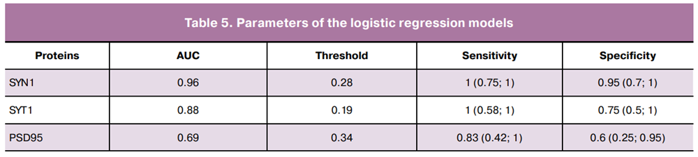
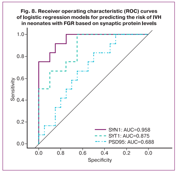
Logistic regression models were developed to assess the probability of IVH development in newborns with growth restriction (Fig. 8), demonstrating high sensitivity and specificity (Table 5).
Discussion
The findings of this study demonstrate that fetal growth restriction (FGR) is associated not only with impaired neurogenesis but also with a protein profile reflecting the dysregulation of synaptogenesis. Synaptogenesis is not completed during the intrauterine period; rather, it is a dynamic process that actively continues after birth, supporting the maturation of neuronal networks [19–21]. Accordingly, the observed changes in the composition of FNVs may reflect not only the acute detrimental effects of chronic placental insufficiency and hypoxia but also the long-term risk of impaired postnatal synaptic network formation [21–23].
Early-onset FGR was associated with decreased SYN1 expression. Given the role of SYN1 in regulating synaptic vesicle transport and neurotransmitter release, altered expression of this protein may indicate reduced presynaptic transmission [24, 25]. The observed increase in SYP expression may represent a compensatory response involving alterations in the composition of secreted FNVs under chronic hypoxia [19, 26, 27].
Notably, there was an association between synaptic protein levels in both early- and late-onset FGR and IVH. In late-onset FGR, alterations affected all assessed synaptic proteins, including SYN1, SYT1, SYNPO, and SYP. These findings may reflect differences in the pathogenesis of FGR phenotypes and the impact of these alterations on the developing fetal brain.
The observed correlations between synaptic protein expression, Doppler parameters, and neonatal complications support their potential clinical significance. Logistic regression and receiver operating characteristic (ROC) curve analyses identified SYN1 and SYT1 as potential biomarkers for predicting IVH in growth-restricted neonates.
Conclusion
These findings highlight the significance of synaptic proteins in FNVs for the noninvasive monitoring of neuroontogenetic processes in fetuses with growth restriction. The use of these biomarkers during pregnancy may complement existing methods of longitudinal fetal surveillance and contribute to risk stratification for neurological complications in newborns.
References
- Lei T.Y., Li D.Z. Perinatal outcome of late-onset fetal growth restriction: etiology matters. Ultrasound Obstet. Gynecol. 2022; 60(5): 707-8. https://dx.doi.org/10.1002/uog.26087
- van de Meent M., Bel E.W., Ganzevoort W., Gordijn S.J., Groenendaal F., Kooi E.M.W. et al.; on behalf of the OPtimal TIming of antenatal COrticosteroids in pregnancies complicated by early‐onset fetal growth REstriction (OPTICORE) study group. Perinatal risk assessment in pregnancies complicated by early-onset fetal growth restriction: development and internal validation of a prediction model for composite adverse perinatal outcome. Ultrasound Obstet. Gynecol. 2025; 66(2): 175-85. https://dx.doi.org/10.1002/uog.29123
- Larsen M.L., Krebs L., Hoei-Hansen C.E., Kumar S. Assessment of fetal growth trajectory identifies infants at high risk of perinatal mortality. Ultrasound Obstet. Gynecol. 2024; 63(6): 764-71. https://dx.doi.org/10.1002/uog.27589
- Министерство здравоохранения Российской Федерации. Клинические рекомендации. Недостаточный рост плода, требующий предоставления медицинской помощи матери (задержка роста плода). М.; 2025. 69 с. [Ministry of Health of the Russian Federation. Clinical guidelives. Insufficient growth of the fetus, requiring the provision of medical care to the mother (fetal growth retardation). Moscow; 2025. 69 p. (in Russian)].
- Adam-Raileanu A., Miron I., Lupu A., Bozomitu L., Sasaran M.O., Russu R. et al. Fetal growth restriction and its metabolism-related long-term outcomes—underlying mechanisms and clinical implications. Nutrients. 2025; 17(3): 555. https://dx.doi.org/10.3390/nu17030555
- Ni W., Gao X., Su X., Cai J., Zhang S., Zheng L. et al. Birth spacing and risk of adverse pregnancy and birth outcomes: A systematic review and dose–response meta‐analysis. Acta Obstet. Gynecol. Scand. 2023; 102(12): 1618-33. https://dx.doi.org/10.1111/aogs.14648
- Sacchi C., O'Muircheartaigh J., Batalle D., Counsell S.J., Simonelli A., Cesano M. et al. Neurodevelopmental outcomes following intrauterine growth restriction and very preterm birth. J. Pediatr. 2021; 238: 135-144.e10. https://dx.doi.org/10.1016/j.jpeds.2021.07.002
- Bahia M.L.R., Velarde G.C., Silva F.C.D., Araujo Júnior E., Sá R.A.M. Adverse perinatal outcomes in fetuses with severe late-onset fetal growth restriction. J. Matern. Fetal Neonatal Med. 2022; 35(25): 8666-72. https://dx.doi.org/10.1080/14767058.2021.1995858
- Юсенко С.Р., Траль Т.Г., Толибова Г.Х., Коган И.Ю. Морфологические особенности плацент при хронической плацентарной недостаточности и задержке роста плода. Вопросы гинекологии, акушерства и перинатологии. 2022; 21(3): 95-101. [Yusenko S.R., Tral T.G., Tolibova G.Kh., Kogan I.Yu. Placental morphology in chronic placental insufficiency and fetal growth restriction. Gynecology, Obstetrics and Perinatology. 2022; 21(3): 95-101. (in Russian)]. https://dx.doi.org/10.20953/1726-1678-2022-3-95-101
- Melamed N., Baschat A., Yinon Y., Athanasiadis A., Mecacci F., Figueras F. et al. FIGO (International Federation of Gynecology and Obstetrics) initiative on fetal growth: best practice advice for screening, diagnosis, and management of fetal growth restriction. Int. J. Gynecol. Obstet. 2021; 152(S1): 3-57. https://dx.doi.org/10.1002/ijgo.13522
- American College of Obstetricians and Gynecologists' Committee on Practice Bulletins—Obstetrics and the Society for Maternal-Fetal Medicine. ACOG Practice Bulletin No. 204: Fetal Growth Restriction. Obstet. Gynecol. 2019; 133(2): e97-e109. https://dx.doi.org/10.1097/AOG.0000000000003070
- Afzal A., Khan M., Gul Z., Asif R., Shahzaman S., Parveen A. et al. Extracellular vesicles: the next frontier in pregnancy research. Reprod. Sci. 2023; 31(5): 1204-14. https://dx.doi.org/10.1007/s43032-023-01434-2
- Goetzl L., Darbinian N., Goetzl E.J. Novel window on early human neurodevelopment via fetal exosomes in maternal blood. Ann. Clin. Transl. Neurol. 2016; 3(5): 381-5. https://dx.doi.org/10.1002/acn3.296
- Salomon C., Nuzhat Z., Dixon C.L., Menon R. Placental exosomes during gestation: liquid biopsies carrying signals for the regulation of human parturition. Curr. Pharm. Des. 2018; 24(9): 974-82. https://dx.doi.org/10.2174/1381612824666180125104428
- Miranda J., Paules C., Nair S., Lai A., Palma C., Scholz-Romero K. et al. Placental exosomes profile in maternal and fetal circulation in intrauterine growth restriction – Liquid biopsies to monitoring fetal growth. Placenta. 2018; 64: 34-43. https://dx.doi.org/10.1016/j.placenta.2018.02.006
- Gusar V., Kan N., Leonova A., Chagovets V., Tyutyunnik V., Khachatryan Z. et al. Non-invasive assessment of neurogenesis dysfunction in fetuses with early-onset growth restriction using fetal neuronal exosomes isolating from maternal blood: A pilot study. Int. J. Mol. Sci. 2025; 26(4): 1497. https://dx.doi.org/110.3390/ijms26041497
- Кан Н.Е., Леонова А.А., Гусар В.А., Чаговец В.В., Тютюнник В.Л., Волочаева М.В., Солдатова Е.Е., Рыжова К.О., Серебрякова А.П. Критерии оценки дисфункции нейрогенеза плода с ранней задержкой роста с использованием внеклеточных везикул. Акушерство и гинекология. 2025; 3: 56-64. [Kan N.E., Leonova A.A., Gusar V.A., Chagovets V.V., Tyutyunnik V.L., Volochaeva M.V., Soldatova E.E., Ryzhova K.O., Serebriakova A.P. Criteria for assessing fetal neurogenesis dysfunction in early-onset growth restriction using extracellular vesicles. Obstetrics and Gynecology. 2025; (3): 56-64 (in Russian)]. https://dx.doi.org/10.18565/aig.2025.30
- Gusar V., Kan N., Leonova A., Chagovets V., Tyutyunnik V., Zolotareva A. et al. Fetal neuronal vesicles in the assessment of perinatal brain dysfunction and late-onset growth restriction: A pilot study. Int. J. Mol. Sci. 2026; 27(2): 679. https://dx.doi.org/10.3390/ijms27020679
- Sarnat H.B. Sequences of synaptogenesis in the human fetal and neonatal brain by synaptophysin immunocytochemistry. Front. Cell. Neurosci. 2023; 17: 1105183. https://dx.doi.org/10.3389/fncel.2023.1105183
- Qi C., Luo L.D., Feng I., Ma S. Molecular mechanisms of synaptogenesis. Front. Synaptic Neurosci. 2022; 14: 939409. https://dx.doi.org/10.3389/fnsyn.2022.939793.
- Fornasiero E.F., Bonanomi D., Benfenati F., Valtorta F. The role of synapsins in neuronal development. Cell. Mol. Life Sci. 2010; 67(9):1383-96. https://dx.doi.org/10.1007/s00018-009-0227-8
- Cesca F., Baldelli P., Valtorta F., Benfenati F. The synapsins: Key actors of synapse function and plasticity. Prog. Neurobiol. 2010; 91(4): 313-48. https://dx.doi.org/10.1016/j.pneurobio.2010.04.006
- Jovanovic J.N., Czernik A.J., Fienberg A.A., Greengard P., Sihra T.S. Synapsins as mediators of BDNF-enhanced neurotransmitter release. Nat. Neurosci. 2000; 3(4): 323-9. https://dx.doi.org/10.1038/73888
- Corradi A., Zanardi A., Giacomini C., Onofri F., Valtorta F., Zoli M. et al. Synapsin-I- and synapsin-II-null mice display an increased age-dependent cognitive impairment. J. Cell Sci. 2008; 121 (18): 3042-3051. https://dx.doi.org/10.1242/jcs.030692
- Tang L.T., Craig T.J., Henley J.M. SUMOylation of synapsin Ia maintains synaptic vesicle availability and is reduced in an autism mutation. Nat. Commun. 2015; 6(1): 7728. https://dx.doi.org/10.1038/ncomms8728
- Cousin M.A. Synaptophysin‐dependent synaptobrevin‐2 trafficking at the presynapse‐Mechanism and function. J. Neurochem. 2021; 159(1): 78-89. https://dx.doi.org/10.1111/jnc.15499
- White D.N., Stowell M.H.B. Room for two: the synaptophysin/synaptobrevin Complex. Front. Synaptic Neurosci. 2021; 13: 740371. https://dx.doi.org/10.3389/fnsyn.2021.740371
Received 22.04.2026
Accepted 08.05.2026
About the Authors
Natalia E. Kan, Professor, Dr. Med. Sci., Honored Scientist of the Russian Federation, Deputy Director of Science, Academician V.I. Kulakov National Medical Research Center for Obstetrics, Gynecology and Perinatology, Ministry of Health of Russia, 117997, Russia, Moscow, Ac. Oparina str., 4, kan-med@mail.ru,Researcher ID: B-2370-2015, SPIN-code: 5378-8437, Authors ID: 624900, Scopus Author ID: 57008835600, https://orcid.org/0000-0001-5087-5946
Vladislava A. Gusar, PhD, Senior Researcher at the Laboratory of Transcriptomic, Department of Systems Biology in Reproduction, Academician V.I. Kulakov National Medical Research Center for Obstetrics, Gynecology and Perinatology, Ministry of Health of Russia, 117997, Russia, Moscow, Ac. Oparina str., 4, v_gusar@oparina4.ru,
https://orcid.org/0000-0003-3990-6224
Anna P. Zolotareva, Applicant, Academician V.I. Kulakov National Medical Research Center for Obstetrics, Gynecology and Perinatology, Ministry of Health of Russia,
117997, Russia, Moscow, Ac. Oparina str., 4; obstetrician-gynecologist at the day hospital department for examination of pregnant women, Primorsky Regional Perinatal Center, 690042, Russia, Vladivostok, Mozhayskaya str., 1B, serebriakovanna@gmail.com, https://orcid.org/0000-0001-7014-2627
Victor L. Tyutyunnik, Professor, Dr. Med. Sci., Honored Doctor of the Russian Federation, Leading Researcher at the Center for Scientific and Clinical Research, Academician V.I. Kulakov National Medical Research Center for Obstetrics, Gynecology and Perinatology, Ministry of Health of Russia, 117997, Russia, Moscow, Ac. Oparina str. 4, tioutiounnik@mail.ru, Researcher ID: B-2364-2015, SPIN-code: 1963-1359, Authors ID: 213217, Scopus Author ID: 56190621500, https://orcid.org/0000-0002-5830-5099
Anastasia A. Leonova, PhD, obstetrician-gynecologist at the Obstetric Department, Academician V.I. Kulakov National Medical Research Center for Obstetrics, Gynecology and Perinatology, Ministry of Health of Russia, 117997, Russia, Moscow, Ac. Oparina str. 4, +7(937)453-54-27, nastena27-03@mail.ru, https://orcid.org/0000-0001-6707-3464
Vitaliy V. Chagovets, PhD, Senior Researcher at the Laboratory of Transcriptomic, Department of Systems Biology in Reproduction, Academician V.I. Kulakov National Medical Research Center for Obstetrics, Gynecology and Perinatology, Ministry of Health of Russia, 117997, Russia, Moscow, Ac. Oparina str., 4, vvchagovets@gmail.com,
https://orcid.org/0000-0002-5120-376X
Leyla E. Alieva, PhD student, Academician V.I. Kulakov National Medical Research Center for Obstetrics, Gynecology and Perinatology, Ministry of Health of Russia,
117997, Russia, Moscow, Ac. Oparina str., 4, leylaalieva00@mail.ru, https://orcid.org/0009-0008-3417-6721
Corresponding author: Anastasia A. Leonova, nastena27-03@mail.ru
Similar Articles

