Среди инфекций, передаваемых половым путем (ИППП), особое значение имеет папилломавирусная инфекция (ПВИ) урогенитального тракта, официальная регистрация которой в виде манифестных проявлений – остроконечных кондилом (ОК) – в соответствии с приказом Минздрава РФ № 286 от 07.12.1993 г. начата с 1993 г. [1]. В РФ в 2014 году аногенитальные бородавки были выявлены с частотой 21,8 на 100 тыс. населения, а в Москве 33,6 на 100 тыс [2]. За период 2004–2014 гг. в общей структуре ИППП увеличилась заболеваемость аногенитальными бородавками – с 6 до 11% [3]. Систематизированный анализ показал, что общая годовая заболеваемость ОК мужчин и женщин (включая новые случаи и рецидивирующие) варьирует от 160 до 289 случаев на 100 000, со средним значением 194,5 на 100 000 населения. По оценкам, средний ежегодный уровень заболеваемости новыми ОК составил 137 случаев на 100 000 среди мужчин и 120,5 на 100 000 среди женщин [4, 5].
Клинико-визуальный метод является ведущим в диагностике проявлений ПВИ гениталий на коже и слизистой. Выраженные клинические проявления ОК видны невооруженным глазом при гинекологическом осмотре. ОК представляют собой разрастания соединительной ткани с сосудами, покрытые плоским эпителием с морфологическими признаками ПВИ. Они выступают над поверхностью кожи и слизистой, могут иметь ножку или широкое основание.
Кольпоскопия/вульвоскопия (КС/ВС) представляет собой высокоинформативный, широкодоступный и относительно недорогой метод диагностики заболеваний вульвы, влагалища и шейки матки, существенно повышающий эффективность обследования женщин. КС/ВС в идеале должен владеть каждый гинеколог на амбулаторном приеме. КС/ВС рассматривается как наиболее чувствительный метод определения субклинической формы ПВИ [6]. Возможности КС/ВС ограничены ложно-позитивными (от 4 до 33%) и ложно-негативными (от 15 до 62%) результатами [7].
Гистологический метод мог бы служить «золотым стандартом» в диагностике субклинических и клинических проявлений ПВИ, однако его использование ограничивают технические сложности и инвазивность. Материал для исследования получают путем биопсии ткани ОК, которую можно выполнить с помощью электро- или радиоволновой петли, либо путем хирургического иссечения [8]. На сегодняшний день одним из новых перспективных направлений в неинвазивной диагностике ОК является применение высокочастотного ультразвукового исследования (ВЧ УЗИ) кожи и слизистых оболочек наружных половых органов. Впервые УЗИ как метод диагностики было предложено в начале 50-х годов прошлого века [9]. Наиболее часто применение ультразвука в диагностике связано с возможностью получения изображения внутренних органов и их структур. Основой метода является взаимодействие ультразвука с тканями человека, обладающими различными акустическими характеристиками. Изображение получают в результате излучения ультразвуковых импульсов, направленных в исследуемые ткани и обработки отраженных сигналов [10].
УЗИ кожи долгое время было недоступным в связи с недостаточной разрешающей способностью изображений, полученных при помощи датчиков, работающих в диапазоне 3–10 МГц. Использование ультразвука для изучения кожи потребовало разработки новых подходов и технических решений для оценки таких тонких структур как отдельные слои кожи [11].
Впервые УЗИ кожи было проведено в 1979 г. С этого времени постоянно происходило усовершенствование оборудования и специальных датчиков, позволяющих визуализировать слои кожи [12–14].
Новые технологии были разработаны в результате масштабных совместных исследований, проведенных университетами Гамбурга, Бохума и Берлина. В начале 90-х годов ХХ века немецкая компания Taberna Pro Medium начала серийное производство аппаратуры для сканирования кожи. Были получены пьезокристаллы, позволяющие генерировать ультразвуковые колебания гораздо более высокой частоты, чем в традиционных ультразвуковых сканерах – это частоты свыше 20 МГц (22, 30, 50, 75 и 100 МГц). Такие характеристики пьезокристаллов предоставили возможность получать детальное изображение структур кожи. Ультразвуковые датчики-аппликаторы значительно отличаются от традиционных датчиков. За счет использования открытой системы исчезают дополнительные препятствия на пути ультразвукового потока, что обеспечивает получение информации с изучаемой области без потерь. Ультразвуковое сканирование кожи дает возможность отображать биологические структуры с разрешением от 90 до 18 микрометров и глубиной проникновения луча от 10 до 0,5 мм [11].
 Принцип работы ультразвуковго сканера Digital Ultraschall Bildsystem (DUB) следующий: генератор производит короткий электрический импульс, который при помощи пьезокристалла преобразуется в ультразвуковй сигнал с частотой от 22 до 100 МГц в зависимости от используемых датчиков. Эти сигналы передаются в кожу и там из-за неоднородности тканей (клеточные структуры, межклеточное пространство и др.) происходит их отражение, поглощение, рассеивание и др. Отраженные эхосигналы принимаются тем же самым преобразователем и трансформируются вновь в электрические импульсы. Принятые сигналы обрабатываются, сохраняются компьютером, производится их цифровая обработка и затем происходит построение изображения, которое и демонстрируется на мониторе. Данный метод позволяет проводить исследование кожи в разные интервалы времени, документально фиксируя все особенности, а также получать количественные параметры: толщину эпидермиса и дермы, размеры сканируемых образований в изучаемой зоне, акустическую плотность эпидермиса, дермы и подкожной клетчатки [11, 15].
При УЗИ эпидермис выглядит как гиперэхогенная однородная структура в виде четкой полосы. Дерма неоднородна по эхогенности [16–18].
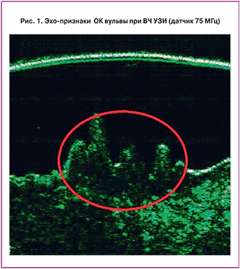
При диагностике ОК ВЧ УЗИ позволяет визуализировать изменения внутренней структуры кожи и слизистых оболочек в виде однородных гипоэхогенных или анэхогенных образований с деформированным наружным контуром в виде пирамид, волн или гребней, четко отграниченных от подлежащих тканей, и снижение ультразвуковой плотности подлежащих тканей, что в совокупности позволяет выявлять субклинические очаги ОК, невидимые невооруженным глазом, а на видимых очагах – определять глубину поражения, проводить более точную деструкцию и, таким образом, снижать число рецидивов [19].
Материал и методы исследования
Для оценки информативности этого метода были обследованы 98 пациенток, обратившихся в ФГБУ НЦАГиП им. В.И. Кулакова Минздрава России в период с 01.10.2013 по 30.08.2014 гг. Женщины разделены на две группы: I – пациентки без видимых проявлений ОК (n=69); II – пациентки с ОК (n=29). Диагноз ОК уставлен на основании клинико-визуальной оценки, ВС с проведением пробы с уксусной кислотой, морфологического исследования биоптата ткани ОК, взятого у 26 пациенток. Всем пациенткам было проведено комплексное обследование, включающее сбор клинико-анамнестических данных в виде специально разработанной нами анкеты-опросника для выявления факторов риска заболеваемости ОК, гинекологический осмотр, ВЧ УЗИ кожи и слизистых оболочек наружных половых органов, расширенная КС. ВЧ УЗИ кожи проводилось на аппарате «DUB – digital Ultraschall Bildsystem – tpm» и программного обеспечения DUB-SkinScan. Оценка структуры кожи осуществлялась контактным ультразвуковым способом. В исследовании использовалась сканирующая головка прибора, генерирующая ультразвуковую волну с частотой 75 МГц. Эта волна проникает в ткани на глубину до 8 мм, что позволяет получить информацию о состоянии эпидермиса и дермы. Датчик располагается перпендикулярно исследуемому участку кожи или слизистой, в процессе исследования используется закрытая водная система и гель для УЗИ.
 Пациенткам II группы дополнительно проведены фоторегистрация ОК, ВС после пробы с уксусной кислотой, 26 пациенткам из II группы проведено морфологическое исследование ткани биоптата ОК. Биопсия ткани ОК проводилась при размерах ОК, позволяющих получить достаточный для гистологического исследования материал. Возраст обследуемых пациенток не различался между исследуемыми группами. В I группе средний возраст был 29,77±7,64 года, во II группе – 26,51±5,97 года. Представленные данные демонстрируют, что в исследуемых группах превалировали женщины репродуктивного возраста, которые имели регулярный менструальный цикл, средний возраст коитархе составил 18±1,5 года. У 10 (10,2%) пациенток был 1 половой партнер за всю жизнь, у 23 (23,44%) – более 2 партнеров за всю жизнь, у 65 (66,32%) – более 5. По результатам цитологического исследования смешанных соскобов из шейки матки с описанием по терминологической системе Бетесда (The Bethesda System – TBS) у 3 (3,06%) пациенток выявлен LSIL (низкая степень плоскоклеточного интраэпителиального поражения), у 8 (8,16%) ASCUS (клетки плоского эпителия с атипией неясного значения), у 87 (88,77%) – NILM (норма, доброкачественные изменения, реактивные изменения). Барьерную контрацепцию применяли 65,5% женщин. Индекс массы тела исследуемых пациенток в среднем составил 22,2±2,8.
Результаты исследования
Всем пациенткам проведена расширенная КС/ВС с высоким разрешением. При оценке результатов учитывались для шейки матки кольпоскопические критерии, такие как наличие ацето-белого эпителия (АБЭ), степень его выраженности, наличие мозаики/пунктации, наличие ОК, степень окрашивания раствором Люголя и наличие йод-негативных участков. Для вульвы – наличие характерной для ОК картины «пальцеобразных выростов» с реакцией последних на пробу с 3% уксусной кислотой. При проведении расширенной КС (n=98) адекватная кольпоскопическая картина наблюдалась у 90 пациенток (91,8%), неадекватная КС – у 8 (8,1%). По полученным результатам было отмечено, что нормальная кольпоскопическая картина наблюдалась у 71 (72,44%) пациенток, аномальная – у 27 (27,55%). Варианты аномальной кольпоскопической картины включали в себя слабовыраженные и выраженные изменения, наиболее важным из которых является наличие АБЭ. Слабовыраженные изменения эпителия шейки матки выявились у 25 (25,51%), выраженные изменения – у 2 (2,04%). Был проведен подробный анализ кольпоскопических картин среди 2 групп пациенток.
У пациенток из II группы при ВС обнаружена характерная картина: белесоватые эпителиальные образования неправильной формы с пальцеобразными выростами. Наиболее важным диагностическим критерием служит наличие правильной капиллярной сети в выростах, которая выявляется после обработки места поражения 3% раствором уксусной кислоты. Сосуды ОК после обработки раствором уксуса обычно сокращаются равномерно, становятся менее видимыми, что придает участку с ОК характерную белесоватость. Полученные данные мы систематизировали и оценили в сравнительном аспекте с результатами ВЧ УЗИ и морфологического исследования.
При обследовании пациенток с ОК при ВЧ УЗИ в этой области нами были выявлены характерные для данной патологии признаки: однородные гипо- или анэхогенных образования с деформированным наружным контуром в виде пирамид, волн или гребней, четко отграниченные от подлежащих тканей, снижение ультразвуковой плотности подлежащих тканей. Ультразвуковая плотность измеряется в условных единицах, при этом нет определенных референсных значений данного эхо-признака, он может варьировать в широких пределах в зависимости от эластичности тканей исследуемой пациентки. Снижение или повышение ультразвуковой плотности выявляется при сравнении с плотностью прилежащих неизмененных участков кожи или слизистой. Совокупность этих признаков позволяет диагностировать ОК. (Рис.1)
 Ниже приведены клинические примеры с описанием данных при выявлении ВЧ УЗИ ОК у обследуемых пациенток.
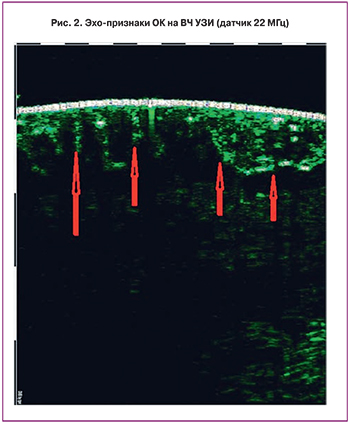
Пример № 1. Пациентка В., 23 года, с множественными ОК, расположенными периуретрально и в области преддверия влагалища. При ВЧ УЗИ на большей площади поля зрения в области преддверия влагалища и вокруг уретры визуализируется деформация и резкое утолщение эпителиальной ткани, деформация наружного контура эпителия в виде пирамидальных волн, максимальная толщина (высота) до 3000 мкм. Отмечается выраженное рассеивание сигнала при использовании датчика 75 МГц, строма не визуализируется. При использовании датчика 22 МГц визуализируется четкая граница измененной эпителиальной ткани и подлежащей стромы, однако из-за выраженного рассеивания сигнала вся толща неизмененной стромы не просматривается (рис. 2). Проведена хирургическая деструкция наиболее крупных ОК с последующим гистологическим исследованием, в ходе которого в препарате выявлены морфологические признаки ОК: койлоцитоз, гиперкератоз.
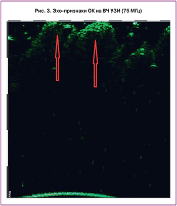
Пример № 2. Обследована пациентка Б., 24 года, с единичными ОК, расположенными на слизистой вульвы: на правой малой половой губе и в области задней спайки малых половых губ. Беспокоят жалобы на зуд, боль в области наружных половых органов. При ВЧ УЗИ на слизистой вульвы визуализируется: множество однородных гипо- и анэхогенных образований, расположенных в пределах эпителия вульвы; наружный контур образований деформирован, выбухает в виде волн и гребней; латеральные границы образования в пределах эпителия не визуализируются. В проекции исследования поперечный размер отдельных образований измерить не удается из-за большого размера последних. Образования четко отграничены от стромы, а также несколько деформируют ее кнутри. Максимальная толщина образований в области исследований достигает 1450 мкм, их внутренняя эхо-структура не просматривается, ультразвуковая плотность составила 2 ед., проведение ультразвукового сигнала в подлежащие ткани было снижено. Строма под образованиями визуализируется менее интенсивно, чем окружающие ткани, толщина стромы под образованиями 1450 мкм, ультразвуковая плотность 4 ед (рис. 3). Проведена хирургическая деструкция наиболее крупных ОК с последующим гистологическим исследованием, в ходе которого в препарате выявлен морфологический признак ОК: койлоцитоз.
Пример № 3. Обследована пациентка Г., 23 года, с ОК, расположенными на коже и слизистой наружных половых органов, с жалобами на психологический дискофорт при половом контакте. В области исследования (преддверие влагалища, периуретральная область) визуализируются однородные гипоэхогенные и анэхогенные образования, расположенные в пределах слизистой; наружный контур образований деформирован, выбухает в виде волн и «гребней»; латеральные границы образования в слизистой не визуализируются. Образования четко отграничены от стромы, несколько деформируют ее кнутри; максимальная толщина в области образования достигает 2500 мкм; внутренняя эхоструктура не просматривается, ультразвуковая плотность составляет 3 ед., проведение ультразвукового сигнала в подлежащие ткани значительно снижено. Структура стромы под образованиями визуализируется не на всю глубину. Проведена ВС, в ходе которой после пробы с уксусной кислотой выявлены кольпоскопические признаки ОК: побеление образований. Далее проведена хирургическая деструкция наиболее крупных ОК с последующим гистологическим исследованием, морфологически подтверждены ОК: койлоцитоз, дискератоз.
Обсуждение
При проведении нашего исследования у 7 (10,14%) пациенток из первой группы (пациентки без видимых признаков ОК) обнаружились эхо-признаки ОК, а у 5 из них диагноз подтвердился при ВС после проведения теста с уксусной кислотой. У этих женщин на слизистой малых половых губ были единичные субклинические очаги ОК, не выявленные при гинекологическом осмотре. У 2 женщин с эхо-признаками ОК диагноз не подтвердился. Напротив, у 3 (10,34%) женщин из второй группы (пациентки с видимыми признаками ОК) эхо-признаков ОК выявлено не было. Оказалось, что у этих пациенток множественные мелкие обрывки девственной плевы были ошибочно расценены как ОК. Таким образом, диагностическая чувствительность метода ВЧ УЗИ составила 94%. Ложноположительный результат отмечен в 6% случаев. Не было отмечено ни одного случая ложноотрицательного результата. Специфичность метода составила 100%. Из 31 пациентки с обнаруженными эхо-признаками ОК у 26 (83,87%) диагноз был верифицирован гистологически. Основными цито- и морфологическими признаками вируса папилломы человека (ВПЧ) являются койлоцитоз и дискератоз, а также трансэпителиальная лимфоцитарная инфильтрация и базально-клеточная гиперплазия и отек дермы с расширением капилляров. В ряде случаев отмечаются только второстепенные признаки ВПЧ: паракератоз и гиперкератоз, которые могут встречаться при других патологических процессах. Койлоцитоз является следствием цитопатогенного эффекта ВПЧ на кератиноциты, особенно на поверхностные слои (шиповатый и зернистый). Койлоциты – оксифилитноокрашенные клетки эпителия с четкими границами, ясно выраженной перинуклеарной зоной просветления и многочисленными вакуолями в цитоплазме [20, 21].
Таким образом, в нашем исследовании удалось обозначить высокий диагностический потенциал метода ВЧ УЗИ, сопоставимый с «золотым стандартом» в диагностике ОК – морфологическим исследованием. Диагностическими критериями ОК при ВЧ УЗИ является совокупность следующих признаков: деформация наружного контура образования, имеющего характерный для данной патологии вид волн или гребней, четко отграниченная от стромы задняя часть, сниженная ультразвуковая плотность образования и снижение проведения ультразвукового сигнала в подлежащие ткани.
Заключение
Метод ВЧ УЗИ позволяет существенно повысить эффективность неинвазивной диагностики ОК: выявлять субклинические поражения, невидимые невооруженным глазом; определять глубину поражения на видимых очагах ОК, что позволяет провести рациональную деструкцию по всей глубине и таким образом, снизить число рецидивов. Преимуществами ВЧ УЗИ кожи являются неинвазивность, безопасность, мгновенный результат, отсутствие противопоказаний к применению, возможность проведения неоднократных исследований любого участка кожного покрова с анализом микроструктуры ткани in situ, метод не требует дополнительной подготовки или обследования пациента. Получен патент РФ на изобретение № 2607956 от 11.01.2017.


