Рак тела матки (РТМ) является наиболее распространенным злокачественным заболеванием органов женской репродуктивной системы в развитых странах и обычно диагностируется на ранней стадии (80% диагностируется на стадии I, при 5-летней выживаемости >95%). Достижение таких высоких показателей выживаемости возможно только в случае своевременной диагностики распространенности опухолевого процесса, адекватного планирования хирургического лечения и определения морфологической стадии заболевания [1].
При этом синхронное поражение яичников, по данным литературы, возникает в широком диапазоне значений – до 70% случаев [2, 3]. Частота метастазов в яичниках при РТМ встречается в 2–11% [4, 5].
Сложность дифференциальной диагностики синхронных и метастатических опухолей яичников при РТМ настолько велика, что правильная интерпретация изменений в яичниках у части больных оказывается невозможной даже во время лапаротомии из-за схожести макроскопической картины. Окончательный диагноз устанавливается лишь после полного гистологического исследования. Это указывает на необходимость дальнейшего изучения и разработки ультразвуковых критериев этих опухолей.
В настоящее время трансвагинальное ультразвуковое исследование (ТВУЗИ) считается высокоинформативным методом для диагностики опухолей тела матки и яичников [6]. Однако ультразвуковые характеристики метастазов в яичниках и синхронных первичных опухолей яичников у больных РТМ на данный момент представлены недостаточно [7]. В данном исследовании мы попытались сравнить особенности ультразвуковой структуры яичников у пациенток с синхронными опухолями яичников и пациенток с метастазами в яичники при РТМ.
Описание
Нами были отобраны 4 пациентки с первичным диагнозом РТМ, у которых при ультразвуковом исследовании (УЗИ) нами были заподозрены метастазы в яичниках и которые в дальнейшем получили лечение в НМИЦО им. Н.Н. Блохина. После комплексного обследования с обязательным проведением всем пациенткам ТВУЗИ выполнено хирургическое лечение в различном объеме. В 4 наблюдениях выполнена экстирпация матки с придатками и подвздошная лимфаденэктомия, помимо этого, 3 – резекция большого сальника и 1 – удаление большого сальника.
При гистологическом исследовании послеоперационного материала в теле матки диагностированы: у 1 пациентки высокодифференцированная эндометриоидная аденокарцинома, у 3 – умеренно дифференцированная эндометриоидная аденокарцинома. У всех 4 больных выявлены патологические образования в обоих яичниках: в одном яичнике метастаз РТМ, а в другом самостоятельная опухоль яичника. Яичники были поражены с обеих сторон: в одном из яичников у всех больных – метастаз эндометриоидной аденокарциномы, в другом: у 1 – муцинозная цистаденокарцинома, в 2 случаях – серозная аденокарцинома и еще в 1 – светлоклеточная аденокарцинома. У 2 пациенток морфологическое исследование подтвердило диссеминированный процесс: метастазы в большом сальнике и канцероматоз брюшины малого таза.
Пациенткам до начала лечения проведено тщательное УЗИ органов брюшной полости, органов малого таза (ТВУЗИ) и забрюшинного пространства. При этом УЗИ состояло из нескольких этапов: оценка эхоструктуры в режиме серой шкалы, анализ васкуляризации с помощью цветового допплеровского картирования (ЦДК), энергетической допплерографии (ЭД) и изучение эластичности ткани в режиме компрессионной эластографии (КЭГ). Исследования проводились на экспертных ультразвуковых аппаратах фирмы «Сименс» Acuson S2000 и «Филипс» EPIQ 5. Результаты компрессионной эластографии проанализированы в зависимости от типа эластографического изображения по 5-балльной шкале, адаптированной для гинекологии [8, 9].
При УЗИ у всех 4 пациенток были выявлены образования яичников, причем у 2 больных мы могли однозначно высказаться о наличии образований в обоих яичниках, у остальных 2 – только в одном яичнике, во втором яичнике имелись неоднозначные признаки образования: небольшие очаги солидной структуры диаметром до 1,5 см или солидная неоднородная структура неувеличенного яичника, что позволило нам заподозрить метастазы.
Сопоставляя данные гистологического и УЗИ, мы обратили внимание на различия между структурой яичника с метастазами и синхронной опухолью в противоположном яичнике. Синхронная опухоль яичника у всех превышала 6,0 см в продольном измерении, имела четкие неровные контуры, структура была солидно-кистозной, при ЦДК регистрировались локусы патологического кровотока в перегородках и в солидных структурах как периферического, так и центрального типа. При метастатическом поражении контралатерального яичника в 3 случаях определялось диффузное изменение структуры неувеличенного яичника (солидная структура) и в 1 визуализировались солидные очаги. Размеры не превышали 6,0 см: в 2 случаях размеры в продольном измерении составили от 3,5 см до 5,7 см, в других 2 – яичники были не увеличены для постменопаузы (до 2,2 см), один яичник имел полностью солидную неоднородную структуру, неровные четкие контуры, во втором имелся отдельный солидный очаг диаметром до 1,5 см. Следует отметить, что серошкальная картина такой метастатической опухоли не отличается спецификой, поскольку аналогичное изображение может быть получено и при доброкачественном новообразовании или неопухолевой патологии. Даже визуализация центральной или периферической патологической васкуляризации при ЦДК и ЭД не может в данном случае служить четким дифференциально-диагностическим критерием.
Использование одной из современных ультразвуковых технологий – КЭГ, безусловно, интересно, поскольку на наших наблюдениях продемонстрировало положительный результат.
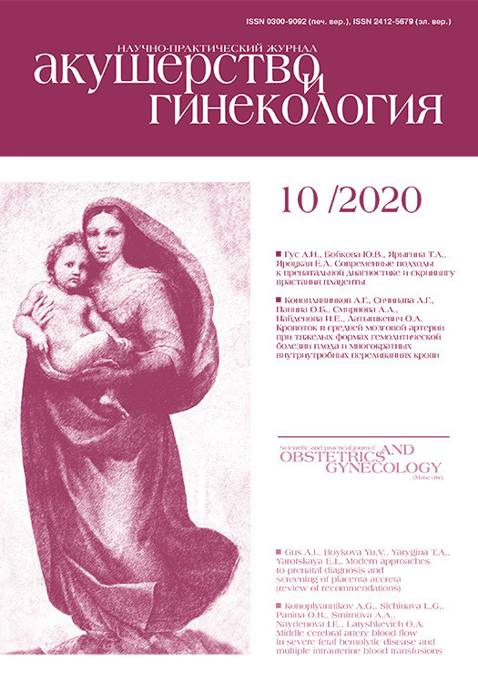
Отметим, что при проведении КЭГ в представленных случаях при метастатическом поражении яичников солидные участки картировались преимущественно 5 типом эластограммы (компонент высокой жесткости) (рис. 1). В солидно-кистозных образованиях в случаях синхронного поражения солидные участки картировались преимущественно 4 типом эластограммы (в равной степени встречались как жесткие, так и эластичные участки).
Клиническое наблюдение
Пациентка А., 54 года. Считала себя больной с августа 2018 г., когда отметила увеличение живота в объеме. За медицинской помощью обратилась в октябре, когда в ГКБ им. М.Е. Жадкевича произведен лапароцентез, цитологически – клетки злокачественной природы, вероятно, аденогенной. Больная направлена в НМИЦ онкологии. При пересмотре готовых цитологических препаратов – 18/21860 – опухолевые клетки (метастаз муцинозной аденокарциномы? пограничная опухоль яичников?). При гистологическом исследовании эндометрия – 47937/18 – аденокарцинома.
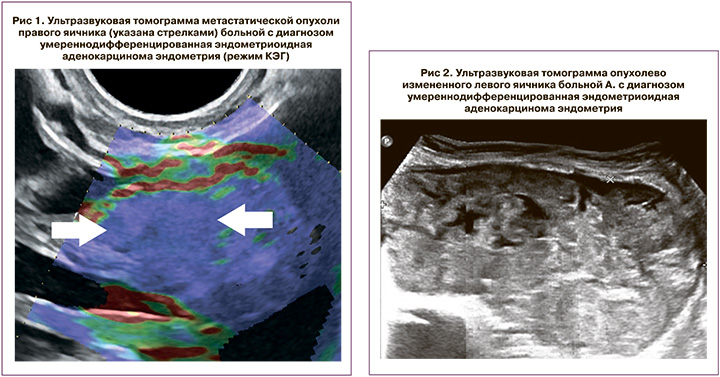
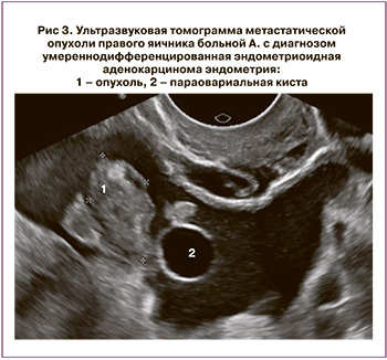
При УЗИ: Печень без очаговых изменений. Большой сальник утолщен, бугристый, метастатически изменен, толщиной 2,2–2,5 см. Матка увеличена, размеры тела 8,0×6,0×8,5 см, контур ровный, с остаточными признаками аденомиоза, из-за которого граница эндометрия отчетливо не визуализируется. В нижней трети тела и просвете цервикального канала определяются солидные структуры толщиной до 2,0 см, нижний полюс которых визуализируется на 1,5 см выше уровня наружного маточного зева. По левому ребру шейки матки определяется образование диаметром 5,0 см (УЗ-картина соответствует шеечной миоме). Над маткой слева определяется солидно-кистозное образование с четкими неровными контурами, размерами 20,0×18,0 см, в его структуре определяются перегородки и кистозные полости, в которых визуализируются множественные солидные сосочковые структуры, в перегородках и сосочковых структурах определяются локусы патологического кровотока при ЦДК и ЭД (рис. 2). Правый яичник не увеличен, солидной неоднородной структуры, с четкими неровными контурами, размерами 2,2×1,2 см, с единичными локусами патологического кровотока при ЦДК и ЭД (рис. 3), рядом определяется параовариальная киста диаметром 2,0 см. Выявлена свободная жидкость в области малого таза (+3,6 см). Заключение: Опухоль яичников (сложно дифференцировать между первичной опухолью и метастатической). Опухоль матки (следует дифференцировать между опухолью цервикального канала и тела матки). Метастазы в большом сальнике. Асцит. Нельзя исключить первично-множественную опухоль.
 22.01.2019. Экстирпация матки с придатками, тазовая лимфаденэктомия. Резекция большого сальника. Результаты гистологического исследования №48981/2018. В теле матки разрастание умеренно дифференцированной эндометриоидной аденокарциномы, врастающей в миометрий на глубину 1 см. Опухоль врастает во внутренний зев шейки матки, стенку цервикального канала. В правом яичнике белые тела, фокус разрастания аденокарциномы. На серозной поверхности яичника папиллярные разрастания реактивного мезотелия. Маточная труба без элементов опухолевого роста. В левом яичнике разрастание муцинозной цистаденокарциномы G2. Маточная труба обычного строения. В большом сальнике множественные кровоизлияния, скопления ксантомных клеток, полнокровие капилляров, участки папиллярного разрастания реактивного мезотелия, очаговые лимфоидные инфильтраты. Элементов опухоли не обнаружено. В жировой клетчатке выделено 5 лимфоузлов с реактивными изменениями, без элементов опухолевого роста.
Представленный нами клинический пример наглядно демонстрирует отличия ультразвуковой картины метастатически пораженного яичника при РТМ и синхронной опухоли контралатерального яичника. Многолетний опыт позволил нам выделить некоторые отличия ультразвуковой картины при метастатическом и синхронном поражении яичников при РТМ, что помогает в повседневной практической деятельности. При синхронном РТМ и яичников структура опухоли яичников чаще солидно-кистозная с неровными четкими контурами, размеры яичников превышают 6,0 см, при проведении эластографии яичники картируются преимущественно 4 типом эластограммы. При метастатическом поражении опухоли яичников чаще представлены солидными образованиями с четкими ровными контурами, размеры яичников не превышают 6,0 см или не отличаются от неизмененных, а при проведении эластографии солидные образования в структуре яичников картируются преимущественно 5 типом эластограммы [10].
Обсуждение
Хирургическое лечение является основным этапом при лечении больных РТМ. Первым этапом лечения большинства больных РТМ является экстирпация матки с придатками [11]. Удаление придатков матки обязательно, так как в них при морфологическом исследовании часто обнаруживаются микрометастазы. По данным Онкогинекологической исследовательской группы (GOG 99), у 38% больных РТМ I клинической стадии во время операции обнаруживаются метастазы в яичниках, маточных трубах, забрюшинных лимфатических узлах, по брюшине и в смывах из брюшной полости [5]. В связи с чем анализ выживаемости позволяет сделать вывод, что прогноз при РТМ определяется в основном морфологической, а не клинической стадией.
Наши результаты согласуются с зарубежными данными об ультразвуковых особенностях опухолей яичников при РТМ. Guerriero S. и соавт. (2012) описали 17 случаев метастазирования РТМ и выявили, что у 12 (76%) пациенток структура яичников была солидной, а у 5 (24%) – кистозно-солидной [12]. Moro F. и соавт. в исследовании 2018 г. (n=239) установили, что при синхронных опухолях образования в яичниках преимущественно характеризовались как солидно-кистозные у 115 пациентов (48,1%), тогда как солидные составляли 34,3% (70 пациенток), чаще были крупными – превышали 10,0 см [13]. Также исследование этих же авторов в 2019 г. (n=196), которое непосредственно было направлено на дифференциальную диагностику синхронных и метастатических опухолей яичников при РТМ, показало, что при метастатическом поражении размеры опухоли не превышали 6,6 см, структура была преимущественно солидной – 66,7%, при синхронном поражении размеры опухоли достигали 9,0 см, а структура была солидно-кистозной в 50% случаев [7].
На данный момент имеется не так много данных о результатах компрессионной эластографии при опухолях яичника, но все сходятся в едином мнении, что эластография выполняет роль надежного дополнительного критерия при дифференциальной диагностике доброкачественных и злокачественных образований. В обзоре 2013 г. Гажонова В.Е. и соавт. указывали, что в солидных опухолях яичника в 96% случает устойчиво регистрируются жесткие 5-й и 6-й типы эластограммы [8].
Заключение
В заключение следует отметить, что синхронные и метастатические опухоли яичников при РТМ имеют, по нашим данным, разные ультразвуковые признаки.
Изучение разных семиотических вариантов опухоли яичников у больных РТМ необходимо для улучшения их визуализации и выявления. В любом случае своевременная диагностика новообразований яичников (вне зависимости от морфологической природы) у больных РТМ имеет принципиально важное значение, поскольку влияет на объем операции и прогноз заболевания.