Опухоли и опухолевидные образования яичников составляют до 14% опухолей женских половых органов; на долю доброкачественных опухолей яичников приходится 75–87% всех истинных опухолей яичников, среди которых опухолевидные ретенционные образования составляют 70,9% наблюдений [1, 2].
Сочетание функциональных кист и беременности встречается в 14–38% всех образований. Во время беременности обычно находят лютеомы, дермоидные и муцинозные опухоли. Отмечается, что простые кисты (без эпителиальной выстилки) во время беременности встречаются у 1–3% наблюдаемых женщин [2].
Частота злокачественных опухолей среди новообразований яичников у беременных не превышает 3–6% [3]; но поскольку они являются одними из самых неблагоприятных по течению и прогнозу для жизни и здоровья, гистологическое исследование оперативно удаленных кистозных новообразований является определяющим для выработки тактики ведения пациентки.
Факторы риска развития ретенционных образований во время беременности: возраст от 20 до 35 лет, курение (повышение частоты выявления функциональных кист в 2 раза по сравнению с некурящими), лечение бесплодия в анамнезе, воспалительные процессы малого таза, эндометриоз, булимия [2]. Гормональным нарушениям в происхождении опухолевидных образований яичников придают ведущее значение, хотя не исключается влияние воспаления.
Инклюзионным кистам отводится ведущее место в гистогенезе наиболее распространенных эпителиальных опухолей яичников. Инклюзионные кисты диаметром свыше 1 см уже относятся к разряду опухолевых кистозных образований, обозначаемых в зависимости от структурных особенностей выстилающего эпителия как серозная, эндометриоидная или муцинозная цистаденома [1, 2, 4–7].
Опухоли яичников независимо от их гистотипа, за исключением гормонпродуцирующих, характеризуются длительным бессимптомным течением, что является одной из причин поздней диагностики [8]. Стандартным рутинным методом, позволяющим выявить новообразования придатков матки у беременной, является ультразвуковое исследование (УЗИ), которое проводится всем пациенткам во время выполнения скринингов в декретированные сроки. При выявлении новообразования оцениваются его размеры, консистенция (кистозная, солидная или кистозно-солидная), особенности строения (наличие перегородок, дополнительных включений, разрастаний, характер содержимого), наличие свободной жидкости в брюшной полости и малом тазу [9]. По показаниям УЗИ дополняется магнитно-резонансной томографией, определением уровня опухолевых маркеров: β-ХГ (бета-хорионический гонадотропин), АФП (α-фетопротеин), СА 15-3 (cancer antigen 15-3 – углеводный антиген 15-3), SCC (squamous cell carcinoma antigen – антиген плоскоклеточной карциномы) и СА-125 (cancer antigen 125 – углеводный антиген 125) [10]. Тактика ведения пациентки определяется совместно с онкологом.
Клиническое наблюдение
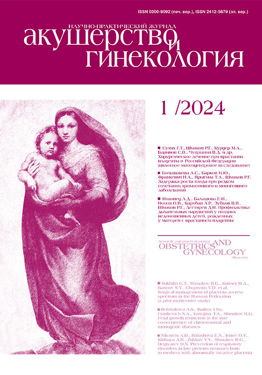
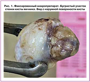
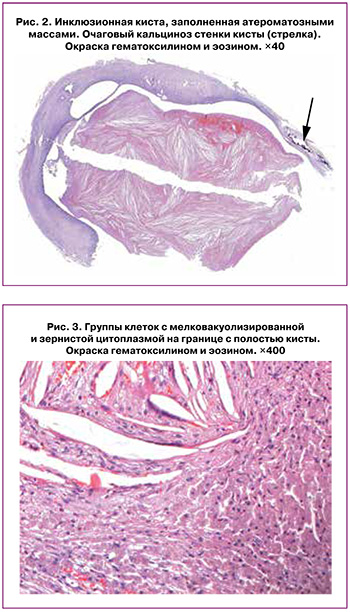
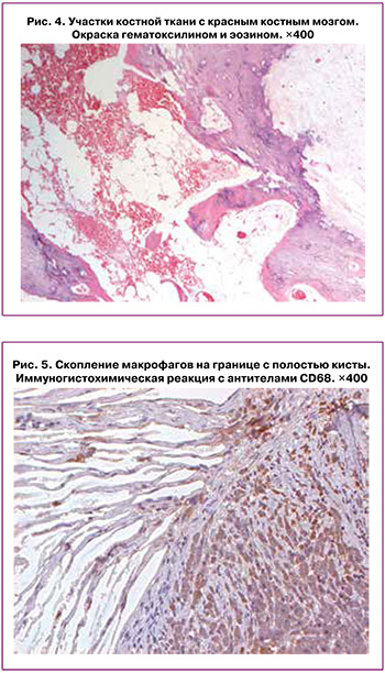
Пациентка К., 41 год, повторно беременная, первородящая, поступила в Перинатальный центр на 39-й неделе беременности. При поступлении определялось наличие смешанного ягодичного предлежания плода. Также у женщины имелся отягощенный акушерско-гинекологический анамнез (несостоявшийся выкидыш раннего срока беременности, аномалия развития полового аппарата – двурогая матка, хронический эндометрит, уреаплазмоз) и отягощенный соматический анамнез (аномалия развития толстого кишечника – левостороннее расположение слепой кишки, отсутствие поперечного и нисходящего отделов ободочной кишки). На скрининговых УЗИ, проводимых во время беременности, дополнительные образования яичников не определялись. Жалобы, связанные с поражением яичников, отсутствовали. Принимая во внимание наличие смешанного ягодичного предлежания плода у первородящей старшего возраста с аномалией развития полового аппарата, показано родоразрешение путем операции кесарева сечения в плановом порядке. Риск венозных тромбоэмболических осложнений средний (2 балла); показано применение низкомолекулярных гепаринов в послеродовом периоде 10 дней. При оперативном родоразрешении была извлечена живая доношенная девочка массой тела 3000 г, длиной 50 см, оценка по Апгар на 1-й минуте – 8 баллов, на 5-й минуте – 9 баллов, без видимых пороков развития. Во время операции кесарева сечения при осмотре придатков в области нижнего полюса правого яичника было обнаружено плотное овальное образование, исходящее из ткани яичника, не спаянное с окружающими тканями, которое было удалено в пределах здоровой ткани яичника. Новообразование яичника, не делая разрезы, целиком поместили в контейнер, содержащий 10% забуференный формалин, и направили в патоморфологическую лабораторию. Левые придатки матки и правая маточная труба не изменены. Произведена ревизия брюшной полости на участках, доступных визуальному осмотру, видимой патологии не обнаружено. Макроскопически образование белесовато-розового цвета, овоидной формы, размерами 1,5×1,1 см с серо-красным участком, плотной консистенции, бугристого вида, размерами 0,7×0,5 см (рис. 1). На разрезе определялась полость, заполненная коричневыми однородными массами; бугристое образование белесоватого цвета, слоистого вида. При микроскопическом исследовании в срезе определялись множественные мелкие инклюзионные кисты с оптически пустым содержимым, выстланные однослойным кубическим эпителием. Стенка самой большой инклюзионной кисты также была выстлана однослойным кубическим эпителием, в ее просвете отмечались множественные, диффузно расположенные, крупные клетки с пенистой и зернистой цитоплазмой, кристаллы холестерина и оксифильные гомогенные массы (рис. 2). С одной стороны, стенка кисты значительно утолщена за счет группы клеток с мелко вакуолизированной и зернистой цитоплазмой, подобных лютеиноцитам (рис. 3), и разрастания соединительной ткани с очагами кальциноза и участком костной ткани (рис. 4). Под базальной мембраной кисты выявлялись единичные макрофаги, содержащие бурые гранулы в цитоплазме. После гистологического исследования было предположение, что киста содержит два компонента. Первый – инклюзионная киста, заполненная гомогенной массой с множественными крупными клетками с пенистой цитоплазмой и кристаллами холестерина. Второй – участок желтого тела с дистрофическими изменениями в виде склероза, кальциноза и оссификации.
Для понимания и уточнения морфогенеза кисты яичника было проведено иммуногистохимическое (ИГХ) исследование с использованием антител к CD68 (маркер макрофагов) (производство Diagnostic BioSystems, США), рецепторам прогестерона (производство Diagnostic BioSystems, США) и эстрогена (производство Diagnostic BioSystems, США). Первоначально при морфологическом исследовании предполагались кистозная трансформация желтого тела и аккумуляция липидных масс и кристаллов холестерина, так как были обнаружены группы крупных пенистых клеток. При этом в кисте группа клеток, напоминающих лютеиноциты, при проведении ИГХ-исследования экспрессировала CD68 (рис. 5), тогда как экспрессия к рецепторам эстрогена и прогестерона в этих клетках отсутствовала. Таким образом, данная группа клеток оказалась очагом холестероза. Экспрессия CD68 определялась в пенистых клетках содержимого кисты и в клетках под базальной мембраной эпителия стенки инклюзионной кисты, единичных клетках стромы яичника.
При ИГХ-исследовании экспрессия рецепторов эстрогена в виде мелких зерен выявлялась в цитоплазме макрофагов, расположенных в содержимом кисты и под базальной мембраной, и единичных клетках выстилки кисты. Положительная реакция к маркеру прогестерона отмечалась в цитоплазме макрофагов, расположенных в содержимом кисты и под базальной мембраной, и в эпителиальных клетках выстилки кист.
После проведенного патоморфологического исследования пациентке был выставлен диагноз: Инклюзионная киста правого яичника с холестерозом и оссификацией.
Обсуждение
Диагностика и дифференциальная диагностика доброкачественных новообразований (опухолей) яичников представляют собой важную клиническую задачу с позиций профилактики возможных осложнений, требующих оказания экстренной стационарной квалифицированной медицинской помощи: перекрут придатков матки, апоплексия яичников, нагноение; а также оценки риска злокачественного процесса, диктующего необходимость правильной маршрутизации пациентки. У беременных частота выявления опухолей придатков матки может достигать 3,2%, а частота злокачественных форм – 6,8% от всех опухолей у беременных [11]. Функциональные или гормонально чувствительные кисты обычно имеют размер 1–3 см и рассасываются к 16 неделям беременности [12]. Консервативное лечение допускается, если у пациентов нет симптомов. Согласно исследованию, максимальная распространенность простых кист ≥3 см составила 5,3% на 8–10-й неделе беременности, которые спонтанно регрессировали через 10 недель, с распространенностью 1,5% на 14-й неделе [13]. Ведение пациенток с объемными образованиями придатков матки, выявленными в I триместре, определяется клинико-анамнестическими и эхографическими данными. Наиболее часто эти яичниковые образования кистозного строения в I триместре представлены кистой желтого тела, которая регрессирует самостоятельно [14]. Опухоли яичников, персистирующие на протяжении беременности, чаще всего являются функциональными кистами с очень низкой частотой озлокачествления [15]. Окончательно судить о характере процесса (доброкачественный/злокачественный) и прогнозе заболевания возможно только после получения результата развернутого морфологического исследования удаленного макропрепарата. Лечебная тактика при доброкачественной опухоли яичника, диагностированной во время беременности, определяется ее сроком и наличием/отсутствием клинических симптомов. Частота осложнений (перекрут придатков матки; разрыв, нарушение питания, нагноение опухоли) во время беременности не превышает 2%. Однако необходимо учитывать, что опухоль яичников может служить причиной механического препятствия в родах [16, 17].
В представленном материале обсуждаются редко встречающиеся патологические изменения ткани яичника – холестероз с накоплением липидов и кристаллов холестерина в инклюзионной кисте, кальциноз и оссификация.
Холестероз проявляется накоплением липидов, особенно сложных эфиров холестерина и триглицеридов, в макрофагах. Он обычно наблюдается в желчном пузыре, но очень редко встречается в яичниках и недостаточно изучен [18].
Предполагается, что при холестерозе скопление гистиоцитов, нагруженных липидами, происходит из-за повышенного синтеза липидов слизистой оболочкой или неспособности макрофагов метаболизировать или выделять холестерин, при перенасыщении сыворотки крови холестерином.
Реактивные гистиоцитарные пролиферации возникают как при нормолипидемических, так и при гиперлипидемических состояниях. Висцеральные скопления пенистых макрофагов, не связанные с воспалением и/или кровоизлиянием, встречаются крайне редко и почти исключительно в желудочно-кишечном тракте, особенно в желудке [19]. Ксантогранулематозное воспаление при холестерозе представляет собой особую форму хронического воспаления, разрушающего нормальные ткани пораженных органов. Чаще всего оно поражает почки, но также сообщается о локализации в желчном пузыре, желудке, аноректальной области, костях, мочевом пузыре, яичках, придатках яичка, влагалище и эндометрии. В ксантогранулеме выявляется плотная инфильтрация тканей гистиоцитами, нагруженными липидами, с примесью воспалительного инфильтрата [20]. В нашем случае воспалительный инфильтрат отсутствовал, что можно объяснить завершением воспалительного процесса.
Зрелые лютеиноциты характеризуются увеличением в объеме цитоплазмы и светлым округло-овальным ядром с крупным ацидофильным ядрышком. Цитоплазма этих клеток эозинофильная, светлая, иногда пенистая или с множественными мелкокапельными суданофильными включениями. Регрессирующие лютеиноциты отличаются кариопикнотичными изменениями и крупновакуольной жировой дистрофией цитоплазмы [4]. Поэтому клетки с пенистой цитоплазмой в составе кисты в первую очередь воспринимаются как лютеиноциты. Однако после проведения ИГХ-исследования неожиданной находкой было обнаружение экспрессии CD68 в цитоплазме клеток, что характерно для макрофагов при отсутствии экспрессии в этих клетках рецепторов прогестерона. Это позволило прояснить патогенез накопления липидов и кристаллов холестерина в инклюзионной кисте.
Глыбки кальция и губчатая кость ткани могли стать следствием разрушения ткани яичника гистиоцитами и последующей репаративной регенерации с окостенением. Очаговая нетератогенная оссификация в яичниках является крайне редкой патологией. В доступной русскоязычной литературе мы не нашли описания данной патологии. При изучении англоязычной литературы найдена статья, где описано 34 случая оссификации в яичниках при различной патологии за многие годы [21]. При этом мы не нашли ни одного описания сочетания холестероза и оссификации в яичнике.
В нашем наблюдении у пациентки образование правого яичника было случайной находкой при оперативном вмешательстве. Это может быть связано с затруднением визуализации яичников, особенно на больших сроках гестации, существенными морфологическими изменениями тканей во время беременности, скудной клинической симптоматикой [22], а также со сравнительно небольшими размерами обнаруженной кисты. После удаления образования в пределах неизмененной ткани яичника дальнейшее клиническое наблюдение женщины не требуется.
Заключение
Представленный случай характеризуется казуистическим сочетанием редких патологических процессов в яичнике. Визуальный осмотр придатков во время операции кесарева сечения является стандартной процедурой для своевременного выявления патологии яичников и маточных труб. Данное наблюдение представляет клинический и научный интерес в связи с редкостью во врачебной практике, а применение ИГХ-исследования явилось информативным методом для детализации патоморфоза кисты яичника.
Изменение гормонального фона во время беременности может индуцировать формирование и рост кистозных образований яичников. Это требует диагностического исследования биопсийного материала с использованием ИГХ-методов, позволяющих понять природу кист яичника, расширить представления о патогенезе данных изменений и определить дальнейшую тактику наблюдения за пациенткой.