В настоящее время заболевания вульвы являются одной из наиболее изучаемых тем в амбулаторно-поликлинической гинекологии. Заболевания вульвы диагностируют гинекологи, дерматологи, урологи, онкологи, поэтому тактика ведения пациенток значительно варьируется. В последнее время наблюдается увеличение распространенности заболеваний вульвы, прежде всего за счет их лучшей выявляемости на приемах гинекологов. Так, регистрируется 1 случай заболевания склерозирующим лихеном вульвы (СЛВ) на 30 женщин менопаузального периода, 1 случай на 59 женщин репродуктивного возраста и всего 1 случай на 300–1000 женщин, обращающихся к дерматологам [1]. Для диагностики заболеваний вульвы используются: цитологическое исследование, вульвоскопия, прицельная биопсия.
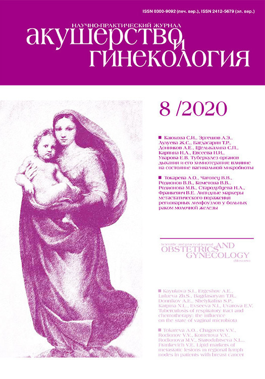
 Биопсия (от греч. bios — жизнь и opsis — зрительное восприятие, рассмотрение) – инвазивный метод диагностики, заключающийся во взятии образцов ткани с целью последующего морфологического исследования [2]. Биопсия проводится по показаниям как в диагностических, так и в лечебных целях, позволяя в некоторых случаях полностью удалить патологический участок. Существуют различные методы проведения биопсии кожи и слизистой вульвы: с использованием скальпеля, радиоволны, щипцов и панча. Выбор метода биопсии зависит от характера и локализации поражения. Тенденция современной медицины направлена на максимальное снижение уровня инвазивности манипуляций и возможности проведения их в амбулаторных условиях. Среди представленных диагностических мероприятий наиболее щадящей, но при этом достаточно информативной, является панч-биопсия (от англ. punch – дырокол, компостер) вульвы, которая обеспечивает забор всех слоев кожи в виде столбца (рис. 1).
Международное общество по изучению заболеваний влагалища и вульвы (ISSVD), в которое входят гинекологи, дерматологи, морфологи, в 2011 г. определило клиническую терминологию, классификацию и методы диагностики дерматозов вульвы. Согласно ISSVD, диагностика заболеваний вульвы происходит в 5 этапов: 1) определение кожного элемента (узелок, папула, трещины, экскориации, эрозии и другие); 2) описание цвета, границ, воспалительного венчика; 3) поиск заболеваний с найденными критериями; 4) дифференциальная диагностика заболеваний вульвы; 5) для постановки окончательного диагноза рекомендуется провести исследование микробиоты влагалища и вульвы и/или биопсию вульвы с последующим гистологическим исследованием [3].
Согласно клиническим рекомендациям ассоциации дерматологов Великобритании, показаниями к проведению биопсии вульвы являются [4]:
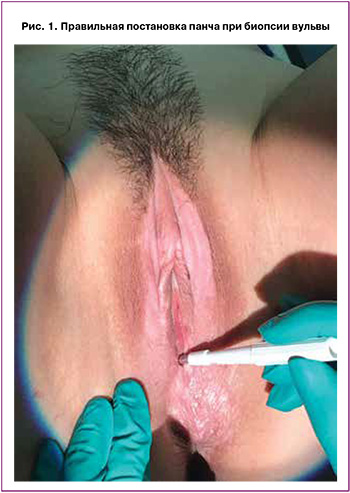
- подозрение на опухолевидные и злокачественные заболевания (особенно участки эрозий и изъязвлений, гиперпигментация, экхимозы, гиперкератоз), рис. 2;
- отсутствие эффекта от проводимой терапии заболеваний вульвы глюкокортикостероидами;
- дифференциальная диагностика (с вульварной интраэпителиальной неоплазией вульвы – VIN), злокачественными новообразованиями, красным плоским лишаем и другими дерматозами вульвы);
- наличие экстрагенитальных очагов склерозирующего лишая;
- биопсия вульвы в ходе хирургического лечения заболеваний наружных половых органов.
В отечественных клинических рекомендациях Российского общества дерматовенерологов и косметологов по локализованной склеродермии, 2016 г. (куда включен СЛВ) рекомендуется проводить гистологическое исследование биоптатов кожи в сомнительных случаях с целью дифференциальной диагностики заболеваний (уровень доказательности – 4D) [5]. В настоящее время данные рекомендации пересматриваются с участием гинекологов.
Панч-биопсия вульвы проводится стерильными одноразовыми или многоразовыми стерилизуемыми стилетами разных размеров. Достаточным для гистологического исследования считается участок кожи, полученный при помощи 3,5–4-миллиметрового биопсийного стилета, панч менее 3 мм в диаметре использовать не рекомендуется. Длительность манипуляции составляет 5–10 минут. Особенности кровоснабжения кожи вульвы заключаются в наличии двух сосудистых сплетений, связанных между собой: верхнее, располагающееся на стыке сосочкового и сетчатого слоев дермы, и нижнее – между сетчатым слоем дермы и подкожножировой клетчаткой (рис. 3). Риск рубцевания после панч-биопсии зависит от техники проведения манипуляции. При взятии биопсии следует углубиться до уровня подкожной жировой клетчатки; в обратном случае, если биопсия заканчивается на середине сетчатого слоя, оставшаяся часть дермы имеет сниженную васкуляризацию, что увеличивает риск вторичного инфицирования и рубцевания [6].
Биопсия вульвы является простой амбулаторной процедурой, однако необходимо учитывать следующие противопоказания:
- нарушение системы свертываемости крови тяжелой степени;
- острая сердечная недостаточность или хроническая сердечная недостаточность в стадии декомпенсации;
- наличие пиодермитов в непосредственной близости от места предполагаемой биопсии;
- противопоказания, относящиеся к применению анестетика артикаина: тяжелые нарушения проводимости (выраженная брадикардия, атрио-вентрикулярная блокада II–III степени), тяжелая артериальная гипотензия.
Для биопсии выбирают участки с наиболее выраженными патологическими изменениями. Наиболее точная диагностика достигается при выполнении биопсии вульвы, содержащей свежие образования, появившиеся не позднее 48 ч. При биопсии очага, не покрытого эпителием, теряется связь между эпителиальными и дермальными клетками, поэтому рекомендуется проводить мультифокальную биопсию из нескольких участков: очага поражения и на границе его перехода в неповрежденную ткань. В случае биопсии сосудистых поражений, таких как гемангиомы, лимфангиомы, не рекомендуется использовать анестетик с адреналином, т.к. последний вызывает сужение сосудов и может повлиять на интерпретацию гистологических результатов. При этом чрезмерная инфильтрация анестетиком создает видимость отека кожи и при гистологическом исследовании может интерпретироваться как крапивница или ангионевротический отек. Перенос биоптата в контейнер с формалином осуществляется с минимальным повреждением ткани. Если ткань сдавливается между браншами пинцета или зажима, она сужается в боковом направлении, образуя артефакт сжатия (рис. 4) [6].
Целью нашего исследования было изучение структуры заболеваемости наружных половых органов у женщин по обращаемости путем проведения панч-биопсии вульвы.

Материалы и методы
На базе отделения эстетической гинекологии и реабилитации (ОЭГиР) ФГБУ «НМИЦ АГП им. академика В.И. Кулакова» МЗ РФ ретроспективно были обследованы 138 пациенток с заболеваниями вульвы, которым в период с 2014 по 2019 гг. была произведена биопсия вульвы. Критериями включения были: заболевания вульвы, возраст от 18 лет и старше, отсутствие применения в течение 30 дней до проведения биопсии топических кортикостероидов на область наружных половых органов. Все женщины заполнили информированное согласие на проведение биопсии вульвы. Биопсия вульвы выполнялась при помощи одноразового стерильного инструмента – дермопанча. Проводилась мультифокальная биопсия нескольких элементов вульвы под местной анестезией 1–2% раствором лидокаина в объеме 0,1–0,15 мл на один участок с помощью иглы калибром 30–33G.
Забранный участок имел диаметр 3,5 мм, длину 4–5 мм (рис. 5). Биоптат переносился в контейнер с 10% формалином, избегая сдавления в боковом направлении и образования артефактов сжатия. После фиксации в 10% нейтральном растворе формалина в течение 24 ч материал заливали в парафин. Далее срезы окрашивались гематоксилином и эозином и проводилось морфологическое исследование по общепринятой методике.
Цифровая обработка данных выполнена на индивидуальном компьютере с помощью электронных таблиц Microsoft Excel и пакета прикладных программ Statistica for Windows v. 8.0, StatSoft Inc. (США).
Включенные в исследование 138 пациенток находились в возрасте от 20 до 72 лет (средний возраст составил 48,0 (13,3) года). Как видно из рис. 6, с 2018 г. пациентки с заболеваниями вульвы стали чаще обращаться за медицинской помощью, что согласуется с общемировой тенденцией роста численности СЛВ [7].
За указанный период отмечен рост частоты обращаемости женщин с заболеваниями вульвы. Так, в 2014 г. заболевания вульвы регистрировались в 6,5% (9/138), в 2019 – уже в 37,0% (51/138), что, возможно, связано с повышением уровня диагностики заболеваний вульвы и квалификации врачей, появлением отделений, специализирующихся по заболеваниям вульвы и влагалища и увеличением обращаемости пациенток в медицинские центры, такие как ФГБУ «НМИЦ АГП им. академика В.И. Кулакова» МЗ РФ.
Анализ структуры дистрофических заболеваний вульвы, согласно результатам гистологического исследования, показал, что лидирующую позицию занимает СЛВ, распространенность которого составляет 62,8%; в 3 раза реже регистрируется гиперпластическая дистрофия вульвы (ГДВ) – 22,5%; в 4,7% – красный плоский лишай (КПЛ); 3,1% приходится на VIN I; по 2,3% – на рак вульвы и другие дерматозы вульвы; VIN III встречается в 1,6% и VIN – II только в 0,8% (рис. 7).
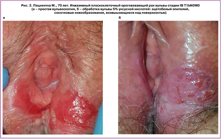
Наибольшая трудность в дифференциальной диагностике прослеживается между ГДВ и СЛВ. Данные заболевания характеризуются гиперплазией верхних слоев кожи и имеют сходные клинические симптомы (жжение, зуд, диспареуния, наличие зоны лихенификации с признаками расчесов). Окончательный диагноз возможен только после проведения гистологического исследования. Так, для СЛВ, в отличие от ГДВ, характерны наличие лимфоцитарно-воспалительной зоны и гомогенизация коллагена в дерме (рис. 8) [8].
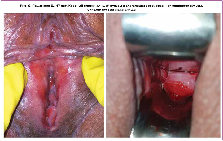
Среди прочих заболеваний вульвы третье место по распространенности приходится на КПЛ, который составляет 4,7% всех дерматозов вульвы (рис. 9). В некоторых случаях довольно трудно выявить различия между СЛВ и КПЛ, как клинически, так и по гистологическим признакам. Данное состояние определяется как «синдром наложения» двух заболеваний и характеризуется низким ответом на проводимое лечение [4]. По данным нашего исследования, VIN I и рак вульвы встречаются в 3,1% и 2,3% случаев соответственно. Среди злокачественных новообразований вульвы в нашем исследовании были диагностированы: плоскоклеточная карцинома с ростом в виде кератоакантомы, интраэпителиальная меланома и плоскоклеточный ороговевающий рак вульвы (рис. 2).
Редкие дерматозы вульвы также составили 2,3% (3/138) и были представлены вульгарной пузырчаткой вульвы, псориазом вульвы и сирингомой кожи вульвы (рис. 10).
При подозрении на дерматоз вульвы пациентку рекомендуется проконсультировать у дерматолога или направить в гинекологическое отделение, специализирующееся на заболеваниях вульвы.

Направительный диагноз не всегда совпадает с окончательным гистологическим. Так, в 98,8% (80/81) клинический (направительный) диагноз СЛВ был подтвержден гистологическим методом, в 1,2% (1/81) вместо VIN III диагностирован СЛВ. Совпадение диагнозов гиперпластической дистрофии вульвы наблюдалось у 8 из 9 женщин. Однако прослеживается тенденция гипердиагностики заболеваний вульвы, поскольку ГДВ была диагностирована у 18/110 женщин с направительным диагнозом СЛВ, у 1/4 и 2/2 женщин – с предварительными диагнозами VIN I и вульвовагинальная атрофия (ВВА), соответственно, диагностическая чувствительность метода составила 44,4% (рис. 11). Следовательно, гистологический метод исследования является, несомненно, важным и необходимым этапом диагностики клинически близких заболеваний вульвы, но имеющих разный патогенез, подходы к терапии и прогнозы излечиваемости, и имеет особенно высокую чувствительность в отношении выявления СЛВ и КПЛ.

Осложнения после панч-биопсии встречаются крайне редко. Гемостаз области, подвергшейся биопсии, достигается путем временной компрессии ватной палочкой, смоченной в 3% растворе перекиси водорода, прикладыванием гемостатической губки и в одном случае (из 138) электро-радиохирургической коагуляцией ложа биопсии в области клитора. Заживление раны обычно происходит вторичным натяжением с минимальным риском формирования рубца, т.к. наружные половые органы имеют хорошо развитую сосудистую сеть.
Заключение
Воспалительные заболевания вульвы аутоиммунного генеза (СЛВ и КПЛ) являются наиболее часто диагностируемыми дерматозами вульвы в практике гинеколога и суммарно составляют 67,5% в общей структуре заболеваний вульвы; из них 62,8% приходится только на СЛВ. Сложность диагностики заболеваний вульвы заключается в близости клинических симптомов на ранних стадиях и в длительном хроническом течении, что затрудняет и смещает по времени постановку точного диагноза и подбор эффективного метода лечения. Данные клинического осмотра не всегда отражают изменения, происходящие на клеточном уровне. Только проведение биопсии вульвы с последующим гистологическим исследованием позволяет установить правильный диагноз уже на ранних стадиях. Панч-биопсия – щадящий способ забора ткани вульвы. Преимущества панч биопсии заключаются в минимальной инвазивности, высокой безопасности, получении ткани однородной формы в виде столбца и простоте выполнения.