
Comparative effectiveness evaluation of resuscitative endovascular balloon occlusion of the aorta (REBOA) and tourniquet hemostasis in organ-sparing surgery in patients with placenta accreta spectrum
Prikhodko A.M., Kan N.E., Amiraslanov E.Yu., Chuprynin V.D., Akinfiev D.M., Ketskalo M.V., Tyutyunnik V.L., Tsukanova A.A.
Objective. To assess the practical feasibility, safety, and changes in the volume of blood loss using REBOA in organ-sparing surgical management of placenta accreta spectrum (PAS).
Materials and methods. This retrospective comparative study evaluated the use of two consecutive clinical hemostasis methods in 32 pregnant women. The main group included 8 patients with PAS who underwent cesarean section using REBOA. The comparison group included 24 patients with PAS who underwent surgery using complex compression hemostasis. To ensure sample homogeneity, patients with histologically confirmed placenta accreta and placenta increta (excluding placenta percreta) were included in the comparison group. Statistical analysis was performed using the Mann-Whitney U test for quantitative variables and Fisher’s exact test for categorical variables. The null hypothesis was rejected at p<0.05.
Results. The following results were obtained in our study: the volume of intraoperative blood loss in the REBOA group was 725.0 [435.0–1100.0] mL versus 2225.0 [1575.0–2862.5] mL in the comparison group (p<0.001). The volume of autologous reinfusion in the REBOA group was 255.0 [250.0–362.5] mL versus 750.0 [537.5–842.5] mL in the comparison group (p=0.003). There were differences between the groups in duration of surgery – 82.50 [58.25–151.8] minutes in the main group and 151.0 [133.5–162.5] minutes in the comparison group (р=0.035). Hemoglobin levels on postoperative day 3 were 102.5 [97.0–108.0] g/L in the REBOA group and 92.5 [87.25–99.5] g/L in the comparison group (p=0.036). Hysterectomy was not performed in the REBOA group – 0/8, while in the comparison group it was performed in 6/24 women (p=0.241). No REBOA-related complications (vascular injury, limb ischemia, thrombosis) were registered.
Conclusion. This study demonstrated the practical feasibility and safety of REBOA. The preliminary findings of our study confirm the potential of REBOA to be used as a technique for pelvic devascularization in organ-sparing surgeries. However, further multicenter prospective studies with a larger sample size are needed to confirm the effectiveness of this technique.
Authors' contributions. Kan N.E., Prikhodko A.M., Tyutyunnik V.L. – study concept and design; Chuprynin V.D., Prikhodko A.M., Amiraslanov E.Yu. – material collection and processing; PrikhodkoA.M., Akinfiev D.M.– statistical analysis; Prikhodko A.M., Tsukanova A.A. – manuscript writing; Kan N.E., Prikhodko A.M., Tyutyunnik V.L., Ketskalo M.V. – manuscript editing.
Conflicts of interest. The authors declare no potential conflicts of interest related to publication of this article.
Funding. The study was conducted without any sponsorship.
Ethical Approval. The study was approved by the local Ethics Committee of V.I. Kulakov National Medical Research Center for Obstetrics, Gynecology and Perinatology, Ministry of Health of Russia (protocol No. 2 of February 19, 2026).
Generative Artificial Intelligence. No generative AI was used in preparing this article.
Patient Consent for Publication. The patients have signed informed consent for publication of their data.
Authors' Data Sharing Statement. The data supporting the findings of this study are available on request from the corresponding author after approval from the principal investigator.
For citation: Prikhodko A.M., Kan N.E., Amiraslanov E.Yu., Chuprynin V.D., Akinfiev D.M., Ketskalo M.V.,
Tyutyunnik V.L., Tsukanova A.A. Comparative effectiveness evaluation of resuscitative endovascular balloon occlusion of the aorta (REBOA) and tourniquet hemostasis in organ-sparing surgery in patients with placenta accreta spectrum.
Akusherstvo i Ginekologiya/Obstetrics and Gynecology. 2026; (8): 83-92 (in Russian)
https://dx.doi.org/10.18565/aig.2026.106
Keywords
PAS
placenta accreta spectrum
REBOA
endovascular balloon occlusion of the aorta
tourniquet hemostasis
hemorrhage
organ-sparing surgery
Placenta accreta (PAS) is a critical obstetric condition characterized by abnormal invasion of chorionic villi into the myometrium, adjacent organs, and parametrial tissues. This pregnancy complication is associated with a high risk of massive hemorrhage and leads to a rise in maternal morbidity and mortality rates. [1]. The increasing trend in the incidence of PAS is observed globally due to the increasing number of abdominal deliveries. The prevalence of PAS ranges from 1 in 300 to 1 in 2,000 pregnancies [2, 3]. The invasion grade of PAS disorders is classified by FIGO into three grades based on the depth of invasion of the placenta into the uterine wall: placenta accreta (Grade 1: the placenta is abnormally adherent to the myometrium); placenta increta (Grade 2: the placental villi invade into the myometrium); and placenta percreta (Grade 3: the placenta penetrates through uterine serosa and may invade adjacent pelvic organs) [2].
In surgical treatment of PAS, it is most important to take into consideration the following aspects: determination to perform organ-sparing surgery and preservation of reproductive function, as well as the control of hemostasis at placental site [3–5]. Complex compression hemostasis (tourniquet hemostasis) is an effective method used during organ-sparing surgery for PAS [6–8]. This method of hemostasis includes mechanical compression of uterine and ovarian vessels, that prevents bleeding during excision of the placental bed and metroplasty.
Resuscitative Endovascular Balloon Occlusion of the Aorta (REBOA) is a modern alternative to complex compression hemostasis. The basic principle of this technique involves temporary blockage of blood flow in the uterine artery basin by occlusion of the arteries distal to the infrarenal aorta [9–11].
The objective of the study was to assess the practical feasibility, safety, and changes in the volume of blood loss using REBOA in organ-sparing surgical approach in PAS.
Materials and methods
The retrospective comparative study was conducted at V.I. Kulakov National Medical Research Center for Obstetrics, Gynecology and Perinatology of the Ministry of Health of Russia in the period from 2024 to 2025. It evaluated the use of two consecutive clinical hemostasis methods. The study was approved by the local Ethics Committee (protocol No. 2 of February 19, 2026).
The study included 32 patients with PAS who underwent elective cesarean sections. Inclusion criteria comprised PAS confirmed by ultrasound examination and/or magnet resonance imaging, elective cesarean section, planning for organ-sparing surgery. Exclusion criteria were emergency delivery, patient refusal to participate in the study.
All pregnant women were divided into 2 groups based on the use of different surgical hemostasis techniques. The sample size in the main group included 8 patients with PAS. In this group 8 surgeries were performed during which REBOA technique was used. Patient distribution by the severity of PAS in this group was the following: placenta accreta (n=2), placenta increta (n=3), placenta percreta (n=3). The comparison group included 24 patients with PAS. In this group, complex compression hemostasis was used during surgery. The minimum sample size in the comparison group was determined using the formula for comparison of two independent groups with unequal sizes at α=0.05 and a target power of 80%:
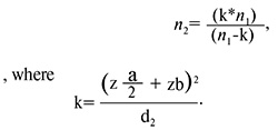
With a standardized difference of d=1.2, the number of patients (n2) was equal to 24. In this group, the structure of PAS was the following: placenta accreta (n=6), placenta increta (n=9), placenta percreta (n=9). The power achieved for this design was 84%.
REBOA technique
Care to women during delivery was provided by a multidisciplinary team, including obstetricians and gynecologists, ultrasound specialist, anesthesiologist, vascular surgeon and neonatologist [12]. The surgery included 3 phases: phase 1 was vascular intervention, phases 2 and 3 included obstetric interventions. Intraoperative reinfusion of autoerythrocytes was used in all patients in this group.
Stage 1 was performed by the vascular surgeon. The right femoral artery was punctured, and 7F introducer sheath was installed. Then the REBOA catheter (MIT) was inserted into the infrarenal segment of the aorta. In stage 2 the obstetrician-gynecologist performed the transverse suprapubic incision. For access to the uterus, we have chosen the transverse incision above the bladder. If the placenta was located near the access to the uterus, the placenta was manually separated with subsequent fetal extraction. Then exteriorization of the uterus was performed. After fetal extraction, the balloon was inflated. The anesthesiologist monitored the REBOA system by tracking systolic blood pressure and oxygen saturation in the toes [13, 14]. The placenta was manually removed by the surgeon. Then the scar was excised together with the area of placental ingrowth and the underlying myometrium within healthy tissues, followed by uterine curettage, if indicated. For devascularization of the lower uterine segment, hemostatic sutures were placed at the internal cervical os. Metroplasty was performed, and the uterine wound was closed with two layers of sutures. After balloon deflation, the catheter was removed, and uterotonics were used to prevent bleeding [15, 16]. Stage 3 included restoration of anterior abdominal wall integrity, femoral access closure using ExoSeal and application of sterile pressure dressing [17, 18].
Tourniquet hemostasis
Complex compression hemostasis as a method of surgical hemostasis and Cell Saver autotransfusion were used during delivery in patients in the comparison group,
Stage 1 of the surgery included incision of the layers of the anterior abdominal wall, sharp or blunt dissection of the bladder and subsequent performance of caesarean section applying fundal pressure, and exteriorization of the uterus. During Stage 2, the windows in the broad ligaments at the uterine isthmus were formed, and tourniquets were applied along the left and right borders of the uterus. The ovaries were transposed laterally, and the third tourniquet fixed with a clamp was placed around the cervix. Maximum cranial uterine traction was performed. The area of adherent placenta along with the myometrium was excised within healthy tissue. The next step of the surgery was performance of curettage in the presence of areas of abnormal placentation in other uterine segments. Two-layer closer was performed to restore the uterine wall defect. Then the tourniquets were removed. Uterotonics were used to prevent bleeding. During Stage 3 the openings in the broad ligaments weree sutured using Vicryl sutures. Bladder integrity check using methylene blue solution and the anterior abdominal wall reconstruction using layered closure were performed. [19, 20].
The quantitative criteria for REBOA performance were the following: successful catheter placement, the time to aortic occlusion, the need for balloon reinsertion, and the frequency vascular complications.
Assessment criteria for the effectiveness of surgical hemostasis techniques were the volume of intraoperative blood loss, the absence of necessity to use additional hemostasis techniques, the level of systolic blood pressure during surgery (not lower than 90 mm Hg), the dynamics of red blood cell counts (erythrocytes, hemoglobin) after surgery and on day 3, no need for relaparotomy, transfusion support and parenteral iron therapy, as well as the length of stay at the Resuscitation and Intensive Care Unit.
The primary endpoint of the study was the volume of intraoperative blood loss (ml), using gravimetric analysis.
The secondary endpoints included the volume of autologous reinfusion (mL), hemoglobin level on the third day after surgery (g/L), the need for transfusion support (%), parenteral iron therapy (%), the rates of hysterectomy (%) and postoperative complications (%). The results of the secondary endpoints were assessed after obtaining the data on the primary endpoints in the study.
Data selection criteria to determine clinical differences between the analyzed surgical hemostasis techniques were prospective and retrospective studies. For the primary endpoint, reduction of intraoperative blood loss of 598.7 mL was considered clinically significant. The obtained results of secondary endpoints were assessed taking into account evaluation of hemostasis parameters: decreased volume of reinfused autoerythrocytes to 300 mL or more, and a decrease in hemoglobin level by 10–15 g/L compared to the baseline data. The indicator of the need for transfusion support in the postpartum period was reduction of the doses of red blood cell suspension by 3.1±5.2 and 416±321 mL of plasma. Additionally, reduction in the rates of hysterectomy and postoperative complications by 28.4% or more was taken into account [10, 11].
Statistical analysis
The data obtained in the study are represented as the median and interguqrtile range (Me [Q1–Q3]). The quantitative data were compared using the Mann–Whitney U test. The qualitative data were compared using Fisher’s exact test. Quantitative indicators were assessed using statistical significance testing (p-value) and the effect size (median difference) with 95% confidence intervals (CI). The qualitative indicators were presented as absolute (n) and relative (%) frequencies. The qualitative indicators were represented as absolute numbers (n) and the percentage (%). The differences were statistically at p<0.05. Statistical processing of data was performed using Python SciPy Stats.
Results
Pregnant women in the studied groups were statistically comparable in age in gestational age (p>0.05) (Table 1). However, body mass index (BMI) was significantly higher in the comparison group (p=0.033). All patients in the groups were multiparous women.

In the comparison group, somatic pathology was in 18/24 (75%) patients and in 7/8 (87.5%) in the main group. In the comparison group, gastrointestinal diseases were detected in 12/24 (50%), urinary system diseases in 3/24 (12.5%), myopia in 4/24 (16.7%), and varicose veins of the lower extremities in 5/24 (20.8%). In the main group, myopia was 3/8 (37.5%) women, varicose veins of the lower extremities in 2/8 (25%), and gastrointestinal diseases in 4/8 (50%).
It should be noted that the signs of PAS were identified by ultrasound examination and magnet resonance imaging (MRI) in all women in the second trimester of pregnancy. The differences were not statistically significant (p=0.456).
In the REBOA group, the volumes of blood loss (p<0.001) and autologous reinfusion (p=0.003) were significantly lower (Fig. 1A, B). Duration of surgery was shorter in the REBOA group than in the comparison group (p=0.035) (Fig. 2). The median time to aortic occlusion was 15.7 [8.0–23.5] minutes. Reinsertion of ballon was required in 1 case. The intraoperative data are represented in table 2.

Additional methods for surgical hemostasis control were used in the comparison group (n=15), including ligation of the internal iliac arteries in 14/24 (58.3%) patients and insertion of the Bakri balloon in 1/24 (4.2%).
There was no difference between the groups in hemoglobin levels before and after operative delivery (p=0.499 and p=0,663, respectively). It was found the on the third postoperative day hemoglobin level was significantly higher in the REBOA group (p=0.036). In the main group, the value of delta hemoglobin (ΔHb) was -2.5. [-5.5–9.5], that reflected the absence of clinically significant reduction in hemoglobin in the postoperative period. In the comparison group, ΔHb was 7.0 [1.5–21.0] g/L, that corresponded to the expected decrease in hemoglobin against the backdrop of larger amounts of blood loss (р=0.064) (Fig. 3). The laboratory data are represented in Table 3.

Transfusion support in the postoperative period was required for 1/8 (12.5%) patient in the REBOA group and for 12/24 (50%) in the comparison group. Respectively, parenteral iron therapy as used in 2/8 (25%) cases in the main group and in 6/24 (25%) cases in the comparison group.
In the REBOA group, the trend toward reduced length of stay at the Resuscitation and Intensive Care Unit, and total length of hospital stay was observed. However, the differences did not rich statistical significance (Table 4).
In operative delivery, hysterectomy was necessary due to massive blood loss, in 6/24 (24%) patients in the comparison group. Hysterectomy was not performed in the REBOA group. However, the differences were not statistically significant (p=0.241), probably due to a small sample size.
The patients had postoperative complications (Table 5). However, there was no statistically significant difference in the frequency of complications between the groups.
In the main group, relaparotomy was performed in one case, in patient with a multiple pregnancy (twins) and severe degree of placenta accreta. According to the results of visual diagnostic methods (ultrasound, MRI), organized hematomas were detected in the uterovesical space, 11.5×9.5×10.4 cm in size, at the left border of the uterys (2.0×2.0×2.2 cm), as well as small hematomas were detected in the Douglas pouch (with a maximum volume of 400 cm3). Due to increasing of inflammatory markers against the backdrop of antibacterial therapy, relaparotomy was performed on the 6th day to open and drain the hematomas.
In the comparison group, relaparotomy was performed in 2 cases due to peritonitis and early postpartum hypotonic bleeding.
It should be noted that higher incidence subgaleal hematomas occurred in the REBOA group – 4/8 (50%) versus 3/24 (12.5%). This difference can be related to the more frequent use of transverse suprapubic incisions in the REBOA group, that could increase the risk of small volume hematomas in the anterior abdominal wall.
Vascular complications associated with the use of REBOA, such as vascular injury, limb ischemia, and arterial thrombosis did not occur.
Assessment of neonatal outcomes showed now statistically significant differences. The obtained data are represented in Table 6. It is important to not that 1 patient in the REBOA group had multiple pregnancy. Due to this, the data are represented for 7 newborns.

Discussion
The study found that REBOA technique is technically feasible. However, we found that the following difficulties can be encounted: balloon catheter placement requires participation of a qualified multidisciplinary team, including endovascular surgeon and ultrasound specialist, to confirm appropriate placement. Currently, this method is not widely used in clinical practice and is performed only in large, specialized centers.
The study demonstrated acceptable level of safety for surgical hemostasis using REBOA, if the protocol and the time of occlusion are followed. In our study, the median time was 15 minutes, that is considered to be a safe time interval. No vascular complications were observed in the REBOA group, that confirms the safety of the method for organ-preserving surgery for PAS. The relaparotomy rate (1/8 (12.5%)) was associated with formation of subgaleal hematoma in patient with severe form of PAS.
The study found preliminary trends toward reduced intraoperative blood loss and operative time using REBOA. The median intraoperative blood loss was 725 mL, compared to 2225 mL in the comparison group. This finding is crucial, since obstetric hemorrhage in PAS is a relevant issue due to its high prevalence and the risk of maternal mortality [2, 6, 20].
Also, in the REBOA group, the volume of autologous reinfusion was lower than in the comparison group (255 mL and 750 mL, respectively). The median hemoglobin level on the 3rd day was 102.5 g/L in the main group, whereas it was 92.5 g/L in the comparison group. The fact that there were no cases of hysterectomy in the REBOA group – 0/8 (0%) is of clinical importance. However, it is noteworthy that hysterectomy rate was higher in patients in the comparison group – 6/24 (25%). It can be assumed that it was due to higher frequency of placenta accreta and placenta percreta in the comparison group, as well as the differences in surgical complexity, intraoperative surgical techniques, organizational factors and their impact on the hysterectomy rate.
The potential of using the REBOA in organ-sparing surgeries in patients with PAS lies in infrarenal aortic occlusion and stopping of blood flow in the uterine arteries, that makes it possible reliably to reduce blood supply to the uterus [9, 11]. The main difference of this method lies in restriction of arterial blood flow not only locally to the uterus, but also distally, that is important in the presence of collateral blood supply or arteriovenous malformations [10]. In tourniquet hemostasis, only uterine devascularization occurs due to compression of uterine and ovarian vessels [12].
A review study of the experience of using REBOA by foreign authors Pinas-Carrillo A. et al. [16], confirms the relevance of using REBOA in organ-sparing surgeries in patients with PAS, but at the same time indicates the need for larger prospective studies. The study by Stensaeth K.H. et al. [9] demonstrated that e REBOA is an effective strategy to control postpartum hemorrhages. A retrospective study by Wei X. et al. [11] reported reduced blood loss in prophylactic balloon occlusion of the aorta in patients with PAS.
Limitations. Our study has a number of limitations. This retrospective study had a small sample size (the REBOA group, n=8), that limits the statistical power of the study and the ability to detect significant differences in a number of parameters. This also results in extremely wide confidence intervals. Due to this, statistical comparisons can be considered primarily as exploratory analysis. Furthermore, due to a small sample size, the study had sufficient power to detect large effects (d≥1.2), while moderate and potentially clinically significant differences could not have been detected. The efficacy assessment is a preliminary evaluation and cannot be extrapolated to clinical practice without confirmation in a randomized clinical trial with a priori sample size calculation.
Another limitation was the volume of intraoperative blood loss as the primary endpoint, due to a high risk of massive obstetric hemorrhage. Maternal mortality was not considered as the primary endpoint, due to extremely low mortality rate associated with PAS in specialized centers.
The results were obtained at one clinical facility and cannot be universal for other healthcare institutions with different levels of equipment and personnel qualifications.
These limitations reduce the universality of the obtained results. Therefore, any conclusions regarding the comparative effectiveness of REBOA are preliminary and require confirmation in a prospective randomized study with a priori sample size calculation and stratified randomization by the severity of PAS
However, the strength of this study include detailed technical description of currently used surgical hemostasis techniques in organ-sparing treatment of PAS, and assessment of the practical feasibility and safety of REBOA.
The issue of wide application of this method in clinical practice remains open due to the need for special equipment and experienced staff.
Conclusion
Over the past decade, a number of innovative techniques have been developed that made it possible to change an approach to patient management with PAS for organ-sparing modality. This study demonstrated the practical feasibility and acceptable safety profile of REBOA in organ-sparing treatment of patients with placenta accreta spectrum.
In the REBOA, the volume of intraoperative blood loss was lower (725/2225 mL), and the volume of autologous reinfusion was lower (255/750 mL), duration of surgery was shorter, and higher hemoglobin levels were on day 3 (102.5/92.5 g/L). Hysterectomy was performed in none of cases in the REBOA group (0/8 (0%) versus 6/24 (25%)). No vascular complications associated with using this hemostasis technique occurred.
However, due to the retrospective study and small sample size, the obtained differences can be regarded as preliminary and cannot be considered as the evidence of the effectiveness of REBOA.
To confirm the obtained results, it is necessary to conduct multicenter prospective randomized studies with a large sample size and to develop standardized protocols for the use of REBOA in management of PAS in obstetric practice.
References
- Jauniaux E., Chantraine F., Silver R.M., Langhoff-Roos J.; FIGO Placenta Accreta Diagnosis and Management Expert Consensus Panel. FIGO consensus guidelines on placenta accreta spectrum disorders: Epidemiology. Int. J. Gynaecol. Obstet. 2018; 140(3): 265-73. https://dx.doi.org/10.1002/ijgo.12407
- Silver R.M., Branch D.W. Placenta accreta spectrum. N. Engl. J. Med. 2018; 378(16): 1529-36. https://dx.doi.org/10.1056/NEJMcp1709324
- Jauniaux E., Ayres-de-Campos D., Langhoff-Roos J., Fox K.A., Collins S.; FIGO Placenta Accreta Diagnosis and Management Expert Consensus Panel. FIGO classification for the clinical diagnosis of placenta accreta spectrum disorders. Int. J. Gynaecol. Obstet. 2019; 146(1): 20-4. https://dx.doi.org/10.1002/ijgo.12761
- Collins S.L., Ashcroft A., Braun T., Calda P., Langhoff-Roos J., Morel O. et al. Proposal for standardized ultrasound descriptors of abnormally invasive placenta (AIP). Ultrasound Obstet. Gynecol. 2016; 47(3): 271-5. https://dx.doi.org/10.1002/uog.14952
- Sentilhes L., Kayem G., Chandraharan E., Palacios-Jaraquemada J., Jauniaux E. for the FIGO Placenta Accreta Diagnosis and Management Expert Consensus Panel. FIGO consensus guidelines on placenta accreta spectrum disorders: Conservative management. Int. J. Gynaecol. Obstet. 2018; 140(3): 291-8. https://dx.doi.org/10.1002/ijgo.12410
- Allen L., Jauniaux E., Hobson S., Papillon-Smith J., Belfort M.A.; FIGO Placenta Accreta Diagnosis and Management Expert Consensus Panel. FIGO consensus guidelines on placenta accreta spectrum disorders: Nonconservative surgical management. Int. J. Gynaecol. Obstet. 2018; 140(3): 281-90. https://dx.doi.org/10.1002/ijgo.12409
- Курцер М.А., Бреслав И.Ю., Барыкина О.П., Скрябин Н.В., Нигматуллина Э.Р. Расползание рубца на матке после кесарева сечения. Акушерство и гинекология. 2022; 2: 59-64. https://dx.doi.org/10.18565/aig.2022.2.59-64 [Kurtser M.A., Breslav I.Yu., Barykina O.P., Skryabin N.V., Nigmatullina E.R. Uterine scar dehiscence following caesarean section. Obstetrics and Gynecology. 2022; (2): 59-64 (in Russian). https://dx.doi.org/10.18565/aig.2022.2.59-64].
- Шмаков Р.Г., Пирогова М.М., Васильченко О.Н., Чупрынин В.Д., Ежова Л.С. Хирургическая тактика при врастании плаценты с различной глубиной инвазии. Акушерство и гинекология. 2020; 1: 78-82. https://dx.doi.org/10.18565/aig.2020.1.78-82 [Shmakov R.G., Pirogova M.M., Vasilchenko O.N., Chuprynin V.D., Ezhova L.S. Surgical management of placenta accreta spectrum with different depths of invasion. Obstetrics and Gynecology. 2020; (1): 78-82 (in Russian). https://dx.doi.org/10.18565/aig.2020.1.78-82].
- Stensaeth K.H., Sovik E., Haig I.N., Skomedal E., Jorgensen A. Fluoroscopy-free resuscitative endovascular balloon occlusion of the aorta (REBOA) for controlling life threatening postpartum hemorrhage. PLoS One. 2017; 12(3): e0174520. https://dx.doi.org/10.1371/journal.pone.0174520
- Peng Z.H., Xiong Z., Zhao B.S., Zhang G.B., Song W., Tao L.X. et al. Prophylactic abdominal aortic balloon occlusion: aneffective method of controlling hemorrhagein patients with placenta previa or accreta. Exp. Ther. Med. 2019; 17(2): 1492-6. https://dx.doi.org/10.3892/etm.2018.7066
- Wei X., Zhang J., Chu Q., Du Y., Xing N., Xu X. et al. Prophylactic abdominal aorta balloon occlusion during caesarean section: a retrospective case series. Int. J. Obstet. Anesth. 2016; 27: 3-8. https://dx.doi.org/10.1016/j.ijoa.2015.12.001
- Xie L., Wang Y., Luo F.Y., Man Y.C., Zhao X.L. Prophylactic use of an infrarenal abdominal aorta balloon catheter in pregnancies complicated by placenta accreta. J. Obstet. Gynaecol. 2017; 37(5): 557-61. https://dx.doi.org/10.1080/01443615.2017.1291588
- Курцер М.А., Григорьян А.М. Временная баллонная окклюзия при врастании плаценты. Эндоваскулярная хирургия. 2020; 7(2): 113-22. https://dx.doi.org/10.24183/2409-4080-2020-7-2-113-122 [Kurtser M.A., Grigoryan A.M. Temporary balloon occlusion in the placenta accreta. Endovascular Surgery. 2020; 7(2): 113-22 (in Russian). https://dx.doi.org/10.24183/2409-4080-2020-7-2-113-122].
- Игнатко И.В., Богомазова И.М., Тимохина Е.В., Белоусова В.С., Федюнина И.А., Карданова М.А., Самара А.Б., Гуцу В. Врастание плаценты: современный взгляд на вопросы этиопатогенеза и акушерской тактики. Акушерство и гинекология. 2024; 1: 5-11. https://dx.doi.org/10.18565/aig.2023.251 [Ignatko I.V., Bogomazova I.M., Timokhina E.V., Belousova V.S., Fedyunina I.A., Kardanova M.A., Samara A.B., Gutsu V. Placenta accreta: a modern view on etiopathogenesis and obstetric tactics. Obstetrics and Gynecology. 2024; (1): 5-11 (in Russian). https://dx.doi.org/10.18565/aig.2023.251].
- Morrison J.J., Galgon R.E., Jansen J.O., Cannon J.W., Rasmussen T.E., Eliason J.L. A systematic review of the use of resuscitative endovascular balloon occlusion of the aorta in the management of hemorrhagic shock. J. Trauma Acute Care Surg. 2016; 80(2): 324-34. https://dx.doi.org/10.1097/TA.0000000000000913
- Pinas-Carrillo A., Bhide A., Moore J., Hartopp R., Belli A.M., Arulkumaran S. et al. Outcomes of the first 50 patients with abnormally invasive placenta managed using the «Triple P Procedure» conservative surgical approach. Int. J. Gynaecol. Obstet. 2020; 148(1): 65-71. https://dx.doi.org/10.1002/ijgo.12990
- Жуков А.И., Заднепровский Н.Н., Иванов П.А., Коков Л.С. О целесообразности применения технологии REBOA для лечения пострадавших с политравмой. Журнал им. Н.В. Склифосовского «Неотложная медицинская помощь». 2024; 13(1): 14-21. https://dx.doi.org/10.23934/2223-9022-2024-13-1-14-21 [Zhukov A.I., Zadneprovsky N.N., Ivanov P.A., Kokov L.S. On the feasibility of using REBOA technology for the treatment of patients with polytrauma. Russian Sklifosovsky Journal "Emergency Medical Care". 2024; 13(1): 14-21 (in Russian). https://dx.doi.org/10.23934/2223-9022-2024-13-1-14-21].
- Ruiz Labarta F.J., Pintado Recarte M.P., Alvarez Luque A., Joigneau Prieto L., Perez Martín L., Gonzalez Leyte M. et al. Outcomes of pelvic arterial embolization inthe management of postpartum haemorrhage: a case series study and systematic review. Eur. J. Obstet. Gynecol. Reprod. Biol. 2016; 206: 12-21. https://dx.doi.org/:10.1016/j.ejogrb.2016.07.510
- Каюмова А.В., Мелкозерова О.А., Башмакова Н.В., Мальгина Г.Б., Чистякова Г.Н., Третьякова Т.Б., Гришкина А.А. Спектр значимых сывороточных биомолекул и генетических предикторов в антенатальной диагностике патологической инвазии плаценты. Акушерство и гинекология. 2023; 12: 5-15. https://dx.doi.org/10.18565/aig.2023.212 [Kayumova A.V., Melkozerova O.A., Bashmakova N.V., Malgina G.B., Chistyakova G.N., Tretyakova T.B., Grishkina A.A. Spectrum of significant serum biomolecules and genetic predictors in antenatal diagnosis of placenta accreta spectrum. Obstetrics and Gynecology. 2023; (12): 5-15 (in Russian). https://dx.doi.org/10.18565/aig.2023.212].
- Баринов С.В., Дикке Г.Б., Шмаков Р.Г. Баллонная тампонада матки в профилактике массивных акушерских кровотечений. Акушерство и гинекология. 2019; 8: 5-11. https://dx.doi.org/10.18565/aig.2019.8.5-11 [Barinov S.V., Dikke G.B., Shmakov R.G. Balloon uterus tamponade in prevention of massive obstetric bleeding. Obstetrics and Gynecology. 2019; (8): 5-11 (in Russian). https://dx.doi.org/10.18565/aig.2019.8.5-11].
Received 01.04.2026
Accepted 22.05.2026
About the Authors
Andrey M. Prikhodko, Dr. Med. Sci., Deputy Director for Research at the Institute of Obstetrics, Head of the Department of Innovative Development, Associate Professor at the Department of Obstetrics and Gynecology, Associate Professor of the Russian Academy of Sciences, obstetrician-gynecologist at the 1st Maternity Department, Academician V.I. Kulakov National Medical Research Center for Obstetrics, Gynecology and Perinatology, Ministry of Health of Russia,117997, Russia, Moscow, Ac. Oparin str., 4, +7(965)344-21-11, andrpri@mail.ru, https://orcid.org/0000-0002-6615-2360
Natalia E. Kan, Professor, Dr. Med. Sci., Deputy Director for Research – Director of the Institute of Obstetrics, Academician V.I. Kulakov National Medical Research Center for Obstetrics, Gynecology and Perinatology, Ministry of Health of Russia, 117997, Russia, Moscow, Ac. Oparin str., 4, kan-med@mail.ru, Researcher ID: B-2370-2015,
SPIN-code: 53788437, Authors ID: 624900, Scopus Author ID: 57008835600, https://orcid.org/0000-0001-5087-5946
Elrad Yu. Amiraslanov, PhD, Head of the Obstetric Department, Academician V.I. Kulakov National Medical Research Center for Obstetrics, Gynecology and Perinatology, Ministry of Health of Russia, 117997, Russia, Moscow, Ac. Oparin str., 4, eldis@mail.ru, https://orcid.org/0000-0001-5601-1241
Vladimir D. Chuprynin, PhD, Head of the Surgical Department, Academician V.I. Kulakov National Medical Research Center for Obstetrics, Gynecology and Perinatology, Ministry of Health of Russia, 117997, Russia, Moscow, Ac. Oparin str., 4, v_chuprynin@oparina4.ru, https://orcid.org/0000-0002-2997-9019
Dmitry M. Akinfiev, endovascular surgeon at the Radiology Department, Academician V.I. Kulakov National Medical Research Center for Obstetrics, Gynecology and Perinatology, Ministry of Health of Russia, 117997, Russia, Moscow, Ac. Oparin str., 4, Author ID: 924170, https://orcid.org/0000-0003-4662-6757
Mikhail V. Ketskalo, PhD, Deputy Director, Director of the National Medical Research Center for Anesthesiology and Reanimatology for Pregnant Women,
Academician V.I. Kulakov National Medical Research Center for Obstetrics, Gynecology and Perinatology, Ministry of Health of Russia, 117997, Russia, Moscow,
Ac. Oparin str., 4, m_ketskalo@oparina4, https://orcid.org/0000-0001-6569-2106
Victor L. Tyutyunnik, Professor, Dr. Med. Sci., Leading Researcher at the Research and Development Service, Academician V.I. Kulakow National Medical Research Center for Obstetrics, Gynecology and Perinatology, Ministry of Health of Russia, 117997, Russia, Moscow, Ac. Oparin str. 4, tioutiounnik@mail.ru, Researcher ID: B-2364-2015,
SPIN-code: 1963-1359, Authors ID: 213217, Scopus Author ID: 56190621500, https://orcid.org/0000-0002-5830-5099
Angelina A. Tsukanova, clinical resident, Academician V.I. Kulakov National Medical Research Center for Obstetrics, Gynecology and Perinatology, Ministry of Health of Russia, 117997, Russia, Moscow, Ac. Oparin str., 4, an-c-00@mail.ru, https://orcid.org/0009-0007-6827-8470
Corresponding author: Andrey M. Prikhodko, andrpri@mail.ru
Similar Articles

