1) Rostov State Medical University, Ministry of Health of Russia, Rostov-on-Don, Russia;
2) Perinatal Center, Rostov-on-Don, Russia
Background: Placenta accreta spectrum (PAS) is an advanced invasive placentation, which is a serious condition associated with high maternal mortality due to massive uterine hemorrhages. These complications can be reduced by early diagnosis of PAS.
Case report: A 35-year-old multiparous patient with a burdened obstetric and gynecological history had indirect signs of abnormally deep invasion of the chorion which were revealed during ultrasound examination at 7+1 weeks gestation. The ultrasound scan showed a heterogeneous structure with the expansion of lacunar spaces and areas of hypervascularization of the myometrium of the anterior uterine wall. The diagnosis of abnormal placental attachment was confirmed by dynamic echography with stereoscopic blood flow imaging (LumiFlow). A planned cesarean section by transverse uterine fundal incision was performed at 37+2 weeks. Metroplasty was performed on both sides after ligation of the internal iliac and ovarian arteries. The diagnosis of PAS was confirmed by a pathology study, and placenta increta (PAS 3a) was verified.
Conclusion: The presented clinical observation clearly demonstrates the real possibility of early ultrasound diagnosis of advanced invasive placentation. The detection of signs of advanced invasive placentation at the earliest possible time of gestation suggests that patients can be referred to a high-risk PAS group for the subsequent search for specific signs of this placental pathology and optimal planning of organ-preserving methods of delivery in these patients.
Authors’ contributions: Volkov A.E. – developing the concept and design of the study, obtaining the data, their analysis and interpretation, writing the text of the article, surgical treatment of the patient; Solonchenko A.S. – ultrasound diagnosis of the case in early pregnancy; Rymashevsky A.N. – developing the concept and design of the study, scientific editing of the text, surgical treatment of the patient, Voloshin V.V. – carrying out morphological research, editing the obtained data, writing the text; Khloponina A.V. – echography of a case in late pregnancy, Kantsurova M.R. – management of the patient in the postoperative period.
Conflicts interest: The authors declare that there are no conflicts of interest.
Funding: The investigation has not been sponsored.
Ethical Approval: The study was approved by the Ethical Review Board of the Rostov State Medical University, Ministry of Health of the Russian Federation, Rostov-on-Don.
Patient Consent for Publication: The patient provided informed consent for the publication of her data and associated images.
For citation: Volkov A.E., Solonchenko A.S., Rymashevsky A.N., Voloshin V.V., Khloponina A.V., Kantsurova M.R. Early diagnosis of abnormally deep invasion of the chorion as a predictor of invasive placentation.
Akusherstvo i Ginekologiya/Obstetrics and Gynecology. 2024; (3): 180-186 (in Russian)
https://dx.doi.org/10.18565/aig.2024.33
abnormal chorionic invasion
placenta accreta spectrum
diagnosis
Аномально инвазивная плацентация (АИП) [врастание плаценты, приращение плаценты, placenta accreta spectrum (PAS)] – патологическое состояние, формирующееся в ранние сроки гестации вследствие имплантации бластоцисты, которое патоморфологически определено полным или частичным отсутствием децидуальной оболочки (decidua basalis), приводящим к приращению или прорастанию ворсинами хориона/плаценты мышечного слоя матки [1].
Актуальность обсуждаемой проблемы определена рядом причин. Во-первых, возросшей в последние годы распространенностью данной аномалии плацентации (примерно 1,7 на 10 000 беременностей) [2, 3], явившейся следствием лавинообразного роста частоты проведения кесарева сечения (КС). Частота операций КС в мире увеличилась более чем в 3 раза за последние 30 лет, и на сегодняшний день составляет около 25%; в некоторых клинических учреждениях третьего уровня Российской Федерации данный показатель превышает 40% [4]. Во-вторых, ассоциированностью PAS с акушерскими кровотечениями, как следствие, – с материнской смертностью (до 52%) [5, 6] и, в заключение, – трудностями диагностики. По данным Гуса А.И. и соавт., до родов PAS не диагностируется в 2/3 случаев, при том, что в 1/3 случаев это происходит в специализированных многопрофильных стационарах [7]; в то же время в 28% случаев пренатально сформулированный диагноз PAS не имеет гистопатологического подтверждения [8].
В настоящее время основным методом диагностики АИП является ультразвуковое исследование (УЗИ) [9–11]. Пионерами ультразвуковой диагностики АИП является группа американских специалистов, которые в 25 недель гестации на фоне маточного кровотечения диагностировали предлежание плаценты с аномально отсутствующей гипоэхогенной ретроплацентарной зоной. Диагноз АИП был подтвержден при морфологическом исследовании удаленной матки [12].
По данным D’Antonio E. et al., при УЗИ у 3907 беременных с риском АИП отмечена высокая эффективность метода с чувствительностью 90,7% (95% ДИ 87,2–93,6) и специфичностью 96,94% (95% ДИ 96,3–97,5) при диагностическом отношении шансов 98,59% (95% ДИ 48,8–199,0) [13].
Долгое время приоритетным сроком ультразвуковой диагностики АИП являлся III триместр беременности [13]. В последнее десятилетие прослеживается тенденция к уменьшению срока обнаружения АИП. Обнаружение предикторов АИП возможно, по мнению Panaitova J. et al., в сроке 12–16 недель [14]. Блинов А.Ю. и соавт. представили описание случая пренатальной диагностики истинного приращения предлежащей плаценты во время скринингового УЗИ в 13 недель и 4 дня у беременной с отягощенным акушерским анамнезом [15]. В 2020 г. Демидов В.Н. и соавт. высказали мнение, что диагноз АИП в большинстве случаев может быть установлен уже в 14–19 недель беременности [16].
Мотивация к «уменьшению сроков» диагностики обсуждаемой проблемы следующая: качественное, успешное лечение АИП в значительной мере предопределено максимально ранней ее диагностикой, позволяющей своевременно сформулировать оптимальный алгоритм действий, определяющий место, сроки, технологии лечения и состав команды специалистов, принимающих участие в родоразрешении такой пациентки. Учитывая сказанное, представляем собственный клинический опыт ранней ультразвуковой диагностики аномально глубокой инвазии хориона – предиктора врастания плаценты, верифицированного в последующем при патологоанатомическом исследовании.
Клиническое наблюдение
Пациентка М., 35 лет. Менархе с 14 лет, менструальная функция без особенностей. Настоящая беременность – пятая. В анамнезе одни физиологические роды per vias naturales в срок (2009 г.), два артифициальных аборта в I триместре (2011 г. и 2021 г.) и одно КС в 2019 г. (отслойка нормально расположенной плаценты).
В сроке 7 недель обратилась к гинекологу для постановки на диспансерный учет по беременности. На данном этапе пациентка жалоб не предъявляла, состояние расценено как удовлетворительное.
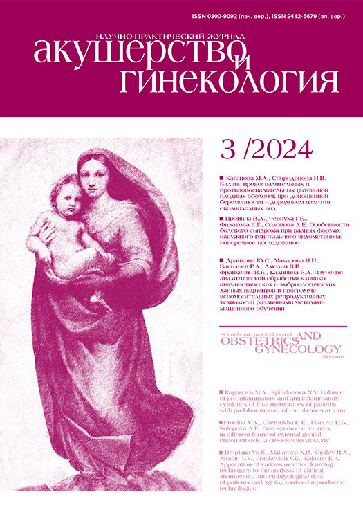
В сроке гестации 7+1 недель при эхографии в ГБУ Ростовской области «Перинатальный центр» было описано следующее: «...локализация хориона по передней стенке матки в области послеоперационного рубца, структура хориона неоднородная с расширенными лакунарными пространствами, визуализируются участки гиперваскуляризации миометрия передней стенки матки, толщина миометрия в области «рубца» 2,1 мм» (рис. 1а, б). Сформулировано заключение: беременность 7+1 недель, эхографические признаки врастания хориона в область послеоперационного рубца.
При эхографии в рамках раннего пренатального скрининга описана «низкая локализация плаценты». Визуализация с применением технологии цветового допплеровского картирования не проводилась.
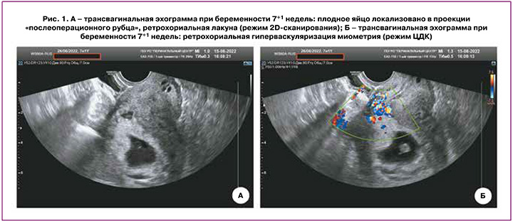
При дальнейшей эхографии, выполненной в отделении ультразвуковой диагностики НИИАП РостГМУ в сроках 17, 19, 22, 25, 30 недель гестации, заключение об аномальном прикреплении хориона в ранние сроки трансформировалось в формулировку о PAS. Так, в сроке 19 недель описано следующее: плацента перекрывает область внутреннего зева, располагаясь по передней стенке матки в проекции послеоперационного рубца, слева визуализируются множественные ретроплацентарные лакуны с ламинарным кровотоком, достигающие зоны истонченного миометрия, в режиме ЦДК – гиперваскуляризация, на участке 50 мм в области «рубца» миометрий отчетливо не лоцируется, субплацентарно в режиме стереоскопической визуализации кровотока (LumiFlow) обнаружены сосуды, достигающие задней стенки мочевого пузыря, не проникающие в него (рис. 2 а, б). На основании проведенного исследования сформулировано заключение о предлежании плаценты, осложненном АИП (placenta increta, PAS 3a по версии Jauniaux E. et al. [10]).
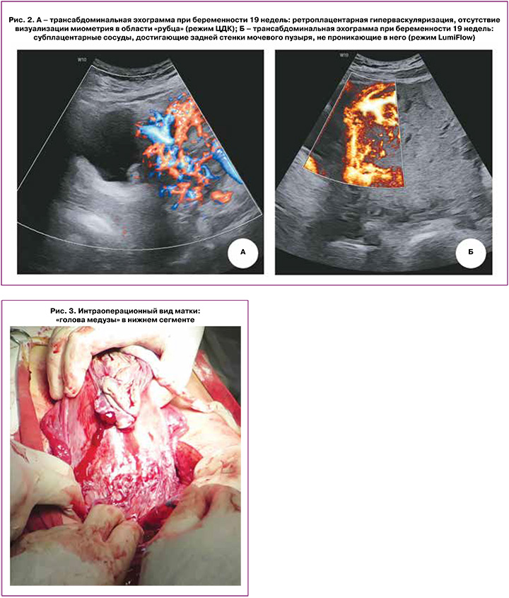
В сроке 37+2 недель в плановом порядке беременность была завершена оперативными родами абдоминальным путем. Под эпидуральной анестезией выполнено донное КС. Родилась доношенная девочка массой 2500 г, длиной 38 см с оценкой по Апгар 7/8 баллов. После рождения ребенка без попыток отделения плаценты осмотрены органы малого таза. Идентифицированы признаки АИП – сосудистая «голова медузы» нижней трети передней стенки тела матки (рис. 3). После лигирования внутренних подвздошных и яичниковых артерий с обеих сторон произведена метропластика. Иссечена часть передней стенки матки с плацентой. Тело матки восстановлено непрерывными швами по авторской методике [17]. Кровопотеря составила 1800 мл. Интраоперационно использована система для реинфузии крови (Sell Saver). Гемотрансфузия в послеоперационном периоде не проводилась. На 7-е сутки в удовлетворительном состоянии пациентка была выписана.
При патологоанатомическом исследовании иссеченной части передней стенки матки выявлены следующие изменения. Большую часть препарата занимала зона волокнистой соединительной ткани толщиной от 1,2 до 0,2 см местами плотной, местами рыхлой. К рубцовой ткани с выраженным ангиоматозом прилежала зона фибриноидного некроза с клетками трофобласта и частично некротизированными терминальными ворсинами. Децидуальная ткань на этом участке истончена, а местами отсутствовала (рис. 4а). На остальном протяжении децидуальная ткань неравномерной толщины с кровоизлияниями, обширными зонами фибриноидного некроза Рора и Нитабух, с образованием глубоких «бухт», приникающих в миометрий до ⅔ толщины, содержащих ворсины и клетки трофобласта (рис. 4б). Между гладкомышечными волокнами и в соединительной ткани были обнаружены клетки синцитио- и цитотрофобласта (рис. 4в). В промежуточных ворсинах плаценты, окруженных фибриноидом, – десквамация эпителия, облитерационная ангиопатия части артерий. Терминальные ворсины хориона с фиброзом стромы, ангиоматозом, образованием синцитиокапиллярных мембран и синцитиальных почек, в гипертрофированном миометрии – гравидарная трансформация артерий (рис. 4г).
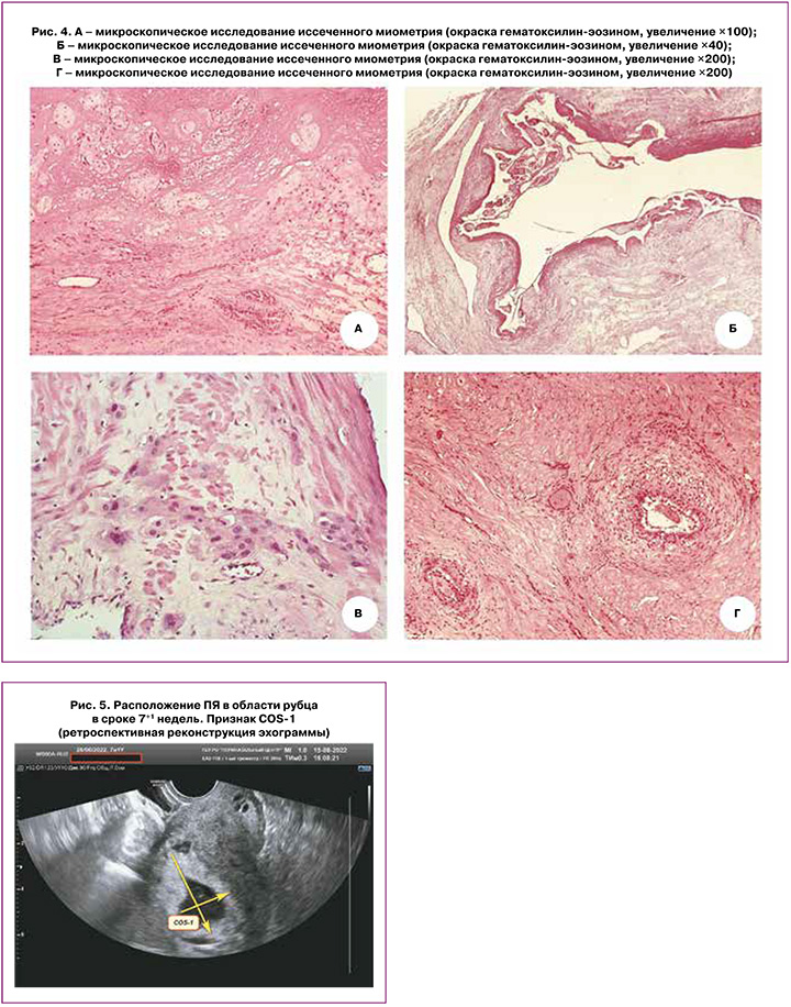
Заключение: послеоперационный рубец в стенке гравидарно трансформированной матки, приращение плаценты (placenta increta), вторичная хроническая плацентарная недостаточность.
Обсуждение
Представленное клиническое наблюдение наглядно демонстрирует реальную возможность ранней ультразвуковой диагностики АИП. Таким ультразвуковым признаком для прогнозирования PAS в ранние сроки беременности в 2017 г. Cali G. et al. [18] определили признак «перекрестка» (crossover sign, COS). В основе его – связь между расположением плодного яйца (ПЯ) при сроках 6–8 недель беременности и рубцом на матке после КС. Для оценки этого признака в сагиттальном сечении проводится прямая линия, соединяющая область внутреннего зева и дно матки и проходящая через эндометрий. Вторая линия проводится перпендикулярно эндометрию через верхний и нижний полюсы ПЯ, имплантированного в область рубца на матке после КС. В зависимости от расположения ПЯ выделяют несколько возможных вариантов, один из которых – COS-1. В этом случае ПЯ расположено на 2/3 своего диаметра выше линии эндометрия по направлению к передней стенке матки. По данным авторов, COS-1 в 100% случаев был ассоциирован с тяжелыми вариантами PAS [18].
Ретроспективно проведенная нами оценка этого признака (рис. 5) продемонстрировала, что в 7+1 недель ПЯ было эктопически расположено на ⅔ своего диаметра выше линии эндометрия по направлению к передней стенке матки в проекции рубца после КС. Таким образом, имел место признак COS-1, предполагающий высокую вероятность PAS 3, что и подтвердилось в дальнейшем.
Заключение
Обнаружение признаков АИП в максимально ранние сроки гестации предполагает отнесение пациенток в группу высокого риска формирования PAS для последующего целенаправленного поиска специфических признаков данной плацентарной патологии, способствующих в дальнейшем оптимальному планированию органосохраняющих приемов родоразрешения этих пациенток. Использование современного ультразвукового оборудования, «заряженного высоким диагностическим потенциалом», в руках подготовленного, мотивированного, ориентированного на целенаправленный поиск данной патологии специалиста – залог успешной ранней диагностики АИП, предполагающей улучшение акушерских и перинатальных исходов.
- Jauniaux E., Collins S., Burton G.J. Placenta accreta spectrum: pathophysiology and evidence-based anatomy for prenatal ultrasound imaging. Am. J. Obstet. Gynecol. 2017; 218(1): 75-87. https://dx.doi.org/ 10.1016/j.ajog.2017.05.067.
- Silver R.M. Abnormal placentation: placenta previa, vasa previa, and placenta accreta. Obstet. Gynecol. 2015; 126(3): 654-68. https://dx.doi.org/10.1097/ AOG.0000000000001005.
- Fitzpatrick K.E., Sellers S., Spark P., Kurinczuk J.J., Brocklehurst P., Knight M. Incidence and risk factors for placenta accreta/increta/percreta in the UK: a national case-control study. PLoS One. 2012; 7(12): e52893. https://dx.doi.org/10.1371/joumal.pone.0052893.
- Шмаков Р.Г., Баев О.Р., Пекарев О.Г.. Пырегов А.В., Приходько А.М., Павлович С.В. Кесарево сечение: показания, хирургическая техника, методы обезболивания. Учебное пособие НЦ акушерства, гинекологии и перинатологии им. акад. В.И. Кулакова. М.: «Радуга»; 2016: 71. [Shmakov R.G., Baev O.R., Pekarev O.G., Pyregov A.V., Prikhod’ko A.M., Pavlovich S.V. Caesarean section: indications, surgical technique, methods of anesthesia. Textbook of the Scientific Centre of Obstetrics, Gynecology and Perinatology named after academician V.I. Kulakov. Moscow: Raduga; 2016: 71. (in Russian)].
- Say L., Chou D., Gemmill A., Tunqalp O., Moller A.B., Daniels J. et al. Global causes of maternal death: a WHO systematic analysis. Lancet Glob. Health. 2014; 2(6): e323-33. https://dx.doi.oiE/10.1016/S2214-IO9X(14)7O227-X.
- Результаты конфиденциального аудита материнской смертности в Российской Федерации в 2016 году (методическое письмо). М.: Департамент медицинской помощи детям и службы родовспоможения Министерства здравоохранения Российской Федерации; 2017: 35. [Results of confidential audit of maternal mortality in the Russian Federation in 2016 (methodological letter). M.: Department of Medical Care for Children and Maternity Services of the Ministry of Health of the Russian Federation, 2017: 35. (in Russian)].
- Гус А.И., Бойкова Ю.В., Ярыгина Т.А., Яроцкая Е.Л. Современные подходы к пренатальной диагностике и скринингу врастания плаценты (обзор рекомендаций). Акушерство и гинекология. 2020; 10: 5-12. [Gus A.I., Boikova Yu.V., Yarygina T.A., Yarotskaya E.L. Modern approaches to prenatal diagnosis and screening of placenta accreta (review of recommendations). Obstetrics and Gynecology. 2020; (10): 5-12. (in Russian)]. https://dx.doi.org/10.18565/aig.2020.10.5-12.
- Eller A., Porter T.F., Soisson P., Silver R.M. Optimal management strategies for placenta accreta. BJOG. 2009; 116(5): 648-54. https://doi.org/10.1111/j.1471-0528.2008.02037.x.
- Jauniaux E., Collins S.L., Jurkovic D., Burton G.J. Accreta placentation: a systematic review of prenatal ultrasound imaging and grading of villous invasiveness. Am. J. Obstet. Gynecol. 2016; 215(6): 712-21. https://dx.doi.org/ 10.1016/j.ajog.2016.07.044.
- Jauniaux E., Chantraine F., Silver R.M., Langhoff-Roos J.; FIGO Placenta Accreta Diagnosis and Management Expert Consensus Panel. FIGO consensus guidelines on placenta accreta spectrum disorders: epidemiology. Int. J. Gynecol. Obstet. 2018; 140(3): 265-73. https://dx.doi.org/10.1002/ijgo.12407.
- Jauniaux E., Bhide A. Prenatal ultrasound diagnosis and outcome of placenta previa accreta after cesarean delivery: a systematic review and meta-analysis. Am. J. Obstet. Gynecol. 2017; 217(1): 27-36. https://dx.doi.org/10.1016/j.ajog.2017.02.050.
- Tabsh K.M., Brinkman C.R., King W. Ultrasound diagnosis of placenta increta. J. Clin. Ultrasound. 1982; 10(6): 288-90. https://dx.doi.org/10.1002/jcu.1870100610.
- D’Antonio F., Iacovella C., Bhide A. Prenatal identification of invasive placentation using ultrasound: systematic review and meta-analysis. Ultrasound Obstet. Gynecol. 2013; 42(5): 509-17. https://dx.doi.org/10.1002/uog.13194.
- Panaitova J., Tokunaka M., Krajewska K., Zosmer N., Nicolaides K.H. Screening for morbidly adherent placenta in early pregnancy. Ultrasound Obstet. Gynecol. 2019; 53(1): 101-6. https://dx.doi.org/10.1002/uog.20104.
- Блинов А.Ю., Гольцфарб В.М., Долгушина В.Ф. Ранняя пренатальная диагностика истинного приращения плаценты. Пренатальная диагностика. 2011; 10(1): 79-84. [Blinov A.Yu., Gol’tsfarb V.M., Dolgushina V.F. Early prenatal diagnosis of true placental increment. Prenatal Diagnosis. 2011; 10(1): 79-84. (in Russian)].
- Демидов В.Н., Гус А.И., Ярыгина Т.А. О возможности высокоточной диагностики врастания плаценты в рубец на матке после кесарева сечения при ультразвуковом исследовании. Пренатальная диагностика. 2020; 19(4): 336-42. [Demidov V.N., Gus A.I., Yarygina T.A. About the possibility of high-precision diagnostics of placenta ingrowth into the uterine scar after cesarean section with ultrasound examination. Prenatal Diagnosis. 2020; 19(4): 336-42. (in Russian)]. https://dx.doi.org/10.21516/2413-1458-2020-19-4-336-342.
- Рымашевский А.Н., Рымашевский М.А., Келлер О.В., Волков А.Е., Канцурова М.Р. Способ ушивания ложа миоматозного узла. Патент на изобретение. Номер патента: RU 2790964 C2. Патентное ведомство: Россия. Номер заявки: 2021108736. Дата регистрации: 30.03.2021. Дата публикации: 28.02.23. Патентообладатели: Ростовский государственный медицинский университет Министерства здравоохранения Российской Федерации. [Rymashevsky A.N., Rymashevsky M.A., Keller O.V., Volkov A.E., Kantsurova M.R. Method of suturing the bed of the myomatous node. Patent for the invention. Patent number: RU 2790964 C2. Patent office: Russia. Application number: 2021108736. Registration date: 30.03.2021. Publication date: 28.02.23. Patent holders: Rostov State Medical University of the Ministry of Health of the Russian Federation. (in Russian)].
- Cali G., Forlani F., Timor-Tritsch I.E., Palacios-Jaraquemada J., Minneci G., D'Antonio F. Natural history of Cesarean scar pregnancy on prenatal ultrasound: the crossover sign. Ultrasound Obstet. Gynecol. 2017; 50(1): 100-4. https://dx.doi.org/10.1002/uog.16216.
Received 12.02.2024
Accepted 13.03.2024
Andrey E. Volkov, PhD (Med.), Associate Professor, Department of Obstetrics and Gynecology No. 1, Rostov State Medical University, Ministry of Health of Russia,
344022, Russia, Rostov-on-Don, Nakhichevan Lane, 29, +7(863)234-24-23,
avolkov@aaanet.ru, https://orcid.org/0000-0002-5899-1252
Alexey S. Solonchenko, ultrasound diagnostics doctor at the Department of Antenatal Fetal Protection, Rostov Regional Perinatal Center, 344068, Russia, Rostov-on-Don, Bodraya str., 90, +7(863)235-50-18,
a-solonchenko@mail.ru, https://orcid.org/0009-0000-9971-5521
Alexander N. Rymashevsky, Dr. Sci. (Med.), Professor, Head of the Department of Obstetrics and Gynecology No. 1, Rostov State Medical University, Ministry of Health of Russia, 344022, Russia, Rostov-on-Don, lane. Nakhichevan Lane, 29, +7(863)234-24-23,
rymashevskyan@mail.ru, https://orcid.org/0000-0003-3349-6914
Vladimir V. Voloshin, PhD (Med.), Associate Professor, Rostov State Medical University, Ministry of Health of Russia, 344022, Russia, Rostov-on-Don, Nakhichevan Lane, 29, +7(863)234-24-23,
voloshinvv2006@yandex.ru, https://orcid.org/0000-0001-8632-082X
Anna V. Khloponina, Dr. Sci. (Med.), Head of the Department of Ultrasound Diagnostics of NIIAP, Rostov State Medical University, Ministry of Health of Russia,
344022, Russia, Rostov-on-Don, Nakhichevan Lane, 29, +7(863)201-44-31,
annakhloponina@yandex.ru, https://orcid.org/0000-0002-2056-5231
Maria R. Kantsurova, Assistant at the Department of Obstetrics and Gynecology No. 1, Rostov State Medical University, Ministry of Health of Russia,
344022, Russia, Rostov-on-Don, Nakhichevan Lane, 29, +7(863)234-24-23,
madlax_san@mail.ru, https://orcid.org/0000-0003-4916-8042
Corresponding author: Andrey E. Volkov,
avolkov@aaanet.ru