
The role of vascular endothelial growth factor receptors (VEGFR1, VEGFR2) in the development of placental insufficiency in women with recurrent miscarriage
Protsenko E.V., Talanova I.E., Malyshkina A.I., Kulida L.V.
Objective. Exploration of the role of vascular endothelial growth factor receptors (VEGFR1, VEGFR2) in formation of placental insufficiency in women with recurrent miscarriage (RM).
Materials and methods. A total of 347 pregnant women were examined: 247 women with RM, and 104 with a history of pregnancy without RM. Of the total number of women, 75 placental samples underwent histological and immunohistochemical examinations with antibodies to VEGFR1 and VEGFR2. Placental samples that underwent pathomorphological examination were divided into 3 groups. Group 1 included placentas of women with RM, who gave birth at term (n=25). Group 2 included placentas of women who gave at 260–366 weeks (n=20). Group 3 (the comparison group) included placentas of 30 patients without a history of RM, who gave birth at 37–40 weeks. Statistical processing of the results was performed using Excel-2010 and STATISTICA 13.0 software packages.
Results. The women with RM had complications during pregnancy. The most common complication of pregnancy was threatened miscarriage in all trimesters of pregnancy that ended with preterm birth. The average gestational age for preterm birth was 311 (260–363) weeks. Immunohistochemical analysis of placental samples showed that in preterm birth, expression of VEGFR1 in decidual cells and VEGFR2 in syncytiotrophoblast of chorial villi was significantly lower (p=0.0002) compared with full-term pregnancy. In addition, preterm birth was associated with premature placental abruption, that commonly occurs with failure of physiologic transformation of spiral arteries.
Conclusion. The most frequent pregnancy complications in women with RM miscarriage were threatened miscarriage with high rates of preterm delivery and placental insufficiency. According to our data, the mechanisms of formation of pregnancy complications in women with RM are associated with low expression of VEGFR1 and VEGFR2 in the placenta, leading to impaired gestational restructuring of the uteroplacental arteries and vascularization of the villous chorion, that should be considered as an element of chronic placental insufficiency and taken into account at the pregravid stage.
Authors' contributions. Protsenko E.V., Kulida L.V. – study concept and design; Talanova I.E. – material collection and processing, statistical data processing; Protsenko E.V., Kulida L.V., Talanova I.E., Malyshkina A.I. – manuscript writing; Malyshkina A.I. – manuscript editing.
Conflicts of interest. The authors confirm that they have no conflict of interest to declare.
Funding. The study was carried out without any sponsorship.
Ethical Approval. The study was approved by the local Ethics Committee of V.N. Gorodkov Ivanovo Research Institute of Maternity and Childhood, Ministry of Health of Russia.
Generative Artificial Intelligence. No generative AI was used in preparing this article.
Patient Consent for Publication. The patients have signed informed consent for publication of their data.
Authors' Data Sharing Statement. The data supporting the findings of this study are available on request from the corresponding author after approval from the principal investigator.
For citation: Protsenko E.V., Talanova I.E., Malyshkina A.I., Kulida L.V. The role of vascular endothelial growth factor receptors (VEGFR1, VEGFR2) in the development of placental insufficiency in women with recurrent miscarriage.
Akusherstvo i Ginekologiya/Obstetrics and Gynecology. 2026; (6): 60-67 (in Russian)
https://dx.doi.org/10.18565/aig.2025.349
Keywords
recurrent miscarriage
preterm birth
placenta. placental insufficiency
VEGFR1
VEGFR2
Recurrent miscarriage (RM), also known as recurrent pregnancy loss is one of the most important medical and social problems worldwide, accounting for 15%–20.0% of reproductive losses in the population. Despite significant advances in medical science in the world, the incidence of RM remains unchanged, affecting 2–5% of married couples of reproductive age [1, 2]. The causes of recurrent miscarriage include chromosomal abnormalities, endocrine and immunological diseases, changes in hemostasis, endometrial receptivity, placental vasculogenesis and angiogenesis, congenital uterine anomalies, and sexually transmitted infections. However, in many cases recurrent miscarriage is an idiopathic recurrent pregnancy loss [3]. It is also important that patients with RM are at risk for obstetric complications, the great obstetrical syndromes (GOS) [4, 5]. The study by Linehan L.A. et al. showed that pregnancy outcomes associated with recurrent miscarriage include high incidence of early or very early preterm birth, fetal growth restriction (FGR), placental abruption, resulting in high incidence of perinatal morbidity and mortality [6]. These complications are undoubtedly associated with impaired placental development, which is a multistage process, where vasculogenesis and angiogenesis play a key role [7]. In recent years, a number of studies have been published that evaluated the impact of placental, transforming, insulin-like, vascular endothelial growth factors on placental development in normal and pathological pregnancies [8]. The role of growth factor receptors in genesis of the placenta remains understudied. The data about location of VEGFR1 and VEGFR2 and mechanisms influencing differentiation of the villous chorion and placental maturity are debatable, including in recurrent miscarriage [9, 10]. Therefore, the objective of the study was exploration of the role of vascular endothelial growth factor receptors (VEGFR1, VEGFR2) in formation of placental insufficiency in women with RM.
Materials and methods
The study was carried out at the Clinic's Obstetric Department, V.N. Gorodkov Ivanovo Research Institute of Maternity and Childhood of the Ministry of Health of Russia. A total of 347 pregnant women were examined from November 2022 to January 2025, who were divided into 2 group. The main group consisted of pregnant women, who had a history of RM, and were followed-up in current pregnancy. The comparison group consisted of 104 pregnant women without a history of RM. Continuous sampling method was used in the study.
Inclusion criteria were the diagnosis of RM; women with a history of 2 or more successive terminations of pregnancy before 22 weeks, prior to current pregnancy; patient’s informed voluntary consent to participate in the study. Non-inclusion criteria in the main group were spontaneous abortion; multiple pregnancy; pregnancy after assisted reproductive technologies; infertility before the onset of current pregnancy; congenital uterine anomalies; structural rearrangements in married couples’ karyotypes; acute or chronic infectious and inflammatory diseases at the time of examination; decompensated external genital pathology; antiphospholipid syndrome, thrombophilia with a high risk of thromboembolic complications; severe allergic reactions at the time of examination.
All patients underwent comprehensive clinical and laboratory examinations according to Order of the Ministry of Health of the Russian Federation of October 20, 2020 No. 1130n "On approval of the procedure for the provision of medical care in the field of obstetrics and gynecology", and clinical recommendations “Recurrent miscarriage” (2022). All patients were followed up during pregnancy and in the postpartum period.
Placental samples from 75 women of the total number of the study participants underwent morthological examination (with observation of delivery and material fixation conditions), including histological and immunohistochemical examinations with VEGFR1 and VEGFR2 antibodies. These placental samples were divided into 3 groups. Group 1 included placentas from women with RM, who had term births (n=25). Group 2 included placentas from women who gave birth at 260–366 weeks (n=20). Group 3 (the comparison group) included placentas from 30 patients without a history of RM, who gave birth at 37–40 weeks.
For morphological study, the fragments (0.5×0.5 cm) containing the basal plate with the villous chorion 0.5×0.5 cm were cut from the provisional organ, fixed in 10% buffered formalin and, after alcohol treatment, embedded in paraffin blocks for microtomy. Parafin-embeded tissue blocks were sectioned into 4-5 μm thick histological sections and stained with hematoxylin and eosin. Immunohistochemical (IHC) examination of 15 placental samples from each group was performed according to the standard protocol. Sections from paraffin blocks were treated with anti-VEGFR1 (Ab-AF6204) and anti-VEGFR2 (Ab-AF628) polyclonal rabbit antibodies at a working dilution of 1:100 (Affinity). The expression of VEGF receptors VEGFR1 and VEGFR2 in decidual cells was assessed using microscope at 400x magnification in 100 cells of the preparation. The results were presented in numerical scores: 0 – no staining, 1 – weak staining, 2 and 3 – moderate and strong staining intensity, respectively. For objective assessment of immunohistochemical analysis of decidual cell reactions, the expression index (EI) was calculated for each biomarker using the formula:
EI=∑P(i) × i/100 conventional units,
where i – the intensity of IHC staining in scores from 0 до 3, Р(i) – the percentage of different staining of cells [11]. The expression of biomarkers in the syncytiotrophoblast (STB) (taking into account its histological structure) was assessed using only semi-quantitative method with staining intensity scoring – negative (0), positive weak (1), moderate (2) and strong (3). Positive and negative controls were used.
Statistical analysis
Statistical data processing was performed using Excel-2010, STATISTICA 13.0, MedCalc 7.4.4.1. software packages. Distribution of the quantitative data was represented as the median and interquartile range Ме (Q1; Q3). Comparison of quantitative data of three independent groups was performed using the Kruskal–Wallis test with subsequent pairwise comparison of the groups using the Mann–Whitney U test. The significance of qualitative data was assessed using Pearson’s chi-squared test with Yate’s correction and Fisher’s exact test. The level of statistical significance was considered at р<0.05. Three groups were compared at significance level of p<0.017 with Bonferroni correction.
Results
Analysis showed that the age of patients in both groups was comparable. The median age of patients in the main group was significantly higher compared with the control group (Table 1). At the same time, among examined women with RM, the number of women of the age group over 30 years prevailed – 190/243 (78%) versus 39/104 (37.5%), р=0.000.
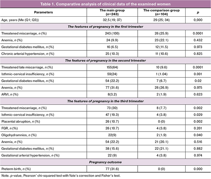
The course of pregnancy in patients in the main group was accompanied by complications (Table 1). In the third trimester, threatened miscarriage occurred more often versus the comparison group. In the second trimester, pregnancy complications, such as threatened late miscarriage, isthmic-cervical insufficiency, were most common. Also, high frequency of gestational diabetes mellitus was observed. Analysis of the course of pregnancy in the third trimester in the main group showed that most common complication was threatened preterm birth. Placental abnormalities, such as placental abruption, oligohydramnios (as a sign of placental insufficiency), and FGR were more common in the group of patients with a history of RM in the third trimester of pregnancy versus the comparison group. The frequency of other complications was similar in both groups. Every third woman in the main group experienced preterm birth, while there were no preterm births in the comparison group. The average gestational age for preterm birth was 311 (260–363) weeks. Therefore, the most common pregnancy complication in women with RM was threatened miscarriage, that occurred in all trimesters of pregnancy and resulted in preterm births, and placental disorders.
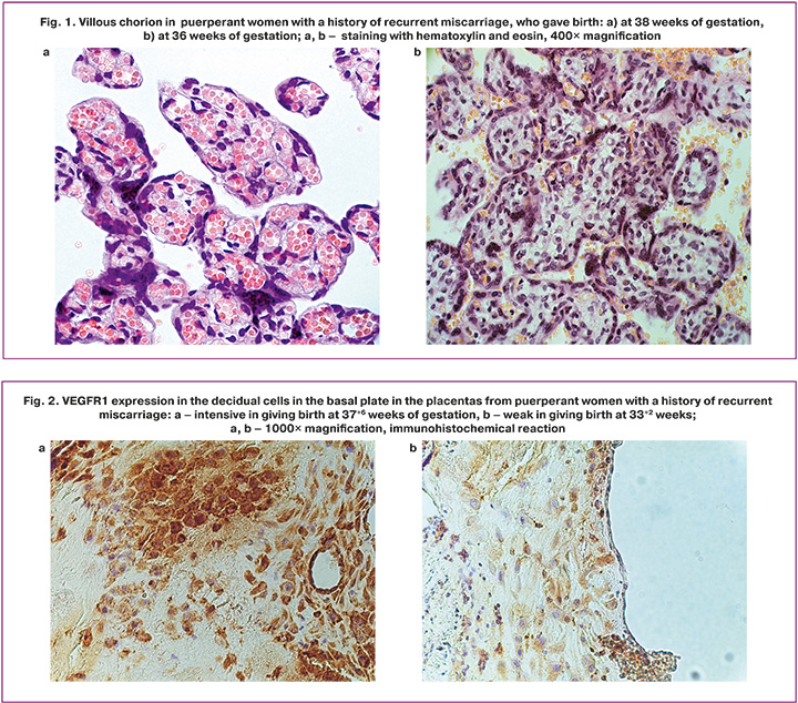
Histological evaluation of the placentas in group 1 (full-term pregnancy in women with RM) showed changes, such as impairment of chorionic villous differentiation related to the dissociative type – 6/25 (24%), regional abnormalities of placental villi associated with perivillous fibrin deposition – 15/25 (60%), chronic inflammation in the basal plate of the placenta – 12/25 (48%). Compensatory processes including hyperplasia in terminal villi, villous capillaries and vasculosyncytial membranes effectively ensuring exchange processes between the maternal and fetal blood flows were observed in 22/25 (88%) women (Fig. 1, a). Different combinations of the above pathological changes formed the structural basis of chronic placental insufficiency (CPI), which was compensated in 19/25 (76%) and subcompensated in 6/25 (24%) women. There was no difference between the groups in CPI (p=0.08). In the control group, similar types of chronic placental insufficiency were diagnosed in 24/30 (80%) and 6/30 (20%) cases, respectively.
Morphological criteria for compensated CPI due to impairment of chorionic villous differentiation and perivillous fibrin deposition included focal localization of these pathological processes and severe compensatory reactions, such as hyperplasia of the capillaries in terminal villi and vasculosyncytial membranes (the markers of specialized terminal villi). In subcompensated CPI, compensatory processes were focal and included hyperplasia in the syncytial knots and hyperemia in the capillaries of intermediate and terminal villi.
In the placentas in group 2 (preterm births in the third trimester in women with RM), impairment of chorionic villous differentiation related to the dissociative type was in 5/20 (25%) cases, immature intermediate villi in 7/20 (35%), infarctions in 7/20 (35%), chronic deciduitis, chorioamnionitis and villusitis that led to inflammatory obliterative vasculopathy (Fig. 1, b) – in 13/20 (65%). Acute chorioamnionitis and membranitis were detected in 2/20 (10%) cases. Compensatory processes were limited to formation of syncytial knots and focal hyperemia in chorionic vessels. Placental insufficiency was diagnosed in all cases of preterm birth, including acute placental insufficiency associated with widespread hemorrhages and damage to basal plate – 13/20 (65%), as well as subcompensated and decompensated CPI – 3/20 (15%) and 4/20 (20%), respectively. CPI occurred due to impaired vascularization of chorionic villi and low number of vasculosyncytial membranes in terminal villi.
Immunohistochemical reaction with anti-VEGFR1 and VEGFR2 antibodies in placental samples was most clearly manifested in the STB in intermediate and terminal villi, as well as in decidual cells (DCs) in the basal plate.
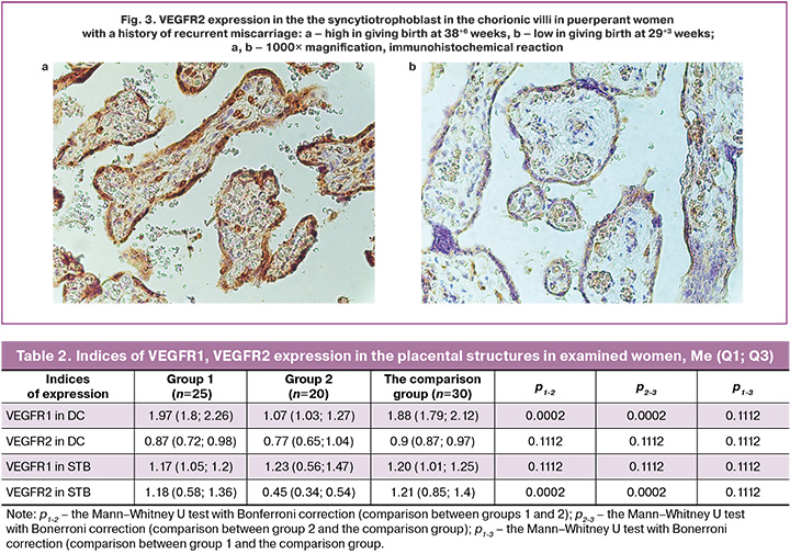
In Group 1 and in the comparison group, VEGFR1 expression in the basal plate of the placentas was seen as distinct, diffuse, fine-grained, membranous-cytoplasmic staining of brown color in the majority of decidual cells (score=2) (Fig. 2, a). Group 2 was characterized by uneven, weak, light brown staining of the majority of DCs (score=1) (Fig. 2, b). Calculation of the indices of biomarker expression in the groups showed that in full-term pregnancy (group 1 and the comparison group), the expression of VEGFR1 in DCs was significantly higher (p1-2=0.0002, p2-3=0.0002) versus preterm birth (Table 2).
VEGFR1 expression in the STB in the placentas in Groups 1, 2 and the comparison group was moderate (++) according to semiquantitative scoring system. It was seen as linear, and in some regions discontinuous staining of the apical membrane of the syncytiotrophoblast and uneven staining of the cytoplasm. No intergroup differences were found in staining intensity of the STB (р1-2=0.1112, р2-3=0.1112, р1-3=0.1112).
Examination of placentas with anti-VEGFR2 antibodies verified strong (+++) immunohistochemical reaction in the STB in chorionic villi in full-term pregnancy (group 1 and the comparison group): continuous linear staining of the apical membrane and diffuse, fine-grained brown staining of the cytoplasm of the STB, and accumulation of DAB-positive inclusions in the region of syncytial knots (Fig. 3, a; Table 2). Biomarker expression in preterm birth exhibited uneven, fine-grained, pale brown staining of the cytoplasm of the STB, that corresponded to weak (+) immunohistochemical reaction. The intergroup differences were statistically significant (Fig. 3, b; Table 2).
Biomarker expression in DCs in the basal plate of the placentas in groups 1, 2, and the comparison group was weak, without significant intergroup differences (р1-2=0.1112, р2-3=0.1112, р1-3=0.1112).
Discussion
It is known that the main mechanism of pregnancy prolongation is normal transformation of the uterine spiral arteries promoting maternal blood flow to the intervillous space [8]. Therefore, the factors promoting adequate maternal vascular remodeling are also important. Given the antiangiogenic properties of VEGFR1 (reduced proliferation and migration of endothelial cells), it is logical to assume that high expression of this biomarker in DCs in the basal plate of the placenta (the expression index = 1.97 (1,7; 2.25) conventional units)) prevents formation of vessels with normal histological structure in the basal plate, which are sensitive to vasopressors. This assumption is consistent with the concept of spiral artery remodeling in pregnancy.
Preterm birth in the third trimester of pregnancy was accompanied by decreased (by 1.5 times) VEGFR1 expression in the DCs compared with full-term pregnancy, and in most cases – 13/25 (65%)) was combined with intradecidual hemorrhages, which are morphologically identical to premature placental abruption. The obtained results indicate relationship between VEGFR1 expression level in the placental basal plate, maternal vascularization of the placenta and the duration of pregnancy.
According to literature data, VEGFR2 is expressed in vascular endothelial cells, stromal cells in chorionic villi. The proangiogenic effect is mediated by interaction with VEGF, promoting endothelial cell proliferation and the formation of new blood vessels [12]. Our study found that in full-term pregnancy (group 1 and the comparison group) VEGFR2 was intensively synthesized in the STB in chorionic villi, that was associated with hyperplasia and the peripheral location of sinusoidal capillaries. It is probable that VEGFR2 promotes not only proliferation, but also endothelial cell migration to the peripheral regions of the chorionic villi, where new blood vessels contribute to the formation of vasculosyncytial membranes that are known to determine placental maturity [13, 14]. It was found that in women with preterm birth with a history of recurrent miscarriages VEGFR2 expression in the STB in chorionic villi was low (+), in combination with impaired vascularization of the villous chorion (inflammatory obliterative vasculopathy). Given the obtained results, we believe that high VEGFR2 expression in the STB in chorionic villi is a necessary condition for adequate angiogenesis in the villous chorion, that is important for pregnancy prolongation.
Conclusion
High rate of pregnancy complications was among with recurrent miscarriage. According to the results of the examinations, threatened miscarriage was the most common pregnancy complication in this cohort of women, that led to preterm births and placental abnormalities.
Expression of endothelial growth factor receptors VEGFR-1 and VEGFR-2 2 in the placentas from women with RM in full-term pregnancy and preterm birth differed significantly.
High expression of VEGFR1 and VEGFR2 was in the placenta from women with RM and term birth, as well as women in the comparison group. This is probably one of the important conditions providing adequate spiral artery remodeling and chorionic angiogenesis necessary for pregnancy prolongation and successful completion. Low expression of VEGFR1 and VEGFR2 in the placenta of patients with RM and preterm birth is associated with impaired utero-placental vascular remodeling during gestation and vascularization of the villous chorion, that should be considered as an element of chronic placental insufficiency. These findings should be considered in preconception care for women with RM when planning subsequent pregnancies, especially in cases of repeated reproductive failures.
References
- Министерство здравоохранения Российской Федерации. Клинические рекомендации. Привычный выкидыш. 2025. [Ministry of Health of the Russian Federation. Clinical guidelines. Recurrent miscarriage. 2025 (in Russian)].
- Тетруашвили Н.К. Привычный выкидыш. Акушерство и гинекология: новости, мнения, обучение. 2017; 4: 70-87. https://dx.doi.org/10.24411/2303-9698-2017-00010 [Tetruashvili N.K. Recurrent abortion. Obstetrics and Gynecology: News, Opinions, Training. 2017; 4: 70-87 (in Russian). https://dx.doi.org/10.24411/2303-9698-2017-00010].
- Vomstein K., Aulitzky A, Strobel L., Bohlmann M., Feil K., Rudnik-Schöneborn S. et al. Recurrent spontaneous miscarriage: a comparison of international guidelines. Geburtshilfe Frauenheilkd. 2021; 81(07): 769-79. https://dx.doi.org/10.1055/a-1380-3657
- Ali N., Elbarazi I., Ghazal-Aswad S., Al-Maskari F., Al-Rifai R., Oulhaj A. et al. Impact of recurrent miscarriage on maternal outcomes in subsequent pregnancy: The Mutaba'ah Study. Int. J. Womens Health. 2020; 12: 1171-9. https://dx.doi.org/10.2147/IJWH.S264229
- Колесникова А.М., Малышкина А.И., Таланова И.Е. Особенности анамнеза, течения гестации и перинатальных исходов у женщин с привычным невынашиванием беременности. Вестник Ивановской медицинской академии. 2024; 29(2): 32-8. https://dx.doi.org/10.52246/1606-8157_2024_29_2_32 [Kolesnikova A.M., Malyshkina A.I., Talanova I.E. Features of the anamnesis, course of gestation and perinatal outcomes in women with habitual miscarriage. Bulletin of the Ivanovo Medical Academy. 2024, 29(2): 32-8 (in Russian). https://dx.doi.org/10.52246/1606-8157_2024_29_2_32].
- Linehan L.A., San Lazaro Campillo I., Hennessy M., Flannery C., O'Donoghue K. Reproductive outcomes following recurrent first-trimester miscarriage: a retrospective cohort study. Hum. Reprod. Open. 2022; 2022(4): hoac045. https://dx.doi.org/10.1093/hropen/hoac045
- Приходько Н.Г. Роль факторов роста в инвазии трофобласта и их ассоциация с патологическим течением беременности. Бюллетень физиологии и патологии дыхания. 2019; 74: 111-8. https://dx.doi.org/10.36604/1998-5029-2019-74-111-118 [Prikhodko N.G. The role of growth factors in trophoblast invasion and their association with the pathological course of pregnancy. Bulletin of Respiratory Physiology and Pathology. 2019; 74: 111-8 (in Russian). https://dx.doi.org/10.36604/1998-5029-2019-74-111-118].
- Хомякова Е.В., Зиганшина М.М., Баев О.Р. Факторы, регулирующие ангио/васкулогенез плаценты при осложненном течении беременности и родов. Акушерство и гинекология. 2023; 9: 12-21. https://dx.doi.org/10.18565/aig.2023.149 [Khomyakova E.V., Ziganshina М.М., Baev O.R. Factors regulating placental angio/vasculogenesis in complications of pregnancy and childbirth. Obstetrics and Gynecology. 2023; (9): 12-21 (in Russian). https://dx.doi.org/10.18565/aig.2023.149].
- Redline R.W., Boyd T.K., Roberts D.J., eds. Placental and gestational pathology. Cambridge University Press; 2018; 350 p. https://dx.doi.org/10.1017/9781316848616
- Brosens I., Puttemans P., Benagiano G. Placental bed research: 1. The placental bed. From spiral arteries remodeling to the great obstetrical syndromes. American Journal of Obstetrics and Gynecology. 2019; 221(5): 437-56. https://doi.org/10.1016/j.ajog.2019. 05.044
- Толибова Г.Х., Траль Т.Г., Клещев М.А., Кветной И.М., Айламазян Э.К. Эндометриальная дисфункция: алгоритм гистологического и иммуногистохимического исследования. Журнал акушерства и женских болезней. 2015; 64(4): 69-77. [Tolibova G.Kh., Tral' T.G., Kleshchov M.A., Kvetnoy I.M., Aylamazyan E.K. Endometrial dysfunction: an algorithm for histological and immunohistochemical studies. Journal of Obstetrics and Women's Diseases. 2015; 64(4): 69-77 (in Russian)].
- Abu-Ghazaleh N., Brennecke S.; Murthi P., Karanam V. Association of vascular endothelial growth factors (VEGFs) with recurrent miscarriage: a systematic review of the literature. Int. J. Mol. Sci. 2023; 24(11): 9449. https://dx.doi.org/10.3390/ijms24119449
- Молчанова О.К., Ордиянц И.М., Луценко И.В., Подставкина В.А. Современные представления о молекулярных механизмах регуляции имплантации. Акушерство и гинекология: новости, мнения, обучение. 2020; 8(3) Приложение: 106-11. https://dx.doi.org/10.24411/2303-9698-2020-13917 [Molchanova O.K., Ordiyants I.M., Lucenko I.V., Podstavkina V.A. Modern concepts of molecular mechanisms of regulation of implantation. Obstetrics and Gynecology: News, Opinions, Training. 2020; 8(3) Supplement: 106-11 (in Russian). https://dx.doi.org/10.24411/2303-9698-2020-13917].
- Зенкина В.Г., Сахоненко В.А., Зенкин И.С. Патоморфологические особенности плаценты на разных этапах гестации. Современные проблемы науки и образования. 2019; 6: 129-38. [Zenkina V.G., Sakhonenko V.A., Zenkin I.S. Pathomorphological features of the placenta at different stages of gestation. Modern problems of science and education. 2019; 6: 129-38 (in Russian)].
Received 03.12.2025
Accepted 14.05.2026
About the Authors
Elena V. Protsenko, Dr. Med. Sci., Head of the Laboratory of Pathomorphology and Electron Microscopy, Ivanovo Research Institute of Maternity and Childhood named after V.N. Gorodkov, Ministry of Health of Russia, 153045, Russia, Ivanovo, Pobedy str., 20, SPIN: 1343-3881, Author ID: 160250, https://orcid.org/0000-0003-0490-5686Iya E. Talanova, PhD, Associate Professor at the Department of Obstetrics, Gynecology, and Medical Genetics, Ivanovo State Medical University, Ministry of Health of Russia, 153000, Ivanovo, Sheremetevsky Ave., 8, +7(910)982-05-48, iya-ta@yandex.ru, SPIN: 6865-5146, Author ID: 884142, https://orcid.org/0000-0003-4950-8174
Anna I. Malyshkina, Professor, Dr. Med. Sci., Director, Ivanovo Research Institute of Maternity and Childhood named after V.N. Gorodkov, Ministry of Health of Russia, 153045, Russia, Ivanovo, Pobedy str., 20; Head of the Department of Obstetrics, Gynecology and Medical Genetics, Ivanovo State Medical University of the Ministry of Healthcare of the Russian Federation, , +74932336263, anna_im@mail.ru, SPIN: 7937-9125, AuthorID:150323, https://orcid.org/0000-0002-1145-0563
Lyudmila V. Kulida, Dr. Med. Sci., Leading Researcher at the Laboratory of Pathomorphology and Electron Microscopy, Ivanovo Research Institute of Maternity and Childhood named V.N. Gorodkov, Ministry of Health of Russia, 153045, Russia, Ivanovo, Pobedy str., 20, SPIN: 6208-4487, Author ID: 160249, https://orcid.org/0000-0001-8962-9048
Corresponding author: Iya E. Talanova, iya-ta@yandex.ru
Similar Articles

