
Comparative evaluation of embryological outcomes following conventional IVF and ICSI in couples with unexplained infertility: a single-center prospective study
Ishchuk M.P., Perminova S.G., Mityurina E.V., Filimonov S.F., Sannikova E.S.
Objective. To evaluate embryological and clinical outcomes in IVF cycles among couples with unexplained infertility following conventional in vitro fertilization (IVF) versus intracytoplasmic sperm injection (ICSI).
Materials and methods. This single-center prospective comparative study included 116 assisted reproductive technology cycles in couples with unexplained infertility, treated between January 2024 and January 2026. Conventional IVF was performed in 58 cycles and ICSI were performed in 58 cycles each. Cycles with split insemination and those involving preimplantation genetic testing for aneuploidy (PGT-A) were excluded from the analysis. The primary outcome was the normal fertilization rate. The secondary outcomes included the blastulation rate, proportion of good-quality blastocysts, implantation rate, pregnancy rate, and clinical pregnancy rate. To account for potential confounding, adjusted analyses were performed using generalized linear models, and pregnancy rates were compared using Fisher's exact test.
Results. Baseline clinical characteristics were generally comparable between the groups, although the starting gonadotropin dose was higher in the ICSI group. Sperm concentration, total motility, and proportion of mature oocytes were higher in the conventional IVF group. No statistically significant between-group differences were observed in the primary outcome, the normal fertilization rate. Likewise, no significant differences were found in the blastulation rate or the proportion of high-quality blastocysts. Implantation, pregnancy, and clinical pregnancy rates were numerically higher in the ICSI group; however, these differences were not statistically significant. After multivariable adjustment, ICSI was not associated with an increased normal fertilization rate (adjusted odds ratio [aOR] 0.96; 95% confidence interval [CI] 0.74–1.25; p=0.763), blastulation rate (aOR 0.77; 95% Cl 0.59–1.01; p=0.063), good-quality blastocyst formation (aOR 0.80; 95% Cl 0.49–1.31; p=0.370), or implantation rate (aOR 1.01; 95% CI 0.003–330; p=0.962).
Conclusion. In patients with unexplained infertility, ICSI was not associated with improved normal fertilization rates or most embryological outcomes compared with conventional IVF. These findings do not support the routine use of ICSI in couples with unexplained infertility and suggest that a selective approach to choosing a fertilization method is warranted.
Authors’ contributions. Perminova S.G., Ishchuk M.P. – conception and design of the study; Ischuk M.P., Mityurina E.V., Filimonov S.F., Sannikova E.S. – material collection and processing; Ishchuk M.P. – statistical analysis, drafting of the manuscript; Perminova S.G., Mityurina E.V. – editing of the manuscript; Filimonov S.F., Sannikova E.S. – embryological stage; Ishchuk M.P., Perminova S.G., Mityurina E.V., Filimonov S.F., Sannikova E.S. – approval of the final version of the manuscript.
Conflicts of interest. The authors have no conflicts of interest to declare.
Funding. There was no funding for this study.
Ethical Approval. The study was reviewed and approved by the Research Ethics Committee of the V.I. Kulakov NMRC for OG&P, Ministry of Health of Russia (Ref. No. 1 from January 25, 2024).
Generative Artificial Intelligence. A generative artificial intelligence tool (ChatGPT, DoTrace) was used during manuscript preparation solely for editorial and technical formatting and for harmonizing the manuscript structure with the journal’s formatting requirements.
Patient Consent for Publication. All patients provided informed consent for the publication of their data.
Authors' Data Sharing Statement. The data supporting the findings of this study are available upon request from the corresponding author after approval from the principal investigator.
For citation: Ishchuk M.P., Perminova S.G., Mityurina E.V., Filimonov S.F., Sannikova E.S.
Comparative evaluation of embryological outcomes following conventional IVF and ICSI
in couples with unexplained infertility: a single-center prospective study.
Akusherstvo i Ginekologiya/Obstetrics and Gynecology. 2026; (6): 163-171 (in Russian)
https://dx.doi.org/10.18565/aig.2026.122
Keywords
unexplained infertility
IVF
ICSI
fertilization
blastulation
implantation
assisted reproductive technology
Despite substantial advances in reproductive medicine, unexplained infertility (UI) remains one of the most challenging conditions for which couples seek assisted reproductive technology (ART) treatment to address. According to the European Society of Human Reproduction and Embryology (ESHRE, 2023), the prevalence of UI among couples undergoing standard infertility workup may reach 30% [1]. UI is most commonly regarded as a diagnosis of exclusion, established when standard investigations fail to identify a cause of infertility in the absence of evident abnormalities in either the female or male reproductive system (patent fallopian tubes, ovulatory menstrual cycles, normal semen parameters, and no significant pelvic pathology detected by laparoscopy or hysteroscopy) [1–3].
The diagnostic complexity of UI lies in its potential to stem from abnormalities that affect the molecular mechanisms of fertilization and early embryogenesis. These can include impaired genomic and epigenetic integrity of gametes, defective acrosome reaction, failure of oocyte activation associated with disrupted calcium-dependent signaling pathways, inadequate cytoplasmic maturation of the oocyte, and disturbances in early embryonic development.
UI appears to represent not a single condition but rather a heterogeneous group of pathophysiological mechanisms that remain undetected by routine clinical and laboratory evaluations [1, 4].
In ART programs for patients with UI, the choice of fertilization method –conventional in vitro fertilization (IVF) or intracytoplasmic sperm injection (ICSI) – remains one of the most debated issues. Importantly, ICSI was originally developed to overcome severe male-factor infertility and has demonstrated high efficacy specifically in this clinical setting [5]. However, the indications for ICSI have expanded considerably over the subsequent decades. According to the ESHRE European Registry, 72.7% of IVF cycles performed in Europe in 2019 utilized ICSI, reflecting the predominance of this technique over conventional IVF methods. A similar trend has been observed in the United States, where ICSI use across all indications increased from 36.4% in 1996 to 76.2% in 2012, with the most pronounced growth occurring in cycles without male factor infertility [3, 6].
The use of ICSI in patients without male factor infertility has traditionally been justified by the desire to reduce the risk of total fertilization failure and bypass potential defects in sperm-oocyte interaction. A systematic review and meta-analysis by Johnson et al. (2013), which included 11 studies and more than 11,700 oocytes, found that the likelihood of fertilization was higher with ICSI than with conventional IVF (OR=1.49; 95% CI 1.35–1.65), while the risk of complete fertilization failure was significantly lower compared with conventional IVF (OR=8.22 for IVF versus ICSI) [7].
However, as evidence has accumulated, support for the routine use of ICSI has become less convincing. A Cochrane review (2023) found no differences between ICSI and IVF among couples with normal sperm concentration and motility, either in fertilization rates (RR 1.00; 95% CI 0.97–1.02) or implantation rates (RR 0.94; 95% CI 0.83–1.06) [8]. Similar conclusions were reported in a large randomized trial by Dang V.Q. et al. (2021), which included 1,064 couples without significant male-factor infertility. The authors found no difference in fertilization failure rates between ICSI and IVF (6% vs. 5%; p=0.60) [9]. Comparable results were obtained in a multicenter randomized study by Wang Y. et al. (2024), which included more than 2,300 patients [10].
This debate is of particular interest in couples with UI, as they lack clear indications for the selection of ICSI. In a study by Bachurin A.V. et al. (2022), fertilization in the UI group was performed using ICSI in 80.3% of cases; however, fertilization rates were comparable between IVF and ICSI (61.2% vs. 76%) [11].
Iwamoto A. et al. (2024) likewise demonstrated no advantage of ICSI over IVF: the ratio of 2PN zygotes to retrieved oocytes was 59.7% with ICSI and 60.9% with IVF, while the proportion of transferred or cryopreserved embryos relative to the number of 2PN zygotes was virtually identical (49.4% vs. 49.6%) [12]. An analysis of the Human Fertilization and Embryology Authority (HFEA) registry by Paffoni A. et al. (2024) showed that ICSI was associated with a higher fertilization rate and a lower cycle cancellation rate; however, among single-embryo transfers, implantation rates were higher following conventional IVF than after ICSI (38.7% vs. 36.9%) [13]. This position is further supported by contemporary systematic reviews emphasizing that, in the absence of significant pathozoospermia, the use of ICSI is frequently not supported by evidence of clinical benefits [8, 14].
An additional argument in favor of ICSI in some ART centers is its use in cycles planned for preimplantation genetic testing for aneuploidy (PGT-A), based on the assumption that it may reduce the risk of contamination by extraneous genetic material. Nevertheless, current evidence does not confirm a clear benefit of ICSI in PGT-A cycles with respect to increasing cumulative live birth rates in the overall ART population, and the concept of its widespread routine use remains a subject of ongoing debate [15, 16].
Thus, whether ICSI offers advantages over IVF in terms of embryological outcomes and pregnancy rates in couples with UI remains a clinically important and unresolved question.
This study aimed to compare the embryological outcomes, implantation, pregnancy, and clinical pregnancy rates following conventional IVF and ICSI in couples with UI.
Materials and methods
A single-center prospective comparative study was conducted among patients with UI undergoing ART at the F. Paulsen Research and Educational Center for ART, Clinical Department, Academician V.I. Kulakov National Medical Research Center for Obstetrics, Gynecology and Perinatology.
Between January 2024 and January 2026, a total of 2,820 ART cycles were analyzed. Of these, 275 cycles were performed in couples with UI. The present study included 116 couples with UI: 58 underwent conventional IVF, and 58 underwent ICSI. Cycles involving a mixed fertilization strategy within the same protocol (n=35) and cycles including PGT-A (n=124) were excluded from the study. Study groups were formed according to the selected fertilization method, and no randomization was performed.
The inclusion criteria were as follows: established diagnosis of UI; female age ≤37 years; anti-Müllerian hormone (AMH) level ≥1.0 ng/mL; retrieval of at least five oocytes in the ART cycle; and a normal karyotype in both partners.
The exclusion criteria were rescue ICSI (re-ICSI), cycles using donor gametes, and cycles with planned PGT. All participants provided written informed consent for study participation and personal data processing prior to their enrollment.
Controlled ovarian stimulation was performed using recombinant follicle-stimulating hormone (rFSH) in a gonadotropin-releasing hormone (GnRH) antagonist protocol. The starting dose of rFSH was individualized according to ovarian reserve parameters and patient body weight, with subsequent dose adjustments based on the ultrasound monitoring findings. To prevent a premature luteinizing hormone (LH) surge, a GnRH antagonist was administered subcutaneously at a dose of 0.25 mg/day once the follicle reached a diameter of 14 mm. Final oocyte maturation was triggered when follicles reached ≥17–18 mm using either human chorionic gonadotropin (hCG) 10,000 IU intramuscularly or 250 μg subcutaneous recombinant hCG (r-hCG). Oocyte retrieval was performed 35–37 hours later by transvaginal ovarian aspiration.
Ejaculate samples were prepared using a double-density gradient technique, followed by centrifugation at 250 g for 10 min. Oocytes were then inseminated conventionally at a concentration of 55,000 spermatozoa per oocyte or fertilized via ICSI.
Following fertilization, zygotes were cultured individually in 25-μL droplets of G-TL medium (Vitrolife) covered with OVOIL culture oil in 60-mm Falcon culture dishes. Embryo culture was performed in a benchtop MINC incubator (USA) under standard low-oxygen conditions (≥6.5% CO₂, 5% O₂, 37°C). Fertilization assessment was performed 16–18 h after insemination. Normal fertilization was defined as the presence of two pronuclei (2PN) in metaphase II (MII) oocytes.
Embryo quality on day 3 was evaluated based on the blastomere number and symmetry, degree of fragmentation, and multinucleation status. Embryos with 6–8 evenly sized blastomeres and minimal fragmentation were classified as high-quality embryos. Blastocyst quality was assessed on day 5 of culture, according to the Gardner classification. High-quality blastocysts were defined as those with an expansion grade ≥3 and inner cell mass and trophectoderm grades of at least B (≥3BB). Poor-quality embryos were defined as blastocysts with an expansion grade ≤2 and/or an inner cell mass and/or trophectoderm grade of C (including 3BC, 3CB, 3CC, and lower grades) [17].
Embryo transfer was performed on either day 3 or day 5, depending on the embryo number and quality. Embryos were cryopreserved when there was a risk of developing ovarian hyperstimulation syndrome. Serum β-hCG levels were measured 12–14 days after the embryo transfer. In cases of positive β-hCG, clinical pregnancy was confirmed by ultrasound examination 21 days after the embryo transfer. Following confirmation of an intrauterine pregnancy, micronized progesterone therapy was continued until 8–10 weeks of gestation, and treatment was discontinued in cases of biochemical pregnancy or absence of pregnancy.
The primary endpoint was the normal fertilization rate. The secondary endpoints included the blastulation rate, rates of good- and excellent-quality blastocyst formation, implantation rate, pregnancy rate, and clinical pregnancy rate. The fertilization rate was calculated as the number of 2PN zygotes divided by the number of MII oocytes ×100%. Blastulation rate was calculated as the number of blastocysts divided by the total number of zygotes ×100%. The rate of good-quality blastocyst formation was defined as the number of good-quality blastocysts divided by the total number of blastocysts ×100%. The implantation rate was calculated as the number of gestational sacs divided by the total number of transferred embryos ×100%. The pregnancy rate was defined as the number of cycles with a positive β-hCG result divided by the number of cycles in which embryo transfer was performed ×100%. The clinical pregnancy rate was calculated as the number of cycles with ultrasound visualization of a gestational sac divided by the total number of embryo transfers ×100%.
Statistical analysis
Statistical analysis was performed using IBM SPSS Statistics version 27.0. The Shapiro–Wilk test was used to assess the normality of the quantitative variables. Normally distributed continuous variables are presented as mean (M) and standard deviation (SD), reported as M (SD). Non-normally distributed variables are presented as median (Me) and interquartile range [Q1; Q3], reported as Me [Q1; Q3]. Between-group comparisons of continuous variables were performed using either Student’s t-test or the Mann–Whitney U test, depending on the data distribution. Categorical variables are presented as absolute counts and frequencies and were compared using Pearson’s χ² test or Fisher’s exact test, as appropriate. Correlations were assessed using Spearman’s rank correlation coefficients. To evaluate the effect of fertilization method on embryological outcomes while accounting for potential confounders, adjusted analyses were performed using generalized linear models with binomial distribution and logit link. For the primary endpoint (normal fertilization rate), the dependent variable was the ratio of 2PN zygotes to MII oocytes. The same approach was applied to the analyses of the blastulation rate and good-quality blastocyst formation. The pregnancy and clinical pregnancy rates were compared between groups using Fisher’s exact test because both outcomes were analyzed as binary variables at the transfer cycle level and were characterized by a small number of events. The covariates included in the models for normal fertilization rate, blastulation rate, and proportion of good- and excellent-quality blastocysts were patient age, AMH level, starting gonadotropin dose, sperm concentration, and total sperm motility. The implantation rate was analyzed using binomial regression adjusted for patient age and day of embryo transfer. The adjusted results are reported as odds ratio (OR) with 95% confidence interval (CI). Statistical significance was set at p<0.05. All available cycles that met the eligibility criteria were included in the analysis.
Results
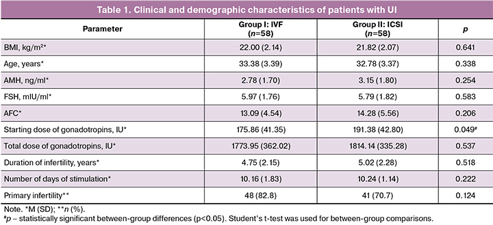
The clinical and laboratory characteristics of the patients and the parameters of the stimulated cycle are presented in Table 1. The mean age of patients in Groups I and II was comparable, at 33.38 (3.39) and 32.78 (3.37) years, respectively (p=0.338). This finding is of particular importance because female age is one of the key determinants of assisted reproductive technology (ART) success. It is well established that, after 34–35 years of age, the likelihood of achieving and maintaining a pregnancy decreases by approximately 10% per year [18]. The conventional IVF and ICSI groups were also comparable with respect to body mass index (BMI), anti-Müllerian hormone (AMH) levels, follicle-stimulating hormone (FSH) levels, antral follicle count (AFC), total gonadotropin dose, duration of infertility, duration of ovarian stimulation, and the proportion of patients with primary infertility. The only statistically significant difference between groups was observed in the starting gonadotropin dose, which was higher in the ICSI group than in the IVF group (191.38 [42.80] IU vs. 175.86 [41.35] IU, respectively; p=0.049). Embryological outcomes are presented in Table 2.
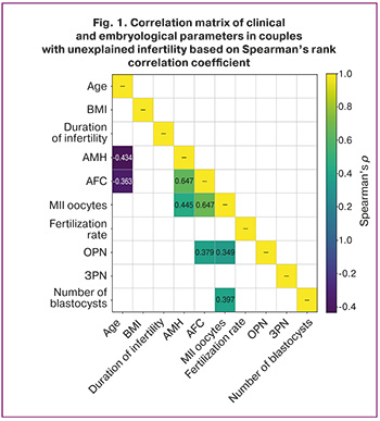
To further investigate the relationships between clinical and embryological parameters, Spearman correlation analysis was performed in the combined patient cohort (Figure 1). Statistically significant positive correlations were identified between AMH levels and AFC (ρ=0.647; p<0.001), AFC and the number of mature MII oocytes (ρ=0.674; p<0.001), and AMH levels and the number of MII oocytes (ρ=0.445; p<0.001). Patient age was negatively correlated with AMH levels (ρ=-0.434; p<0.001) and AFC (ρ=-0.363; p<0.001). The number of MII oocytes was positively correlated with the total number of blastocysts (ρ=0.397; p<0.001). These findings highlight the close association between embryo yield and quality and the baseline ovarian reserve.
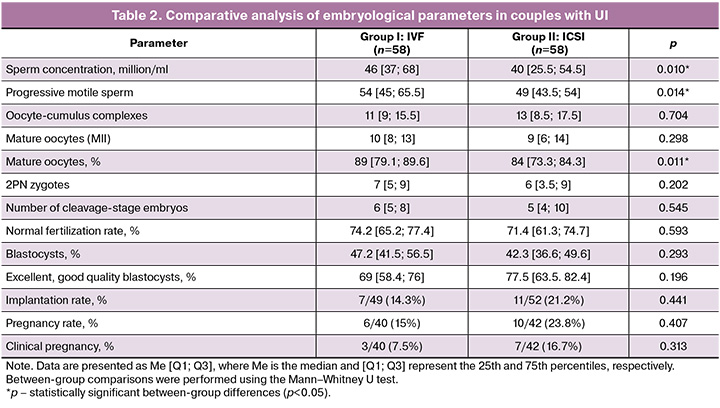
The conventional IVF group demonstrated significantly higher sperm concentration and a greater number of progressively motile spermatozoa than the ICSI group (p=0.010 and p=0.014, respectively). The proportion of mature oocytes was significantly higher in the IVF group than in the ICSI group, reaching 89 [79.1; 89.6] versus 84 [73.3; 84.3], respectively (p=0.011). No statistically significant differences were observed between groups in the number of cumulus–oocyte complexes, MII oocytes, 2PN zygotes, or cleaving embryos (all p>0.05).
Similarly, no statistically significant difference was found for the primary outcome, the normal fertilization rate, which was 74.2% in the IVF group and 71.4% in the ICSI group (p=0.593). No significant between-group differences were observed in the blastulation rate (47.2% vs. 42.3%, p=0.293) or in the proportion of good-quality blastocysts (69.0% vs. 77.5%, p=0.196). Embryo transfer was performed in 40 patients in the IVF group and 42 patients in the ICSI group. The pregnancy rate was 15.0% (6/40) in the IVF group and 23.8% (10/42) in the ICSI group; however, the difference did not reach statistical significance (Fisher’s exact test, p=0.407). Ultrasound-confirmed clinical pregnancy occurred in 7.5% (3/40) of patients in the IVF group and 16.7% (7/42) of patients in the ICSI group, with no statistically significant difference between groups (Fisher’s exact test, p=0.313).
A total of 52 embryos were transferred in the ICSI group and 49 in the IVF group. The number of gestational sacs visualized on ultrasound was 11 and 7, respectively. Although the implantation rate was higher in the ICSI group than in the IVF group (21.2% [11/52] vs. 14.3% [7/49]), the difference was not statistically significant (p=0.441).
Multivariable analysis revealed no statistically significant differences between the ICSI and conventional IVF groups.
ICSI was not associated with an increased normal fertilization rate (adjusted odds ratio [aOR] 0.96; 95% CI 0.74–1.25; p=0.763). A trend toward a lower blastulation rate was observed in the ICSI group; however, the difference did not reach statistical significance (aOR 0.77; 95% CI 0.59–1.01; p=0.063). Likewise, no significant difference was found in the formation of good-quality blastocysts (aOR 0.80; 95% CI 0.49–1.31; p=0.370). Implantation rates did not differ between groups (aOR 1.01; 95% CI 0.003–330; p=0.962). Of particular note is the extremely wide confidence interval for the implantation rate estimate (Fig. 2).
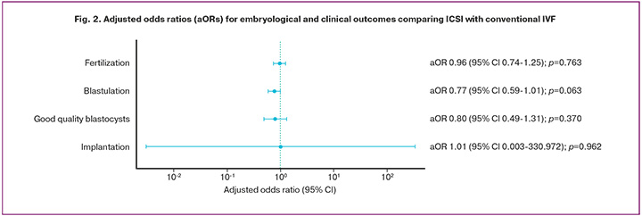
This finding is likely attributable to the limited number of embryo transfers and the substantial variability of the implantation outcome, both of which contribute to instability in model estimates. Consequently, the odds ratio estimate is characterized by low precision, as reflected by the markedly wide confidence interval. Nevertheless, the absence of statistical significance and an odds ratio close to 1 suggest that the fertilization method had no clinically meaningful effect on implantation rates. From a clinical perspective, these findings support a more selective use of ICSI in patients with UI.
Discussion
In the present study, no statistically significant differences were observed between conventional IVF and ICSI in patients with UI regarding the primary outcome, the normal fertilization rate. This parameter was comparable between the two groups, reaching 74.2% in the IVF group versus 71.4% in the ICSI group (p=0.593). Similar findings were observed for other embryological outcomes: the blastulation rate was 47.2% in the IVF group compared with 42.3% in the ICSI group (p=0.293), while the proportion of good-quality blastocysts was 69.0% versus 77.5%, respectively (p=0.196). The pregnancy rate was 15.0% in the IVF group and 23.8% in the ICSI group (p=0.407), whereas clinical pregnancy occurred in 7.5% and 16.7% of patients, respectively (p=0.313). In other words, the use of ICSI was not associated with a clinically or statistically meaningful improvement in key embryological outcomes.
Notably, comparable results for the main outcomes were obtained despite certain between-group differences in laboratory characteristics. Specifically, sperm concentration, total sperm motility, and the proportion of mature oocytes were higher in the IVF group, with mature oocytes accounting for 89% versus 84% in the ICSI group (p=0.011). Conversely, the starting gonadotropin dose was higher in the ICSI group. Given this background, the absence of any advantage of ICSI with respect to either normal fertilization or subsequent embryo development is particularly noteworthy. These findings suggest that, in this clinical setting, the choice of fertilization method is unlikely to be a major determinant of cycle outcome.
After adjustment for potential confounders, the results remained largely unchanged. The likelihood of normal fertilization in the ICSI group did not differ substantially from that in the IVF group (aOR 0.96; 95% CI 0.74–1.25; p=0.763). A trend toward a lower blastulation rate was observed in the ICSI group (aOR 0.77; 95% CI 0.59–1.01; p=0.063), although statistical significance was not reached. Similarly, the probability of obtaining good-quality blastocysts did not differ between groups (aOR 0.80; 95% CI 0.49–1.31; p=0.370). The stability of the findings after adjustment further strengthens the robustness of the conclusions.
Our results are consistent with those reported by Dang V.Q. et al. (2021), in which ICSI was associated with a higher fertilization rate than conventional IVF (75% vs 67%); however, this did not translate into a greater number of embryos available for transfer [9]. Likewise, the secondary analysis of the INVICSI study (Berntsen S. et al., 2025), which included 388 patients in the ICSI group and 378 in the IVF group, demonstrated no advantage of ICSI in terms of the number of cleaving embryos. On the contrary, ICSI was associated with fewer day-2 embryos, fewer blastocysts overall, and fewer good-quality day-5 blastocysts. These findings are in good agreement with our data, in which ICSI also failed to demonstrate benefits with respect to either blastulation rate or blastocyst quality [19]. Similar results were reported by Wang Y. et al. (2024), who found a lower implantation rate following ICSI than following IVF (34.3% vs 37.7%) [10].
ICSI is often perceived as a technology capable of reducing the risk of total fertilization failure and providing greater control over gamete interaction. However, in couples with UI, the underlying problem is likely to be considerably more complex than merely overcoming the mechanical barrier of sperm penetration into the oocyte. Although the implantation rate was higher in the ICSI group than in the conventional IVF group (21.2% [11/52] vs 14.3% [7/49]), the difference was not statistically significant (p=0.441) (Table 2). In the adjusted analysis, the odds ratio was 1.01, with an extremely wide 95% CI ranging from 0.003 to 330 (p=0.962) (Fig. 2). This imprecision is likely attributable to the limited number of embryo transfers. Therefore, the observed numerical difference should not be interpreted as evidence of a clinical advantage of ICSI but rather as a reflection of insufficient statistical power for this secondary outcome.
Importantly, within the pooled cohort, embryological outcomes appeared to be more strongly associated with markers of ovarian reserve than with the fertilization technique itself. AMH levels were positively correlated with AFC and the number of mature oocytes, while the number of MII oocytes was, in turn, correlated with the total number of blastocysts obtained. These findings indirectly shift the focus from the fertilization technique to oocyte quality and embryological potential. From a clinical perspective, this suggests that, in patients with UI, the outcome of assisted reproductive technology (ART) treatment is determined to a greater extent by oocyte quality and subsequent early embryonic development than by the choice of fertilization method.
From a practical standpoint, the findings of the present study do not support the routine use of ICSI in couples with UI. Given the comparable normal fertilization rates (74.2% vs 71.4%) and the absence of differences in blastulation, blastocyst quality, and implantation, the routine use of a more invasive technique as a standard strategy for this population does not appear justified. A selective approach may be more appropriate, whereby the choice of fertilization method is guided by the individual clinical and embryological characteristics of each couple.
Conclusion
The use of ICSI in patients with UI did not demonstrate convincing advantages over conventional IVF with respect to key embryological outcomes and therefore does not support its routine use in this clinical population. These findings underscore the biological heterogeneity of UI and highlight the need for further research aimed at identifying markers that could facilitate a personalized approach to fertilization method selection.
References
- Romualdi D., Ata B., Bhattacharya S., Bosch Е., Costello М., Gersak K. et al. Evidence-based guideline: unexplained infertility. Hum. Reprod. 2023; 38(10): 1881-90. https://dx.doi.org/10.1093/humrep/dead150
- Министерство здравоохранения Российской Федерации. Клинические рекомендации. Женское бесплодие. 2021. [Ministry of Health of the Russian Federation. Clinical guidelines. Female infertility. 2021 (in Russian)].
- Practice Committees of the American Society for Reproductive Medicine and the Society for Assisted Reproductive Technology. Intracytoplasmic sperm injection (ICSI) for non-male factor indications: a committee opinion. Fertil. Steril. 2020; 114(2): 239-45. https://dx.doi.org/10.1016/j.fertnstert.2020.05.032
- Zoltan M. The signal that stimulates mammalian embryo development. Front. Cell Dev. Biol. 2024; 12: 1474009. https://dx.doi.org/10.3389/fcell.2024.1474009
- Palermo G., Joris H., Devroey P. Pregnancies after intracytoplasmic injection of single spermatozoon into an oocyte. Lancet. 1992; 340: 17-8. https://dx.doi.org/10.1016/0140-6736(92)92425-F
- Smeenk J., Wyns C., De Geyter C., Kupka M., Bergh C., Cuevas Saiz I. et al. ART in Europe, 2019: results generated from European registries by ESHRE. Hum. Reprod. 2023; 12(38): 2321-38. https://dx.doi.org/10.1093/humrep/dead197
- Johnson L.N.C., Sasson I.E., Sammel M.D., Dokras A. Does intracytoplasmic sperm injection improve the fertilization rate and decrease the total fertilization failure rate in couples with well-defined unexplained infertility? A systematic review and meta-analysis. Fertil. Steril. 2013; 100(3): 704-11. https://dx.doi.org/10.1016/j.fertnstert.2013.04.038
- Cutting E., Horta F., Dang V., van Rumste M.M., Mol B.W.J. Intracytoplasmic sperm injection versus conventional in vitro fertilisation in couples with males presenting with normal total sperm count and motility. Cochrane Database Syst. Rev. 2023; 8(8): CD001301. https://dx.doi.org/10.1002/14651858.CD001301.pub2
- Dang V.Q., Vuong L.N., Luu T.M., Pham T.D., Ho T.M., Ha A.N. et al. Intracytoplasmic sperm injection versus conventional in-vitro fertilisation in couples with infertility in whom the male partner has normal total sperm count and motility: an open-label, randomised controlled trial. Lancet. 2021; 397(10284): 1554-63. https://dx.doi.org/10.1016/S0140-6736(21)00535-3
- Wang Y., Li R., Yang R., Zheng D., Zeng L., Lian Y. et al. Intracytoplasmic sperm injection versus conventional in-vitro fertilisation for couples with infertility with non-severe male factor: a multicentre, open-label, randomised controlled trial. Lancet. 2024; 403(10430): 924-34. https://dx.doi.org/10.1016/S0140-6736(23)02416-9
- Бачурин А.В., Киракосян Е.В., Назаренко Т.А., Павлович С.В. Анализ эмбриологического этапа программ экстракорпорального оплодотворения у пациентов с бесплодием неясного генеза. Акушерство и гинекология. 2022; 9: 81-6. https://dx.doi.org/10.18565/aig.2022.9.81-86 [Bachurin A.V., Kirakosyan E.V., Nazarenko N.A., Pavlovich S.V. Analysis of the embryonic stage of in vitro fertilization programs in patients with unexplained infertility. Obstetrics and Gynecology. 2022; (9): 81-6 (in Russian). https://dx.doi.org/10.18565/aig.2022.9.81-86].
- Iwamoto A., Summers K.M., Sparks A., Mancuso A.C. Intracytoplasmic sperm injection versus conventional in vitro fertilization in unexplained infertility. F. S. Reports. 2024; 5(3): 263-71. https://dx.doi.org/10.1016/j.xfre.2024.06.003
- Paffoni A., Vitagliano A., Corti L., Somigliana E., Viganò P. Intracytoplasmic sperm injection versus conventional in vitro insemination in couples with non-male infertility factor in the 'real-world' setting: analysis of the HFEA registry. J. Transl. Med. 2024; 22(1): 687. https://dx.doi.org/10.1186/s12967-024-05515-x
- Somigliana E., Barbara G., Micci L.G., Di Stefano G., Paffoni A., Vigano P. ICSI for non-male infertility: from ineffectiveness to gender bias? Reprod. Biomed. Online. 2025; 50(3): 104706. https://dx.doi.org/10.1016/j.rbmo.2024.104706
- Cornelisse S., Zagers M., Kostova E., Fleischer K., van Wely M., Mastenbroek S. Preimplantation genetic testing for aneuploidies (abnormal number of chromosomes) in in vitro fertilization. Cochrane Database Syst. Rev. 2020; 2020(9): CD005291. https://dx.doi.org/10.1002/14651858.CD005291.pub3
- Gleicher N., Barad D.H., Patrizio P., Orvieto R. We have reached a dead end for preimplantation genetic testing for aneuploidy. Hum. Reprod. 2022;37(Suppl. 1): 2730-4. https://dx.doi.org/10.1093/humrep/deac052
- Gardner D.K., Schoolcraft W.B. Blastocyst score affects implantation and pregnancy outcome: towards a single blastocyst transfer. Fertil. Steril. 2000; 73(6): 1155-8. https://dx.doi.org/10.1016/S0015-0282(00)00518-5
- Wang X., Tian P.Z., Zhao Y.J., Lu J., Dong C.Y., Zhang C.I. The association between female age and pregnancy outcomes in patients receiving first elective single embryo transfer cycle: a retrospective cohort study. Sci. Rep. 2024; 14(1): 19216. https://dx.doi.org/0.1038/s41598-024-70249-1
- Berntsen S., Zedeler A., Grøndahl M.L., Vestergaard Gabrielsen A., Rønn Petersen M., Frøding Skipper D. et al. Early embryo developmental kinetics following IVF versus ICSI in patients without severe male factor infertility: a secondary analysis of a multicentre, randomized controlled trial (INVICSI). Hum. Reprod. 2025; 40(10): 1877-85. https://dx.doi.org/10.1093/humrep/deaf157
Received 16.04.2026
Accepted 10.06.2026
About the Authors
Maria P. Ishchuk, PhD Student, F. Paulsen Research and Educational Center for ART with the Clinical Department, Academician V.I. Kulakov National Medical Research Center for Obstetrics, Gynecology and Perinatology, Ministry of Health of Russia, 117997, Russia, Moscow, Ac. Oparina str., 4, marumariyaish@gmail.com,https://orcid.org/0000-0002-9577-1777
Svetlana G. Perminova, Dr. Med. Sci., Professor, Leading Researcher, F. Paulsen Research and Educational Center for ART with the Clinical Department, Academician V.I. Kulakov National Medical Research Center for Obstetrics, Gynecology and Perinatology, Ministry of Health of Russia, 117997, Russia, Moscow, Ac. Oparin str., 4,
s_perminova@oparina4.ru, https://orcid.org/0000-0003-44
Elena V. Mituyrina, Dr. Med. Sci., Senior Researcher, F. Paulsen Research and Educational Center for ART with the Clinical Department, Academician V.I. Kulakov National Medical Research Center for Obstetrics, Gynecology and Perinatology, Ministry of Health of Russia, 117997, Russia, Moscow, Ac. Oparin str., 4, e_mityurina@oparina4.ru, https://orcid.org/0000-0001-8830-2158
Sergey F. Filimonov, Head of the Laboratory of Clinical Embryology, F. Paulsen Research and Educational Center for ART with the Clinical Department,
Academician V.I. Kulakov National Medical Research Center for Obstetrics, Gynecology and Perinatology, Ministry of Health of Russia, 117997, Russia, Moscow,
Ac. Oparin str., 4, s_felimonov@oparina4.ru
Elena S. Sannikova, Embryologist, F. Paulsen Research and Educational Center for ART with the Clinical Department, Academician V.I. Kulakov National Medical Research Center for Obstetrics, Gynecology and Perinatology, Ministry of Health of Russia, 117997, Russia, Moscow, Ac. Oparin str., 4, el.sannikowa2013@yandex.ru,
https://orcid.org/0000-0002-9577-1777
Corresponding author: Maria P. Ishchuk, marumariyaish@gmail.com
Similar Articles

