
Clinical and anamnestic characteristics of couples referred for whole-exome sequencing in the context of assisted reproductive technology programs
Martirosyan Ya.O., Nazarenko T.A., Tsabai P.N., Pavlova N.S.
Objective. To evaluate the diagnostic accuracy of whole-exome sequencing in identifying genetic causes of recurrent embryonic developmental arrest in IVF/ICSI cycles and assess its potential to guide treatment strategies.
Materials and methods. This retrospective, single-center study was conducted between January 2021 and May 2025. Among 3,779 couples who underwent 6,674 ART cycles, a subgroup with ≥3 IVF/ICSI attempts was identified (n = 1,750). The phenotype of recurrent early embryonic arrest, defined as the absence of embryos available for transfer or cryopreservation by day 5, was observed in 782 couples. A target subgroup of women younger than 36 years with total arrest across all cycles was selected for in-depth genetic analysis (n = 125). All participants underwent standardized clinical assessments, embryological evaluations, and whole-exome sequencing.
Results. Early embryonic arrest was present in 44.7% (782/1,750) of couples with ≥3 ART attempts, representing 20.7% of the entire initial cohort. In the target subgroup (n=125), the mean age was 32.1 (2.8) years, the mean duration of infertility was 5.2 (1.4) years, and primary infertility was documented in 85.4% of the cases. Ovarian reserve parameters were preserved (AMH 2.31 ng/mL; AFC 14.2 (4.0), and the mean fertilization rate was 92.7 (4.4)%; however, embryonic development was uniformly arrested in all cycles.
Conclusion. Recurrent early embryonic arrest, in the presence of preserved ovarian reserve and normal fertilization, is a clinically significant problem affecting approximately one in five couples undergoing multiple ART attempts. Among women < 36 years of age presenting with this phenotype (who accounted for 16% of the arrest subgroup), a genetic etiology is highly probable. This supports the use of whole-exome sequencing as a tool for etiological stratification and individualized treatment planning.
Authors' contributions. Martirosyan Y.O., Nazarenko T.A. – conception and design of the study, data collection and analysis, review of the relevant literature, drafting of the manuscript; Tsabai P.N. – conducting and supervising the embryological phase of the research, interpreting embryological data; Pavlova N.S. – organizing and conducting genetic studies, bioinformatic analysis of whole-exome sequencing data, interpreting molecular genetic results.
Conflicts of interest. The authors have no conflicts of interest to declare.
Funding. Agreement No. 25-65-00040 with the Russian Science Foundation, dated May 22, 2025, on the topic: “Development of genetically informed approaches to realizing the reproductive potential of patients and their families in cases of abnormalities in the development and functioning of the reproductive system”.
Ethical Approval. The study was reviewed and approved by the Research Ethics Committee of the V.I. Kulakov NMRC for OG&P.
Generative Artificial Intelligence. No artificial intelligence tools were used in the preparation of this manuscript.
Patient Consent for Publication. All patients provided informed consent for the publication of their data.
Authors' Data Sharing Statement. The data supporting the findings of this study are available upon request from the corresponding author after approval from the principal investigator.
For citation: Martirosyan Ya.O., Nazarenko T.A., Tsabai P.N., Pavlova N.S. Clinical and anamnestic characteristics of couples referred for whole-exome sequencing in the context of assisted reproductive technology programs.
Akusherstvo i Ginekologiya/Obstetrics and Gynecology. 2026; (6): 153-162 (in Russian)
https://dx.doi.org/10.18565/aig.2025.354
Keywords
unexplained infertility
genetics
genetic testing
assisted reproductive technologies
exome sequencing
Poor outcomes of assisted reproductive technology (ART), particularly recurrent IVF failure, remain among the most challenging and clinically unresolved problems in contemporary reproductive medicine. A substantial proportion of these failures is attributable to disturbances in oocyte and embryo development, including the recurrent absence of viable embryos, which manifests as a failure to obtain mature oocytes, failed fertilization, or cleavage-stage arrest.
The embryological phase of IVF encompasses a sequence of critically important processes that determine both the embryonic developmental potential and overall treatment success. Despite the progressive refinement of ovarian stimulation protocols and laboratory procedures for IVF/intracytoplasmic sperm injection (ICSI), preimplantation embryonic arrest remains frequent, underscoring the need for a deeper understanding of the underlying pathogenesis.
Published data indicate that developmental arrest during the cleavage stage occurs in 25–30% of embryos produced in vitro through ART [1]. Empirical evidence suggests that arrest most commonly occurs on days 2–3 of culture, coinciding with the transition from maternal genomic control to embryonic genome activation.
The determinants of early embryonic arrest have not been fully elucidated; however, the pivotal role of oocyte quality is widely accepted [2]. Early preimplantation development is largely governed by maternal factors inherited from the oocyte [3], which include the following:
- maternal RNAs and proteins sustaining essential cellular processes prior to embryonic genome activation.
- energy-supplying organelles, primarily mitochondria, are responsible for ATP production and metabolic homeostasis until implantation [4].
Dysfunction or deficiency of these maternal components is the leading hypothesis to explain the high rate of early developmental arrest, which is supported by extensive clinical and experimental evidence [5–9].
Elucidating the specific molecular mechanisms underlying embryonic arrest is one of the most pressing objectives in reproductive biology. Patients with idiopathic infertility pose particular diagnostic challenges, as the etiology of embryogenic failure frequently remains unidentified [5–7, 10].
Standard genetic testing algorithms, including karyotyping and targeted mutation screening, establish a causative diagnosis in only a minority of patients. Current evidence increasingly implicates monogenic disorders arising from both de novo and inherited variants of genes critical for gametogenesis, fertilization, and early embryonic development. Mutations in TUBB8 have been associated with meiotic oocyte arrest, confirming the contribution of monogenic causes to the earliest stages of reproductive failure [11]. Systematic reviews and case series have highlighted the role of WEE2 and other regulators of post-fertilization oocyte activation [12–14]. Variants in genes such as TLE6 and NLRP5 illustrate the direct link between defects in oogenesis-derived maternal proteins and recurrent embryonic arrest in the offspring [15].
Taken together, early embryonic arrest in IVF represents a multifactorial condition rooted in a complex of disturbances predominantly related to oocyte quality and its molecular components. Future research should focus on identifying novel candidate genes and molecular pathways governing early embryogenesis using next-generation sequencing (NGS) and functional analyses. Such an approach would enable a transition from phenotypic characterization to the development of predictive diagnostic tools and, ultimately, pathogenetically targeted therapeutic interventions.
This study aimed to evaluate the diagnostic accuracy of whole-exome sequencing in identifying the genetic causes of recurrent embryonic developmental arrest in IVF/ICSI programs and to assess its potential to inform and individualize treatment strategies.
Materials and methods
Study design. This was a single-center, retrospective, observational cohort study conducted at the F. Paulsen Research and Educational Center for Assisted Reproductive Technologies (ART), V.I. Kulakov National Medical Research Center for Obstetrics, Gynecology and Perinatology, Ministry of Health of the Russian Federation. The study period was from January 2021 to May 2025. This study was conducted and reported in accordance with the Strengthening the Reporting of Observational Studies in Epidemiology (STROBE) guidelines.
Data were obtained from the medical information systems and embryology records of the ART department. The initial cohort consecutively included all married couples who underwent ART procedures (IVF/ICSI) during the study period and for whom key embryological parameters were available. These parameters included the number of retrieved oocytes, number of metaphase II (MII) oocytes, number of normally fertilized zygotes with two pronuclei (2PN), day-5 culture outcomes, and blastocyst quality. For clinical and anamnestic characteristics, the unit of observation was a married couple. Embryological parameters were analyzed at the ART cycle level, with subsequent descriptions of parameter distributions across predefined groups.
Embryology procedures. Following follicular aspiration, cumulus–oocyte complexes were transferred to the culture medium. Oocyte maturity was assessed 2–4 h after retrieval. Oocytes at the metaphase II (MII) stage were considered mature and suitable for fertilization. Fertilization was performed using conventional IVF or intracytoplasmic sperm injection (ICSI) according to clinical indications. The presence of two pronuclei (2PN) was assessed 16–18 h after insemination as evidence of normal fertilization.
Embryos were cultured in sequential media within mini-incubators at 37°C, 6% CO₂, and 5% O₂. Embryo assessment was performed daily on culture days 1–5 using the Istanbul Consensus grading criteria. On day 5, the blastocysts were evaluated according to their developmental stage (grades 1–6), inner cell mass quality (grades A–C), and trophectoderm quality (grades A–C). Good- and excellent-quality blastocysts (≥3BB according to the Gardner grading system) were considered suitable for transfer or cryopreservation procedures. The absence of embryos suitable for transfer or cryopreservation on day 5 was defined as no embryo reaching the blastocyst stage or all resulting blastocysts being graded below 3BB.
Embryo vitrification was performed using the Cryotop system (Kitazato, Shiga, Japan) according to the standard protocol of the Center. All procedures were performed by certified embryologists using Class II biological safety equipment.
Inclusion criteria. The phenotypic group included couples who had undergone three or more IVF/ICSI attempts and demonstrated adverse embryological outcomes in every cycle. This was defined as the recurrent absence of embryos suitable for transfer or cryopreservation on day 5 of culture, or the production of only isolated blastocysts of extremely poor quality. Additional criteria for inclusion in the target subgroup for in-depth analysis were as follows: female age ≤36 years at the time of ART treatment; a normal 46,XX karyotype based on peripheral blood lymphocyte karyotyping; absence of genetically related children; no contraindications to ART according to Order No. 803n of the Ministry of Health of the Russian Federation; and the presence of a recurrent total embryogenesis arrest phenotype, defined as the complete absence of embryos suitable for transfer or cryopreservation in all completed cycles.
Exclusion criteria. Couples were excluded if the male partner had severe pathological sperm abnormalities, including cryptozoospermia, azoospermia, or severe oligoasthenoteratozoospermia with a sperm concentration <1 million/mL and progressive motility <5% (except in cycles using the donor sperm). Couples were also excluded if the female partner had marked endocrine abnormalities, such as follicle-stimulating hormone (FSH) levels >20 IU/mL during the early follicular phase.
All participating couples underwent comprehensive clinical and laboratory evaluations in accordance with the current regulatory requirements of the Russian Federation.
A retrospective analysis was performed on 6,674 ART cycles conducted between January 2021 and May 2025 in 3,779 married couples. From the overall cohort, a subgroup of 1,750 couples (46.3%) who had undergone three or more IVF/ICSI attempts was identified.
A history of recurrent failure to obtain embryos suitable for implantation was the criterion for inclusion in the target subgroup for in-depth genetic analyses. The phenotypic group (n=782) comprised couples who had undergone ≥3 IVF/ICSI cycles and in whom adverse embryological outcomes were recorded in every cycle, namely the absence of embryos suitable for transfer or cryopreservation on day 5 or the production of only isolated blastocysts of extremely poor quality.
The most common causes of complete embryo developmental arrest during cleavage were diminished ovarian reserve, resulting in a low number of oocytes retrieved at aspiration, and advanced female reproductive age. Subsequent analyses were therefore restricted to a subgroup of couples in which the female partner was no older than 36 years at the time of ART treatment; the male partner’s age was not used as a selection criterion.
Ovarian stimulation was performed in patients from the study group (n=782) in accordance with the inclusion criteria. Standard stimulation protocols employing gonadotropin-releasing hormone (GnRH) antagonists or agonists combined with recombinant FSH and/or luteinizing hormone (LH) were used in all cycles. Human chorionic gonadotropin (hCG) and/or GnRH agonist was administered to trigger final oocyte maturation once dominant follicles reached a diameter of 17–18 mm. Follicular aspiration was performed 35–37 h after the trigger administration.
Statistical analysis
Statistical analyses were performed using TIBCO Statistica (version 13.3). Given the skewed distribution of several variables, all continuous variables were treated as non-normally distributed and are presented as median and interquartile range, Me [Q1; Q3]. Categorical variables are presented as numbers (n) and percentages (%). Between-group comparisons of continuous variables were performed using the Mann–Whitney U test, whereas proportions were compared using Pearson's χ² test or Fisher's exact test when expected cell frequencies were <5. Statistical significance was set at p<0.05.
Results
A retrospective analysis was conducted on 6,674 ART cycles from January 2021 to May 2025, involving 3,779 couples. Of these, 1,750 couples (46.3%) had undergone three or more IVF/ICSI attempts. An adverse embryological outcome phenotype, defined as the absence of usable embryos or the retrieval of only a small number of poor-quality blastocysts, was identified in 782 of these couples (44.7%; 20.7% of the original cohort).
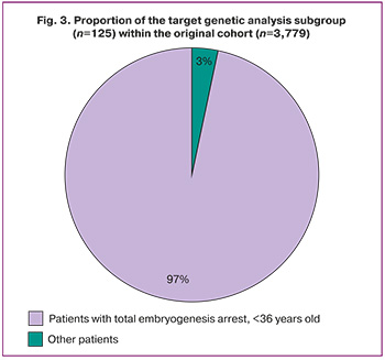
For subsequent in-depth genetic analysis, a target subgroup (n=125) was selected from this phenotypic group based on a more stringent criterion: recurrent total embryonic arrest, defined as the complete absence of embryos suitable for transfer or cryopreservation across all cycles, in patients <36 years of age. This subgroup represented 3.3% of the total original cohort (125/3,779). The following data pertain to the phenotypic group (n=782) and the subgroup of patients <36 years of age (n=125).
In the first phase of the study, the clinical and obstetric characteristics of couples who had experienced three or more failed IVF cycles were evaluated. Data from 782 couples who received ART-based infertility treatment at the F. Paulsen Research and Educational Center for Assisted Reproductive Technologies, V.I. Kulakov National Medical Research Center for Obstetrics, Gynecology and Perinatology, Ministry of Health of the Russian Federation, between 2021 and 2025, were analyzed (Fig. 1). Embryonic developmental arrest, despite an adequate oocyte yield, was documented in 20.7% of couples (782/3,779).
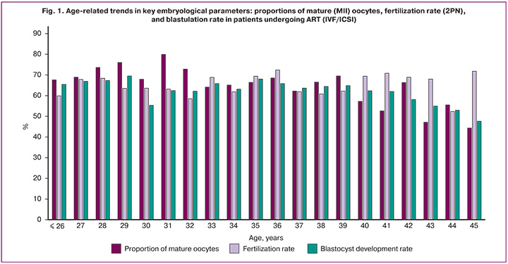
The results of the clinical and obstetric assessment of the selected patients are presented in Table 1. Median anti-Müllerian hormone (AMH) levels progressively declined with advancing age, from 3.7 [1.6; 4.7] ng/mL in patients aged ≤26 years to 0.8 [0.3; 2.01] ng/mL in the 45-year age group. To achieve an adequate ovarian response in older age groups, a higher median total gonadotropin dose was required, increasing from 1,800 [1,350; 2,025] IU in patients aged ≤26 years to 2,250 [1,425; 2,300] IU in those aged 45 years.
The distribution of fertilization methods varied across age groups. In younger patients (≤26 years), intracytoplasmic sperm injection (ICSI) was used in 48.1% of cycles (13/27), whereas in patients >40 years, the proportion exceeded 70%, reaching 83.3% (20/24) in the 45-year group. The gonadotropin-releasing hormone (GnRH) antagonist stimulation protocol was predominant across all age groups, with utilization ranging from 62.5% (15/24) in the 45-year group to 100% (17/17) in the 28-year group.
The prevalence of male-factor pathozoospermia showed no clear age-related pattern, ranging from 30.9% (17/55) in the 36-year group to 81.8% (18/22) in the 44-year group. Body mass index (BMI) remained stable across all age groups, with a median of approximately 23.5–24.5 kg/m².
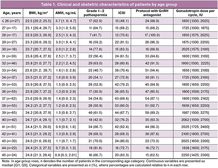
Analysis of age-related trends in key embryological parameters (Fig. 1) revealed divergent patterns. The proportion of mature (metaphase II, MII) oocytes was approximately 75–85% in patients aged 26–38 years, with a moderate subsequent decline to 65–70% in patients >40 years. The normal fertilization rate (two pronuclei, 2PN) similarly remained high (approximately 80–90%) up to age 37, after which a progressive decline was observed, falling to approximately 60% by age 45.
The most pronounced adverse trend was observed for blastulation rate. In patients without recurrent cleavage-stage embryonic arrest, the blastulation rate was 65–70% before age 30; it began to decline substantially after age 35, exhibiting a steep fall from approximately 50% at age 37 to below 20% in patients aged 43–45 years.
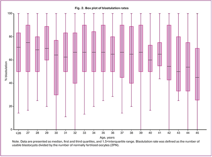
Data analysis demonstrated that a subgroup with comparatively low blastulation rates was also identifiable among patients <36 years of age (Fig. 2). Even within this younger age group with formally preserved ovarian reserve, a distinct pathological phenotype characterized by unexplained poor blastocyst development was apparent. This finding provides direct justification for investigating specific genetic causes – including maternal-effect gene variants and meiotic defects – that are independent of general age-related decline, through whole-exome sequencing.
The proportion of patients <36 years of age with recurrent total embryonic arrest accounted for 3.3% of all couples in the original cohort (125/3,779) (Fig. 3). This subgroup defines the key clinical profile for which high-throughput sequencing is considered warranted.
Notably, although some patients had established and prevalent causes of infertility – including male factor, tuboperitoneal factor, and endometriosis – the majority (60%) of patients in the study group had unexplained infertility (Table 2). Male-factor infertility was the second most common etiology, identified in 14.4% of cases, followed by tuboperitoneal factor (12.0%).
The principal clinical and laboratory characteristics of the enrolled patients are presented in Table 3. The median duration of infertility in the target subgroup (n = 125) was 5.2 [4.2; 6.3] years. Primary infertility predominated, accounting for 107 of 125 cases (85.4%); secondary infertility was diagnosed in 18 of 125 couples (14.6%). Conventional IVF was used in 224 of 375 cycles (59.7%), and ICSI in 301 of 375 cycles (80.3%). These characteristics describe the subgroup of patients with complete embryonic arrest.
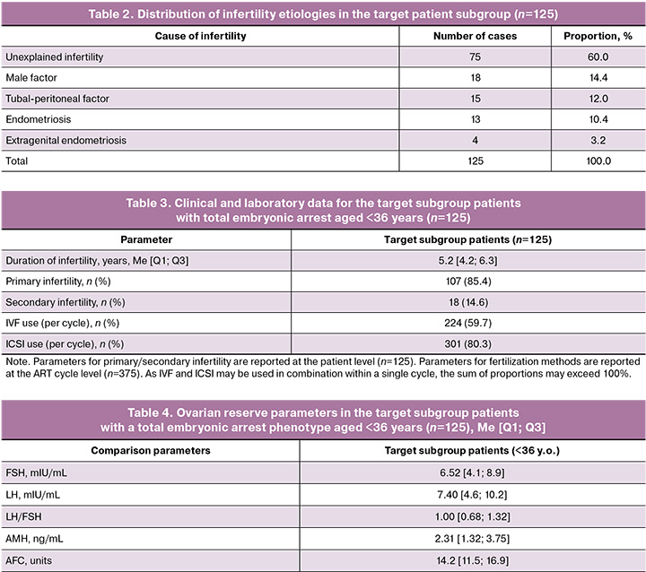
For contextual reference in subsequent comparisons, ovarian reserve parameters – including FSH, LH, AMH, and antral follicle count (AFC) – are reported separately for this subgroup (Table 4). These values were within the normal reference ranges for the patients concerned.
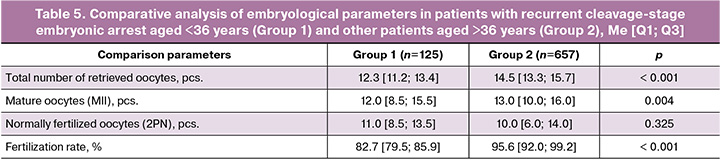
Quantitative analysis of ovarian reserve parameters in the target subgroup (n=125) demonstrated that median FSH, LH, and AMH values were within established reference ranges. However, when compared with patients >36 years of age with embryonic arrest attributable to other causes (Group 2, n=657), the target subgroup exhibited statistically significantly lower ovarian reserve values. This finding suggests that, even in younger patients, the phenotype of early embryonic arrest may be associated with incipient ovarian reserve decline – a factor that nevertheless does not fully account for the complete block in embryonic development.
These findings confirm a dissociation between the relatively preserved oocyte pool and fertilization parameters on the one hand, and markedly impaired subsequent embryological outcomes on the other – a pattern consistent with the proposed pathogenetic mechanism of the phenotype under investigation (Table 4). The identified profile supports the rationale for whole-exome sequencing to detect genetic determinants influencing embryonic cleavage and blastulation.
Between-group comparisons of continuous variables were performed using the Mann–Whitney U test (Table 5); p-values are reported for the comparison between Group 1 and Group 2.
In summary, a substantial attrition of developmentally competent embryos occurs during the period of early embryonic development—from oocyte retrieval following transvaginal follicle aspiration through to the formation of viable, high-implantation-potential embryos.
Discussion
In the present study, detailed clinical, anamnestic, and embryological stratification was performed based on a retrospective analysis of a large single-center ART cohort (3,779 couples; 6,674 cycles). This approach enabled the identification of a key phenotypic group of patients (n=782) with recurrent adverse embryological outcomes and, within this group, a clinically significant subgroup (n=125) of women aged <36 years who exhibited recurrent total embryogenesis arrest despite apparently preserved ovarian reserve and fertilization parameters. This subgroup accounted for 3.3% of the overall cohort, indicating a relatively common, yet previously insufficiently characterized, clinical problem.
The principal finding of this study was the identification of a distinct dissociation between the early and later stages of embryonic development. Despite satisfactory ovarian response parameters (AMH and AFC) and high rates of normal fertilization (2PN), embryo development in these patients consistently failed to reach the blastocyst stage. This pattern, the preservation of early developmental processes accompanied by complete arrest during late cleavage and blastulation, is a classic hallmark of impaired maternal-to-embryonic transition, during which developmental control shifts from maternally derived factors accumulated within the oocyte to the activation of the embryonic genome [3, 8, 9].
The detailed characterization of this carefully selected cohort – young age, prolonged primary infertility (85.4%), and preserved ovarian reserve – effectively excludes the most common causes of ART failure, such as age-related deterioration in oocyte quality and markedly poor ovarian response, thereby focusing attention on qualitative and likely genetic defects affecting the oocyte and/or embryo. In this specific patient population, conventional diagnostic approaches, including karyotyping and ovarian reserve assessment, provide limited clinical insight, creating a diagnostic dead end and resulting in repeated unsuccessful IVF cycles.
The stratification strategy employed in this study provides a direct clinical rationale for modifying the diagnostic pathway. Couples matching the identified profile – recurrent complete developmental arrest by day 5 in women younger than 36 years with preserved fertilization capacity and ovarian reserve – should be considered candidates for comprehensive genetic evaluation, particularly whole-exome sequencing. The primary objective of such testing is to identify pathogenic variants in genes whose products play critical roles in oogenesis, meiotic completion, oocyte activation, zygotic division, and embryonic genome activation, including maternal-effect genes such as TUBB8, WEE2, TLE6, NLRP5, PADI6, and others [5, 10–25].
The identification of a monogenic cause has the potential to fundamentally alter ART management and warrants separate consideration.
Genetic counseling and reproductive risk assessment. Establishing a molecular diagnosis enables a more accurate estimation of the likelihood of obtaining viable embryos from the couple’s gametes and facilitates an informed discussion of reproductive risks.
Modification of the treatment strategy. Depending on the affected gene and the functional consequences of the identified variant, clinical management may be adjusted as follows:
- in cases of confirmed mutations resulting in absolute oocyte defects, early consideration of donor-oocyte programs may be justified, thereby avoiding multiple futile attempts using autologous gametes.
- when a pathogenic variant is identified and embryo formation remains possible, preimplantation genetic testing for monogenic disorders may be used to select embryos free of inherited mutations.
- for certain types of defects, targeted in vitro activation approaches or other experimental corrective strategies may become available in the future.
- establishing a genetic diagnosis may also justify the discontinuation of empirical IVF/ICSI cycles performed according to standard protocols that are unlikely to be effective for a given couple, thereby reducing both psychological and financial burdens.
Adjunctive embryological approaches. Even in the absence of an identified monogenic cause, alternative laboratory strategies may be considered for couples exhibiting the described phenotype, including:
- blastocyst culture under reduced oxygen conditions (5% O₂) to minimize oxidative stress;
- use of sequential culture media optimized to overcome developmental arrest;
- time-lapse imaging to improve the selection of embryos with normal cleavage kinetics.
In the present study, ovarian reserve indicators in the target subgroup, although remaining within reference ranges, were statistically lower than those observed in women older than 36 years with other causes of developmental arrest. This finding suggests that the phenotype of early embryonic arrest may be associated with an earlier decline in oocyte quality rather than solely with a reduction in oocyte quantity. Nevertheless, the primary underlying mechanism is likely related to functional (molecular) defects in the ooplasm, further supporting the need for a genetic diagnostic approach. Previous Russian studies on infertility of unknown origin have demonstrated that reduced blastulation rates in IVF programs are primarily attributable to developmental arrest occurring within the first 3 days of embryo culture. These studies have also demonstrated the practical feasibility of whole-exome sequencing in married couples and summarized its potential applications in reproductive medicine [26–29].
Conclusion
In this large single-center retrospective ART cohort (3,779 couples; 6,674 cycles), the phenotype of recurrent adverse embryological outcomes following ≥3 IVF/ICSI attempts was identified in 44.7% of couples (782/1,750), corresponding to 20.7% of the original cohort. Within this phenotype, a clinically significant subgroup of women aged ≤36 years with recurrent total embryogenesis arrest (125/3,779; 3.3%) was identified, despite apparently preserved ovarian reserve markers and relatively preserved fertilization outcomes. This clinical profile is consistent with the hypothesis that defects affecting oocyte and/or early embryonic quality, including monogenic causes, predominantly contribute to reproductive failure. These findings support the targeted referral of such patients for molecular genetic counseling and the consideration of whole-exome sequencing as a diagnostic tool within a clinically selected population. Dedicated studies specifically designed to evaluate the clinical utility of whole-exome sequencing, including diagnostic yield, variant spectrum, impact on clinical decision-making, and reproductive outcomes, are required.
References
- Yang Y., Shi L., Fu X., Ma G, Yang Z., Li Y. et al. Metabolic and epigenetic dysfunctions underlie the arrest of in vitro fertilized human embryos in a senescent-like state. PLoS Biol. 2022; 20(6): e3001682. https://dx.doi.org/10.1371/journal.pbio.3001682
- Anderson R.A., Marston A.L., Telfer E.E. Oocyte development: it's all about quality. Reprod. Biomed. Online. 2025; 50(4): 104804. https://dx.doi.org/10.1016/j.rbmo.2025.104804
- Wilkinson A.L., Zorzan I., Rugg-Gunn P.J. Epigenetic regulation of early human embryo development. Cell Stem Cell. 2023; 30(12): 1569-84. https://dx.doi.org/10.1016/j.stem.2023.09.010
- Mitchell L.E. Maternal effect genes: update and review of evidence for a link with birth defects. HGG Adv. 2021; 3(1): 100067. https://dx.doi.org/10.1016/j.xhgg.2021.100067
- Solovova O.A., Chernykh V.B. Genetics of oocyte maturation defects and early embryo development arrest. Genes (Basel). 2022; 13(11): 1920. https://dx.doi.org/10.3390/genes13111920
- Conti M., Franciosi F. Acquisition of oocyte competence to develop as an embryo. Hum. Reprod. Update. 2018; 24(3): 245-66. https://dx.doi.org/10.1093/humupd/dmy002
- Rienzi L., Vajta G., Ubaldi F. Predictive value of oocyte morphology in human IVF: a systematic review. Hum. Reprod. Update. 2011; 17(1): 34-45. https://dx.doi.org/10.1093/humupd/dmq029.
- Kojima M.L., Hoppe C., Giraldez A.J. The maternal-to-zygotic transition: reprogramming of the cytoplasm and nucleus. Nat. Rev. Genet. 2025; 26(4): 245-67. https://dx.doi.org/10.1038/s41576-024-00792-0
- Taubenschmid-Stowers J., Rostovskaya M., Santos F., Ljung S., Argelaguet R., Krueger F. et al. 8C-like cells capture the human zygotic genome activation program in vitro. Cell Stem Cell. 2022; 29(3): 449-59.e6. https://dx.doi.org/10.1016/j.stem.2022.01.014
- Feng R., Shao L., Feng Y., Wang L., Jin L. Mutations in TUBB8 and human oocyte meiotic arrest. N. Engl. J. Med. 2016; 374(3): 223-32. https://dx.doi.org/10.1056/NEJMoa1510791
- Xue Y., Cheng X., Xiong Y., Li K. Gene mutations associated with fertilization failure after IVF/ICSI: a review. Front. Endocrinol. 2022; 13: 1086883. https://dx.doi.org/10.3389/fendo.2022.1086883
- Cardona Barberán A., Reddy Guggilla R., Colenbier C., Van der Velden E., Rybouchkin A., Stoop D. et al. High rate of detected variants in male PLCZ1 and ACTL7A genes causing failed fertilization after ICSI. Hum. Reprod. Open. 2024; 2024(4): hoae057. https://dx.doi.org/10.1093/hropen/hoae057
- Torra-Massana M., Rodríguez A., Vassena R. Exonic genetic variants associated withunexpected fertilization failure and zygoticarrest after ICSI: a systematic review. Zygote. 2023; 31(4): 316-41. https://dx.doi.org/10.1017/S096719942300014X
- Li R., Mei M., Zhou L., Zhao H., Yang M., Li Y. et al. Biallelic recessive mutations in TLE6 and NLRP5 cause female infertility characterized by human early embryonic arrest. Hum. Mutat. 2024; 2024: 9278518. https://dx.doi.org/10.1155/2024/9278518
- Shoubridge E.A., Wai T. Mitochondrial DNA and the mammalian oocyte. Curr. Top. Dev. Biol. 2007; 77: 87-111. https://dx.doi.org/10.1016/S0070-2153(06)77004-1
- Wei Y., Wang J., Qu R., Zhang W., Tan Y., Sha Y. et al. Genetic mechanisms of fertilization failure and early embryonic arrest: a comprehensive review. Hum. Reprod. Update. 2024; 30(1): 48-80. https://dx.doi.org/10.1093/humupd/dmad026
- Zhang Z., Mu J., Zhao J., Zhou Z., Chen B., Wu L. et al. Novel mutations in WEE2: expanding the spectrum of mutations responsible for human fertilization failure. Clin. Genet. 2019; 95(4): 520-4. https://dx.doi.org/10.1111/cge.13505
- Zhou X., Zhu L., Hou M., Wu Y., Li Z., Wang J. et al. Novel compound heterozygous mutations in WEE2 causes female infertility and fertilization failure. J. Assist. Reprod. Genet. 2019; 36(9): 1957-62. https://dx.doi.org/10.1007/s10815-019-01553-3
- Zeng Y., Chen B., Sun Y., Yang A., Wu L., Li B. et al. Bi-allelic variants in ASTL cause abnormal fertilization or oocyte maturation defects. Hum. Mol. Genet. 2023; 32(14): 2326-34. https://dx.doi.org/10.1093/hmg/ddad070
- Zhao L., Xue S., Yao Z., Shi J., Chen B., Wu L. et al. Biallelic mutations in CDC20 cause female infertility characterized by abnormalities in oocyte maturation and early embryonic development. Protein Cell. 2020; 11(12): 921-7. https://dx.doi.org/10.1007/s13238-020-00756-0
- Zhao L., Guan Y., Meng Q., Wang W., Wu L., Chen B. et al. Identification of novel mutations in CDC20: expanding the mutational spectrum for female infertility. Front. Cell. Dev. Biol. 2021; 9: 647130. https://dx.doi.org/10.3389/fcell.2021.647130
- Li M., Jia M., Zhao X., Shi R., Xue X. A new NLRP5 mutation causes female infertility and total fertilization failure. Gynecol. Endocrinol. 2021; 37(3): 283-4. https://dx.doi.org/10.1080/09513590.2020.1832069
- Alazami A.M., Awad S.M., Coskun S., Al-Hassan S., Hijazi H., Abdulwahab F.M. et al. TLE6 mutation causes the earliest known human embryonic lethality. Genome Biol. 2015; 16(1): 240. https://dx.doi.org/10.1186/s13059-015-0792-0
- Pascal C., Zonszain J., Hameiri O., Gargi-Levi C., Lev-Maor G., Tammer L. et al. Human histone H1 variants impact splicing outcome by controlling RNA polymerase II elongation. Mol. Cell. 2023; 83(21): 3801-17.e8. https://dx.doi.org/10.1016/j.molcel.2023.10.003
- Wu X., Tian Y., Yu Y., He X., Tang X., Li S. et al. Novel MEI1 mutations cause chromosomal and DNA methylation abnormalities leading to embryonic arrest and implantation failure. Mol. Genet. Genomics. 2024; 299(1): 18. https://dx.doi.org/10.1007/s00438-024-02113-w
- Бачурин А.В., Киракосян Е.В., Назаренко Т.А., Павлович С.В. Анализ эмбриологического этапа программ экстракорпорального оплодотворения у пациентов с бесплодием неясного генеза. Акушерство и гинекология. 2022; 9: 81-6. https://dx.doi.org/10.18565/aig.2022.9.81-86 [Bachurin A.V., Kirakosyan E.V., Nazarenko T.A., Pavlovich S.V. Analysis of the embryonic stage of in vitro fertilization programs in patients with unexplained infertility. Obstetrics and Gynecology. 2022; (9): 81-6 (in Russian). https://dx.doi.org/10.18565/aig.2022.9.81-86].
- Киракосян Е.В., Померанцева Е.А., Павлович С.В. Полноэкзомное секвенирование супружеских пар с бесплодием неясного генеза (пилотное исследование). Акушерство и гинекология. 2022; 12: 115-21. https://dx.doi.org/10.18565/aig.2022.247 [Kirakosyan E.V., Pomerantseva E.A., Pavlovich S.V. Whole exome sequencing in couples with unexplained infertility (pilot study). Obstetrics and Gynecology. 2022; (12): 115-21 (in Russian). https://dx.doi.org/10.18565/aig.2022.247].
- Глотов О.С., Чернов А.Н., Глотов А.С., Баранов В.С. Перспективы применения экзомного секвенирования для решения проблем в репродукции человека (часть II). Акушерство и гинекология. 2022; 12: 40-5. https://dx.doi.org/10.18565/aig.2022.220 [Glotov O.S., Chernov A.N., Glotov A.S., Baranov V.S. Prospects for using exome sequencing to solve problems in human reproduction (Part II). Obstetrics and Gynecology. 2022; (12): 40-5 (in Russian). https://dx.doi.org/10.18565/aig.2022.220].
- Chen B., Wang W., Shi J., Sun X., Guan Y., Hao G. et al. Genetic landscape of human oocyte/embryo defects. Cell. Genom. 2025: 101012. https://dx.doi.org/10.1016/j.xgen.2025.101012
Received 04.12.2025
Accepted 30.04.2026
About the Authors
Yana O. Martirosyan, PhD, Obstetrician-Gynecologist, Researcher at the F. Paulsen Research and Educational Center for ART with the Clinical Department,Academician V.I. Kulakov National Medical Research Center for Obstetrics, Gynecology and Perinatology, Ministry of Health of Russia, 117997, Moscow, Russia,
Ac. Oparina str., 4, marti-yana@yandex.ru, https://orcid.org/0000-0002-9304-4410
Tatiana. A. Nazarenko, Professor, Dr. Med. Sci., Director of the Institute of Reproductive Medicine, Academician V.I. Kulakov National Medical Research Center for Obstetrics, Gynecology and Perinatology, Ministry of Health of Russia, 117997, Moscow, Russia, Ac. Oparina str., 4, t.nazarenko@mail.ru,
https://orcid.org/0000-0002-5823-1667
Polina N. Tsabai, Geneticist at the Department of Clinical Genetics, Academician V.I. Kulakov National Medical Research Center for Obstetrics, Gynecology and Perinatology, Ministry of Health of Russia, 117997, Moscow, Russia, Ac. Oparina str., 4, polinatsabai@gmail.com, https://orcid.org/0000-0001-5110-0827
Nadezhda S. Pavlova, Junior Researcher at the Department of Clinical Genetics at the Institute of Reproductive Genetics, Academician V.I. Kulakov National Medical Research Center for Obstetrics, Gynecology and Perinatology, Ministry of Health of Russia, 117997, Moscow, Russia, Ac. Oparina str., 4, pav.nad.ser@gmail.com,
https://orcid.org/0000-0001-5619-2695
Corresponding author: Yana O. Martirosyan, marti-yana@yandex.ru
Similar Articles

