
Endocrine and metabolomic profile and the effectiveness of melatonin as a preconception intervention in women with recurrent assisted reproductive technology failure
Biryukova D.A., Berdnikova A.I., Patskova P.O., Aksenenko A.A., Lapina V.S., Novoselova A.V., Chagovets V.V., Frankevich V.E., Gavisova A.A.
Objective. To characterize the endocrine and metabolomic profile of women with recurrent assisted reproductive technology failure and to evaluate the effectiveness of oral melatonin administration as a preconception intervention for improving ART outcomes in this patient population
Materials and methods. This study enrolled 100 women aged 25–40 years who presented at the V.I. Kulakov National Medical Research Center for Obstetrics, Gynecology, and Perinatology for infertility treatment. Oocyte retrieval was performed during IVF cycles conducted either under controlled ovarian stimulation or in a natural cycle. Participants were stratified into four groups: group 1 comprised 37 women with recurrent assisted reproductive technology failure and diminished ovarian reserve (anti-Mullerian hormone [AMH] <1.2 ng/mL; antral follicle count [AFC] <5); group 2 included 18 women with recurrent assisted reproductive technology failure but preserved ovarian reserve (AMH≥1.2 ng/mL; AFC≥5); group 3 (melatonin group) consisted of 18 women with recurrent assisted reproductive technology failure and diminished ovarian reserve (AMH<1.2 ng/mL; AFC<5) who received oral melatonin 3 mg daily for one month before initiation of the ART cycle; and group 4 (control group) included 27 oocyte donors with preserved ovarian reserve (AMH ≥1.2 ng/mL; AFC ≥5). Peripheral blood samples were collected from all participants on days 2–3 of the menstrual cycle before ART initiation, and follicular fluid was collected on the day of transvaginal oocyte retrieval. Amino acid profiles, melatonin and 6-hydroxymelatonin (6-OH-melatonin) concentrations were quantified using liquid chromatography–mass spectrometry (LS–MS). The effectiveness of melatonin treatment was evaluated by comparing the total number of retrieved oocytes, number of mature metaphase II (MII) oocytes, number of normally fertilized zygotes (2PN), fertilization rate, number of blastocysts obtained and cryopreserved, and clinical pregnancy rates between groups 1 and 3.
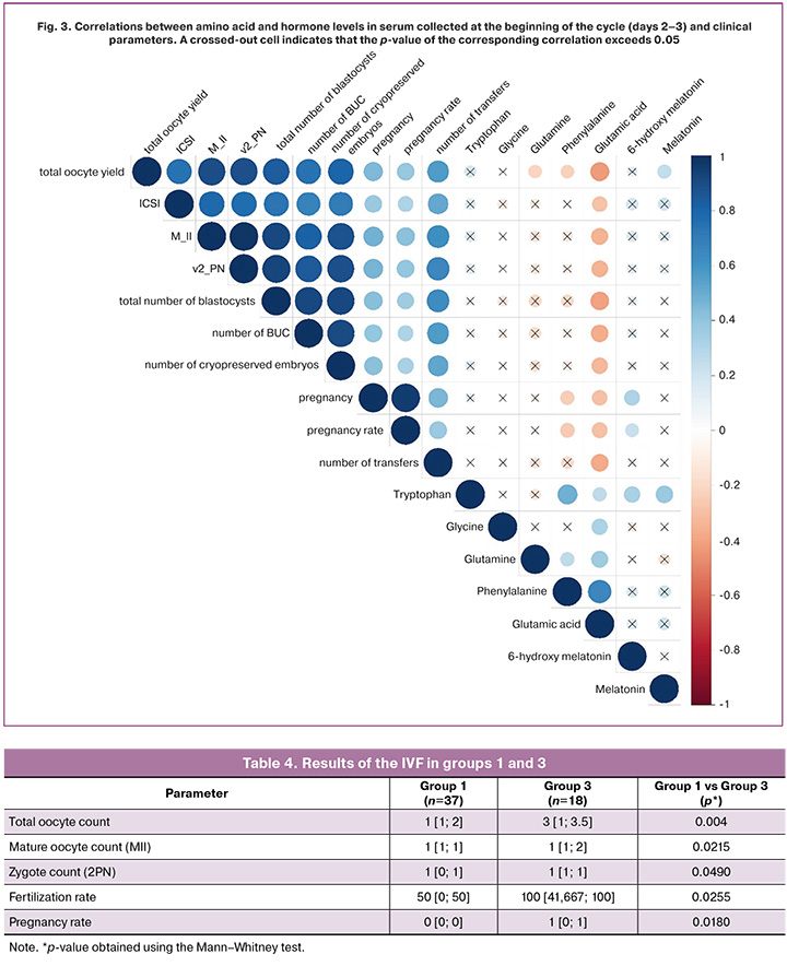
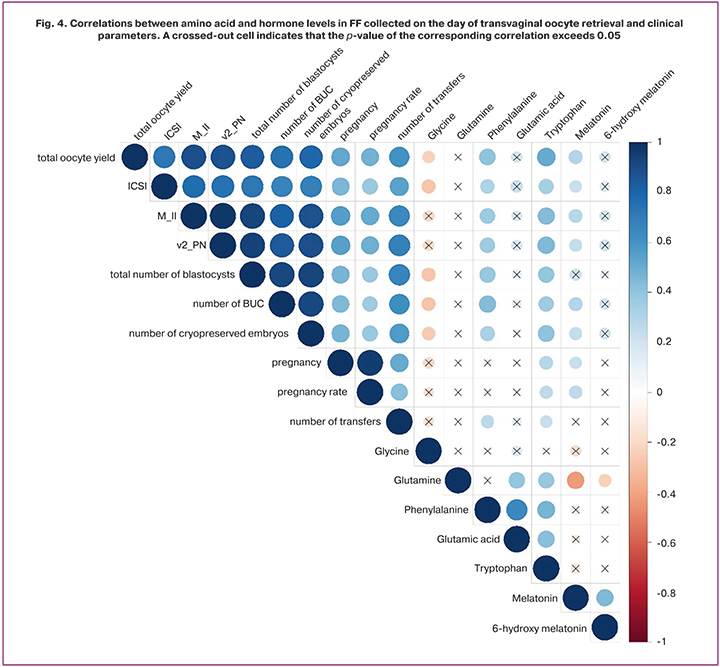
Results. Compared with the control group, women in group 1 demonstrated lower relative concentrations of phenylalanine, tryptophan, melatonin, and 6-OH-melatonin, and higher concentrations of glycine, glutamine, and glutamic acid (glutamate) in peripheral blood. Follicular fluid obtained from group 1 was characterized by lower relative levels of phenylalanine, glutamine, glutamic acid, tryptophan, melatonin, and 6-OH-melatonin, as well as elevated glycine concentrations, a pattern that was not observed in oocyte donors (group 4). Comparative analysis of endocrine and metabolomic profile of serum and clinical parameters revealed moderate negative correlations between glutamine, glutamic acid, and phenylalanine concentrations and both the total number of oocytes retrieved during ART and clinical pregnancy rates. Moderate positive correlations were identified between serum melatonin levels and the total number of retrieved oocytes, and between serum 6-OH-melatonin concentrations and clinical pregnancy rates. Correlation analysis of endocrine and metabolomic profile of the follicular fluid demonstrated moderate negative associations between glycine concentrations and indicators of oogenesis and early embryogenesis, whereas phenylalanine levels showed moderate positive correlations with the total number of mature oocytes and the number of blastocysts obtained and cryopreserved. Strong positive correlations were observed between follicular fluid tryptophan and melatonin concentrations and the parameters of oogenesis, early embryonic development, and clinical pregnancy.
Conclusion. This study evaluated the effectiveness of melatonin as a preconception intervention in women with recurrent assisted reproductive technology failure. These findings indicate that amino acid and melatonin concentrations are closely associated with key parameters of folliculogenesis and early embryogenesis, suggesting that melatonin supplementation may influence the metabolic pathways involved in amino acid metabolism.
Authors’ contributions. Biryukova D.A., Gavisova A.A. – collection and processing of material, the search and analysis of literature, drafting of the manuscript, editing and final approval of the manuscript, design of the study; Berdnikova A.I., Patskova P.O., Aksenenko A.A., V.S. Lapina – material collection and reviewing; Novoselova A.V. – development of the method for LC–MS analysis of amino acids and hormones in serum and follicular fluid, preparation of blood and follicular fluid samples, conducting analysis, processing of experimental data; V.V. Chagovets, V.E. Frankevich – statistical analysis, reviewing, final approval of the manuscript.
Conflicts of interest. The authors have no conflicts of interest to declare.
Funding. Grant of the Russian Science Foundation No. 24-64-00006, https://rscf.ru/project/24-64-00006/
Ethical Approval. The study was reviewed and approved by the Research Ethics Committee of the V.I. Kulakov NMRC for OG&P.
Generative Artificial Intelligence. No artificial intelligence tools were used in the preparation of this manuscript.
Patient Consent for Publication. All patients provided informed consent for the publication of their data.
Authors' Data Sharing Statement. The data supporting the findings of this study are available upon request from the corresponding author after approval from the principal investigator.
For citation: Biryukova D.A., Berdnikova A.I., Patskova P.O., Aksenenko A.A., Lapina V.S., Novoselova A.V.,
Chagovets V.V., Frankevich V.E., Gavisova A.A. Endocrine and metabolomic profile and the effectiveness of melatonin as a preconception intervention in women with recurrent assisted reproductive technology failure.
Akusherstvo i Ginekologiya/Obstetrics and Gynecology. 2026; (7): 102-113 (in Russian)
https://dx.doi.org/10.18565/aig.2026.74
Keywords
melatonin
amino acids
oxidative stress
diminished ovarian reserve
recurrent assisted reproductive technology failure
follicular fluid
endocrine and metabolomic profile
Assisted reproductive technology (ART) has revolutionized infertility management. According to global statistics, more than one million children have been born through in vitro fertilization (IVF) worldwide [1]. According to the official 2023 report of the Russian Association of Human Reproduction, 443,411 live births have been achieved in Russia since 1995 through the use of ART [2]. Behind these encouraging outcomes lie decades of continuous scientific progress and an ongoing search for innovative therapeutic approaches aimed at preserving and restoring reproductive health.
Despite remarkable technological advances and extensive clinical experience, reproductive specialists are increasingly confronted with the challenge of treatment failure in routine clinical practices. Within the reproductive medicine community, the term failed ART cycles is most commonly associated with recurrent implantation failure (RIF). However, this concept encompasses a broader spectrum of clinical conditions and is frequently associated with advanced reproductive age and a diminished ovarian reserve. Multiple failed ART cycles refer to a clinical scenario in which a woman fails to achieve a clinical pregnancy after two or more consecutive ART cycles, including frozen embryo transfer cycles, despite the transfer of good or excellent quality embryos [3]. Unfortunately, the number of women experiencing repeated ART failures continues to increase. This patient population represents a distinct subgroup within the broader infertility population and is associated with poorer ART outcomes and an increased risk of obstetric and perinatal complications. Failure to achieve pregnancy following ART is often emotionally devastating for patients, and the psychological burden experienced by both the couple and the treating physician intensifies with each subsequent failed treatment cycle. Consequently, the management of these patients presents substantial clinical challenges and underscores the need for individualized multidisciplinary approaches tailored to each clinical situation.
Female age remains the principal determinant of reproductive potential and the success of ART. It is well established that female fertility declines rapidly after the age of 35 years, with the probability of achieving pregnancy decreasing progressively thereafter [4]. Nevertheless, ART failure is also observed in younger women, highlighting the need to identify additional mechanisms underlying impaired reproductive outcomes and potential strategies to overcome these. Over the past decade, considerable research has focused on characterizing the metabolomic and hormonal composition of follicular fluid (FF) using multi-omics technologies [4]. Follicular fluid, secreted primarily by granulosa cells, constitutes a specialized microenvironment surrounding the developing oocyte and provides the optimal conditions required for its growth and maturation. Owing to its unique biochemical composition, even subtle alterations in follicular fluid induced by external or internal stimuli may trigger a cascade of molecular events, leading to oocyte apoptosis. Oxidative stress is one of the principal mechanisms implicated in this process.
Advanced maternal age is accompanied by reduced sex steroid production, increased ovarian hypoxia, impaired endogenous antioxidant defense mechanisms, and excessive accumulation of reactive oxygen species (ROS), ultimately resulting in oxidative damage to DNA, proteins, and lipids. Elevated concentrations of oxidative stress metabolites in the follicular fluid have consistently been associated with impaired oocyte quality and reduced reproductive potential [5,6]. The degree of oxidative stress within the follicular microenvironment can be assessed by measuring the levels of reactive oxygen species in the follicular fluid [7].
According to the available evidence, the extent of oxidative damage may correlate with the concentrations of specific amino acids in the follicular fluid (FF) [8, 9]. Amino acids are the fundamental building blocks of proteins, play a central role in cellular metabolism, maintain redox homeostasis, and participate in every stage of folliculogenesis. Altered metabolism of several amino acids, including tryptophan, glycine, glutamine, glutamic acid, and phenylalanine, has been associated with impaired folliculogenesis and, consequently, with a range of disorders affecting the female reproductive system, including diminished ovarian reserve, endometriosis, and other reproductive pathologies [1, 8, 10–13].
For example, Kurdi C. et al. reported that elevated glycine concentrations in FF, a key substrate for glutathione biosynthesis, are associated with oxidative stress in oocytes. Increased glycine secretion appears to be a compensatory response to excessive reactive oxygen species exposure in women with endometriosis [8]. A similar metabolic pattern has been observed in women of advanced reproductive age (>35 years), reflecting adaptive metabolic responses to the decline in the antioxidant capacity of oocytes. Glutamine, another functionally important amino acid, serves as a major energy substrate, is indispensable for mitochondrial ATP synthesis, and exhibits anti-inflammatory and antioxidant properties. Wang L. et al. demonstrated that reduced glutamine concentrations in FF are associated with impaired antioxidant defense mechanisms and mitochondrial dysfunction in oocytes, both of which are characteristic features of reproductive aging [14]. In 2024, Gavisova A.A. et al. reported a statistically significant reduction in follicular fluid phenylalanine concentrations in women with diminished ovarian reserve [13]. Phenylalanine serves as a substrate for protein biosynthesis, contributes to cellular energy metabolism, and possesses antioxidant properties [14]. Together with tyrosine, phenylalanine participates in the synthesis of benzoquinone, an essential precursor for coenzyme Q10 production, which is one of the most important endogenous antioxidants. These biological functions underscore the importance of phenylalanine in female reproductive physiology and support its potential role as a prognostic biomarker of oocyte competence and ART success.
Tryptophan and its metabolites exert a wide range of biological activities, including potent antioxidant effects. Serotonin, synthesized from tryptophan, is one of the principal neurotransmitters of the central nervous system and regulates steroid hormone secretion while influencing oocyte growth and maturation [15]. According to Li Q. et al., another key tryptophan metabolite, melatonin, possesses powerful antioxidant properties that are critical during oocyte maturation, fertilization, implantation, embryogenesis, and pregnancy [16]. Therefore, melatonin has been widely investigated as a pre-treatment before ART initiation and has also been incorporated into embryo culture media to improve oocyte quality and enhance embryological outcomes [5,17]. In addition, accumulating evidence suggests that the principal melatonin metabolite, 6-hydroxymelatonin (6-OHM), exhibits antioxidant activity [18]. Accordingly, both melatonin and 6-OHM were included in this study.
Taken together, these findings suggest that the endocrine and metabolomic profiles of serum and follicular fluid (FF) may reflect molecular alterations occurring within the female reproductive system and provide valuable insights into the mechanisms underlying repeated ART failure in women with a history of multiple failed treatment cycles.
The aim of the present study was to characterize the endocrine and metabolomic profiles of serum and follicular fluid in women with multiple failed ART cycles and to evaluate the effectiveness of oral melatonin administration in this patient population as a preconception intervention to improve ART outcomes.
Materials and methods
This study included women of reproductive age (25–40 years) presenting to the V.I. Kulakov National Medical Research Center for Obstetrics, Gynecology, and Perinatology for treatment of infertility using ART between 2023 and 2025. A total of 100 patients were enrolled and stratified into four groups. Group 1 comprised 37 women with a history of recurrent ART failure (in vitro fertilization [IVF]/intracytoplasmic sperm injection [ICSI]) cycles, anti-Müllerian hormone (AMH) <1.2 ng/mL, and antral follicle count (AFC) <5; Group 2 comprised 18 women with a history of recurrent ART failure, AMH≥1.2 ng/mL, and AFC≥5; Group 3 (treatment group) comprised 18 patients with a history of recurrent ART failure who received oral melatonin 3 mg daily for one month before initiating the ART protocol, with AMH<1.2 ng/mL and AFC<5; Group 4 (control group) comprised 27 healthy oocyte donors with no somatic comorbidities, AMH≥1.2 ng/mL, and AFC≥5. To evaluate the efficacy of melatonin supplementation, we assessed the total oocyte yield, number of mature (MII) oocytes, number of zygotes (2PN), fertilization rate, number of blastocysts obtained and cryopreserved, and pregnancy rate in groups 1 and 3.
Inclusion criteria were infertility for at least one year of regular unprotected intercourse, age 25–40 years, and provision of written informed consent to participate in the study. Exclusion criteria were contraindications to ART, prior ovarian surgery, immunodeficiency (HIV infection), immunoinflammatory or oncological disease, chromosomal or genetic abnormalities, participation in a donor oocyte/embryo or gestational surrogacy program, and severe male-factor infertility.
Before entering the ART program, all patients underwent a complete clinical and laboratory work-up in accordance with the Russian clinical guidelines "Female Infertility" (2024) [19] and Order No. 803n of the Russian Ministry of Health, dated July 31, 2020, "On the Procedure for the Use of Assisted Reproductive Technologies, and Contraindications and Restrictions to Their Use" [20]. Folliculogenesis was monitored by ultrasound. Oocytes were retrieved during a stimulated or natural IVF cycle. Ovarian stimulation was performed using a gonadotropin-releasing hormone (GnRH) antagonist protocol. Final oocyte maturation was triggered with human chorionic gonadotropin (5000–10,000 IU) or a GnRH agonist (0.2 mg). The ovulation trigger was administered 35–36 hours before transvaginal oocyte retrieval, per protocol, once follicles reached a diameter ≥17 mm. Peripheral blood samples were collected on cycle day 2–3. Follicular fluid (FF) was also collected and pooled from all punctured follicles for each patient.
Sample preparation was performed as follows: 480 μL of chloroform-methanol solution (2:1, v/v) was added to 100 μL of sample at 4°C; the sample was sonicated for 10 minutes; 150 μL of water was added; the mixture was vortexed for 5 minutes; the resulting solution was centrifuged at 13,000 g for 5 minutes at room temperature; 200 μL of the upper aqueous-methanolic layer was collected and dried under a stream of nitrogen for 30 minutes at 60°C; 200 μL of 3N hydrochloric acid in butanol was added and vortexed for 3 minutes; the sample was centrifuged at 13,000 g for 15 seconds at room temperature and incubated at 60°C for 15 minutes to allow derivatization; it was then centrifuged again at 13,000 g for 15 seconds at room temperature and dried under nitrogen for 30 minutes at 60°C; the residue was reconstituted in 200 μL of acetonitrile/water (1:1, v/v), vortexed for 5 minutes, and centrifuged at 13,000 g for 15 seconds at room temperature; and 120 μL of the resulting sample was transferred to a vial with an insert for analysis.
Amino acid, melatonin, and 6-hydroxymelatonin (6-OH-melatonin) concentrations in serum and FF were determined by high-performance liquid chromatography with mass spectrometric detection (HPLC-MS), using an Agilent 1260 II liquid chromatograph coupled with an Agilent 6460 mass spectrometric detector (Agilent, USA). Chromatographic separation was performed on an Agilent Zorbax Eclipse XDB-C18 column (100×2.1 mm, 1.8 μm; Agilent, USA). Mobile phase A consisted of 10 mM aqueous ammonium acetate, and mobile phase B consisted of 100% acetonitrile. The flow rate was 150 μL/min, and the injection volume was 3 μL. The following optimized gradient elution program was used: 5% B at 0 minutes, held for 0.1 minutes; increased linearly to 25% by 0.2 minutes; 30% B by 5 minutes; 55% B by 15 minutes; 70% B by 20 minutes; 95% B by 20.5 minutes, held until 25 minutes; returned to the initial 5% B by 25.5 minutes; and column re-equilibration until 40 minutes. Optimized mass spectrometer settings were as follows: drying gas temperature, 150°C; drying gas flow rate, 10 L/min; nebulizer gas pressure, 2.76 bar; sheath gas temperature, 400°C; sheath gas flow rate, 10 L/min; and capillary voltage, 2000 V.
Statistical analysis
Before statistical analysis, data were normalized to the total signal of all analytes and standardized using the transformation zi = (xi − x̄)/SD(x), where zi is the standardized value of the parameter, xi is the raw value of the parameter, x̄ is the mean value of the parameter, and SD(x) is the standard deviation of the population.
Statistical analysis of the experimental data was performed using scripts written in R [R Core Team (2018). R: A Language and Environment for Statistical Computing. R Foundation for Statistical Computing, Vienna, Austria. URL: https://www.R-project.org/] in RStudio [RStudio Team (2016). RStudio: Integrated Development for R. RStudio, Inc., Boston, MA. URL: http://www.rstudio.com/].
In the first stage of statistical analysis, the distribution of quantitative variables in the compared samples was assessed using the Kolmogorov–Smirnov test and graphical analysis. Levene's test was used to assess the equality of variances. Levels of amino acids, melatonin, and 6-OH-melatonin were determined semi-quantitatively, with the relative abundance of each analyte calculated as the ratio of its chromatographic peak area to the sum of all peak areas detected in the sample.
Normally distributed quantitative variables are presented as M (SD), where M is the mean and SD is the standard deviation; pairwise comparisons were performed using Student's t-test with Bonferroni correction, and comparisons among multiple groups were performed using ANOVA. Non-normally distributed data are presented as Me [Q1; Q3], where Me is the median and Q1 and Q3 are the lower and upper quartiles, respectively; these data were analyzed using the Mann–Whitney U test with Bonferroni correction for pairwise comparisons and the Kruskal–Wallis test for comparisons among multiple groups.
Correlations between the studied parameters were assessed using Spearman's rank correlation coefficient. A p-value <0.05 was considered statistically significant.
Results
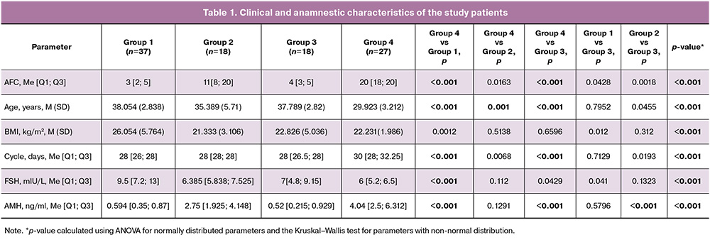
The clinical and anamnestic characteristics of the patients included in the study are presented in Table 1. Women in group 1 had a statistically significantly higher BMI than those in groups 2, 3, and 4. The mean age of patients in groups 1, 2, and 3 differed significantly from the mean age of the control group. groups 1 and 3 demonstrated significantly lower AFC, lower AMH levels, a shorter menstrual cycle, and elevated FSH levels compared with the control group (oocyte donors).
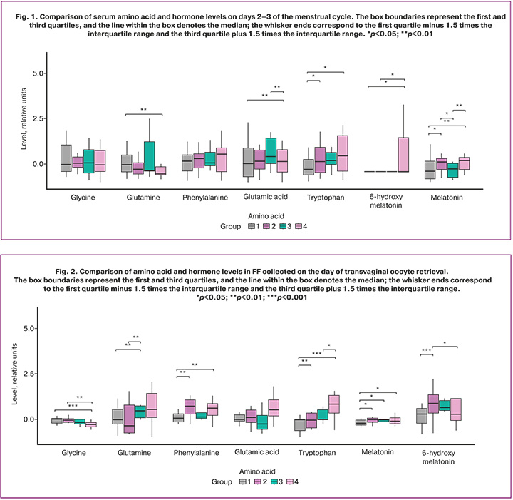
Analysis of the serum amino acid profile obtained on days 2–3 of the menstrual cycle (Fig. 1, Table 2) revealed a statistically significant decrease in the relative tryptophan content and an increase in the relative glutamine content in group 1 compared with the control group. Comparative analysis also showed a statistically significant increase in the relative glutamic acid content in patients from groups 1 and 3 compared with the oocyte donor group (Table 2). A non-significant decrease in the relative phenylalanine content was observed in groups 1, 2, and 3 relative to the control group (Table 2). Assessment of the peripheral blood hormonal profile showed a statistically significant decrease in the relative melatonin content in patients from groups 1 and 3, with a non-significant decrease in group 2 compared with the oocyte donors (Table 2). A statistically significant decrease in the relative content of 6-OH-melatonin was found in groups 1 and 2, whereas the decrease in this metabolite in group 3 compared with group 4 was not statistically significant (Table 2). No significant differences in the relative serum glycine content on days 2–3 of the menstrual cycle were observed among the study groups.
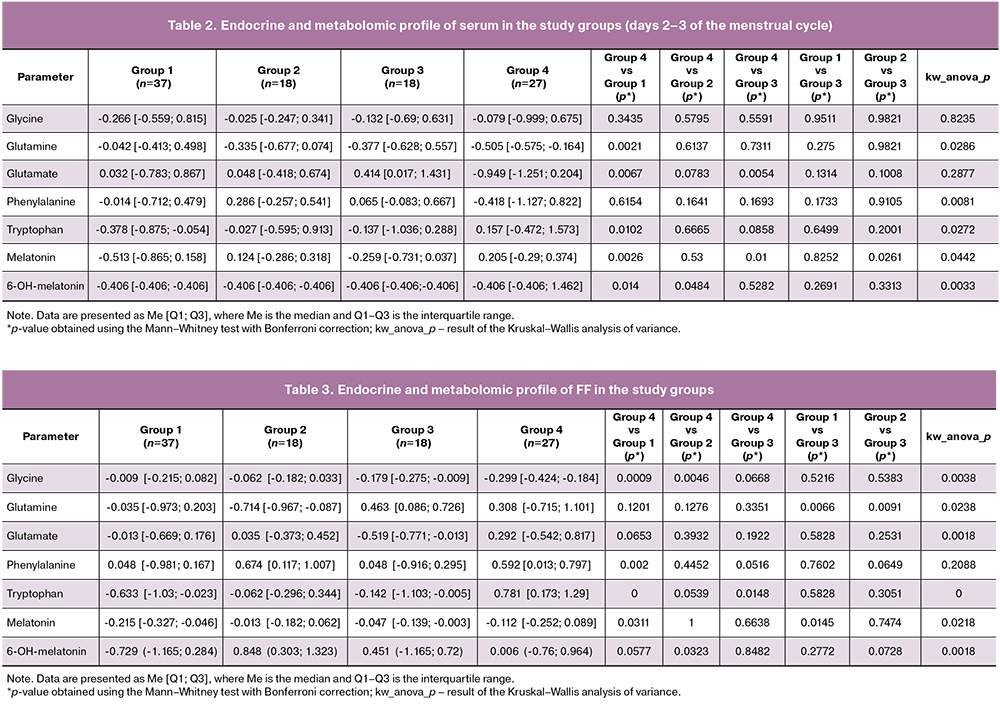
Analysis of the endocrine and metabolomic profile of follicular fluid (FF) (Fig. 2, Table 3) revealed a statistically significant decrease in the relative tryptophan content in groups 1 and 3 compared with group 4, as well as a decrease in phenylalanine in group 1 compared with the control group (Table 3). A statistically significant increase in the relative glycine content in FF was observed in groups 1 and 2, with a non-significant increase in group 3 compared with the donor group (Table 3). A significant decrease in the relative glutamine content in FF was found in patients from groups 1 and 2 compared with group 3, along with a non-significant decrease in this amino acid in groups 1 and 2 relative to the control group (Table 3). The relative glutamic acid content was non-significantly decreased in patients from groups 1, 2, and 3 compared with group 4 (Table 3). A statistically significant decrease in the relative melatonin content and a non-significant decrease in 6-OH-melatonin were also observed in patients from group 1 compared with the control (group 4) and with group 3, which received oral melatonin supplementation (Table 3).
Comparative correlation analysis of clinical parameters and the endocrine and metabolomic profile of blood collected on days 2–3 of the menstrual cycle (Fig. 3) revealed moderate negative correlations between glutamine and the total number of oocytes retrieved during the ART program, between phenylalanine and both the total oocyte number and pregnancy rate, and between glutamic acid and oogenesis parameters, early embryogenesis parameters, and pregnancy rate. Moderate positive correlations were found between serum melatonin levels on days 2–3 of the cycle and the total number of oocytes retrieved, as well as between 6-OH-melatonin levels and pregnancy rate.
Comparative correlation analysis of the endocrine and metabolomic profile of FF and clinical parameters (Fig. 4) revealed moderate negative correlations between glycine levels and oogenesis and early embryogenesis parameters, as well as moderate positive correlations between phenylalanine and the total number of mature oocytes and the number of blastocysts obtained and cryopreserved. Strong positive correlations were established between tryptophan and melatonin levels in FF and oogenesis parameters, early embryogenesis parameters, and pregnancy rate.
Statistically significant differences were found in the total number of oocytes (p=0.004), the number of mature oocytes (MII) (p=0.0215), the number of zygotes (2PN) (p=0.0490), the fertilization rate (p=0.0255), and the pregnancy rate (p=0.0180) between groups 1 and 3.
Discussion
According to our findings, patients in group 1 were characterized by decreased relative concentrations of phenylalanine, tryptophan, melatonin, and 6-hydroxymelatonin (6-OH-melatonin) in blood and FF, a finding corroborated by several investigators [5, 8, 10, 12–16]. Phenylalanine and tryptophan are aromatic amino acids that serve as precursors of neurotransmitters, including serotonin, melatonin, and catecholamines. Deficiency of these amino acids and their metabolites is associated with increased accumulation of reactive oxygen species (ROS), which exert toxic effects on oocyte, follicular, and embryonic development [12, 13].
As noted above, melatonin is one of the key tryptophan metabolites relevant to reproductive function. This hormone is detectable in nearly all body tissues, including the ovaries. Its functional versatility enables melatonin to exert both direct and indirect effects on cellular physiology and molecular biology through a range of mechanisms. Within the central nervous system, melatonin regulates the hypothalamic–pituitary–ovarian axis via melatonin receptors located in the suprachiasmatic nucleus and preoptic area of the hypothalamus, inhibiting or inducing GnRH secretion depending on prevailing physiological conditions [21]. Recent evidence indicates that disruption of circadian rhythms impairs the physiological pulsatile release of luteinizing hormone, resulting in ovulatory dysfunction [22]. Within the ovaries, melatonin inhibits apoptosis and neutralizes ROS, directly influences mitochondrial metabolism and function, modulates signaling pathways, and stimulates the production of anti-inflammatory factors as well as cellular and humoral immune responses involved in folliculogenesis, ovulation, oocyte maturation, and corpus luteum formation [23]. The melatonin metabolite 6-OH-melatonin, described earlier, exhibits similar antioxidant properties. Notably, women who received oral melatonin supplementation at a dose of 3 mg (group 3) showed increased relative concentrations of melatonin and 6-OH-melatonin in FF compared with group 1¹. Comparative analysis of folliculogenesis and embryogenesis parameters revealed a statistically significant increase in oocyte yield, including mature oocyte count – early embryonic development indices, and pregnancy rate in group 3. Sadeghpour S. et al. reported that oral melatonin administration exerts a beneficial effect on folliculogenesis and significantly improves outcomes of ART programs, consistent with our findings [5].
We also observed increased relative concentrations of glycine in blood and FF among patients in group 1, a finding consistent with the work of Kurdi C. et al. This observation may be attributable to the elevated oxidative stress characteristic of diminished ovarian reserve, which stimulates increased synthesis of glutathione – a key antioxidant for which glycine serves as a substrate [8].
In an observational study, Wang L. et al. reported that decreased glutamine levels in FF may be associated with elevated oxidative stress and mitochondrial dysfunction, mechanisms directly implicated in oogenesis and folliculogenesis [14]. Glutamic acid (glutamate), the precursor of glutamine, plays a role in reproduction by regulating the hypothalamic–pituitary–ovarian axis through modulation of gonadotropin release. Accordingly, reduced concentrations of glutamine and glutamic acid may be associated with impaired reproductive function and reduced efficacy of ART programs, as demonstrated in our study. The increased relative concentrations of glutamine and glutamate observed in peripheral blood among patients in group 1 may represent a compensatory response to increased glutathione consumption as an antioxidant.
These findings indicate that amino acids and melatonin are directly involved in oocyte, follicular, and early embryonic development, underscoring the need for further investigation of the amino acid and hormonal composition of serum and FF. Given the evolving clinical profile of patients with reproductive failure, characterized by arrested embryonic development and the absence of good- or excellent-quality blastocysts – these findings suggest that culture media composition warrants reconsideration. Future studies should explore the potential enrichment of culture media with amino acids directly involved in oocyte and follicular metabolism to improve the efficacy of ART programs.
Conclusion
In patients with multiple failed IVF/ICSI cycles, the observed differences in amino acid and melatonin levels in blood and FF may reflect distinct features of folliculogenesis and early embryogenesis, related in part to impaired antioxidant defense mechanisms, as well as the influence of melatonin supplementation on amino acid metabolism pathways. These findings support the potential value of incorporating melatonin into pregravid preparation protocols for patients with a history of reccurent ART failure, with the aim of improving treatment outcomes.
_________________________________________________________________________________
¹ At baseline, prior to treatment initiation, blood melatonin levels in group 3 patients were lower than after one month of daily supplementation. However, compared with group 4, serum melatonin levels on cycle days 2–3 were significantly lower in group 3, potentially reflecting age-related metabolic changes in this patient population. These results were not reported in the article.
References
- Lee S.H., Lira-Albarrán S., Rinaudo P.F. Proteomic and metabolomic insights into oxidative stress response activation in mouse embryos generated by in vitro fertilization. Hum. Reprod. Open. 2025; 2025(2): hoaf022. https://dx.doi.org/10.1093/hropen/hoaf022
- Корсак В.С., Смирнова А.А., Шурыгина О.В. Регистр ВРТ Общероссийской общественной организации «Российская Aссоциация Репродукции Человека». Отчет за 2023 год. Проблемы репродукции. 2025; 31(6): 6-22. https://dx.doi.org/10.17116/repro2025310616 [Korsak V.S., Smirnova A.A.,Shurygina O.V. ART Registry of the All-Russian Public Organization “Russian Association of Human Reproduction”. Report for 2023. Russian Journal of Human Reproduction. 2025; 31(6): 6-22 (in Russian). https://dx.doi.org/10.17116/repro2025310616].
- Ma J., Gao W., Li D. Recurrent implantation failure: A comprehensive summary from etiology to treatment. Front. Endocrinol. (Lausanne). 2023; 13:1061766. https://dx.doi.org/10.3389/fendo.2022.1061766
- Jia Z.C., Li Y.Q., Zhou B.W., Xia Q.C., Wang P.X., Wang X.X. et al. Transcriptomic profiling of human granulosa cells between women with advanced maternal age with different ovarian reserve. J. Assist. Reprod. Genet. 2023; 40(10):2427-37. https://dx.doi.org/10.1007/s10815-023-02915-8
- Sadeghpour S., Ghasemnejad-Berenji M., Maleki F., Behroozi-Lak T., Bahadori R., Ghasemnejad-Berenji H. The effects of melatonin on follicular oxidative stress and art outcomes in women with diminished ovarian reserve: a randomized controlled trial. J. Ovarian Res. 2025; 18(1): 5. https://dx.doi.org/10.1186/s13048-024-01584-0
- Yan F., Zhao Q., Li Y., Zheng Z., Kong X., Shu C. et al. The role of oxidative stress in ovarian aging: a review. J. Ovarian Res. 2022; 15(1): 100. https://dx.doi.org/10.1186/s13048-022-01032-x
- Begum I.A. Oxidative stress: oocyte quality and infertility. Reprod. Toxicol.2025; 137: 109011. https://dx.doi.org/10.1016/j.reprotox.2025.109011
- Kurdi C., Hesszenberger D., Csabai D., Lajtai A., Lakatos Á., Jakabfi-Csepregi R. et al. Follicular fluid amino acid alterations in endometriosis: evidence for oxidative stress and metabolic dysregulation. Biomedicines. 2025; 13(11): 2634. https://dx.doi.org/10.3390/biomedicines13112634
- Wu D., Liu C., Ding L. Follicular metabolic dysfunction, oocyte aneuploidy and ovarian aging: a review. J. Ovarian Res. 2025; 18(1): 53. https://dx.doi.org/10.1186/s13048-025-01633-2
- Wang L., Liu Y., Li J., Liu X., Wu S., Jiang A. et al. Glutamine protects porcine granulosa cell from oxidative stress-induced apoptosis by inhibiting JNK activation. Reprod. Domest. Anim. 2026; 61(2): e70179. https://dx.doi.org/10.1111/rda.70179
- Bao S., Yin T., Liu S. Ovarian aging: energy metabolism of oocytes. J.Ovarian Res. 2024; 17(1): 118. https://dx.doi.org/10.1186/s13048-024-01427-y
- Liu A., Shen H., Li Q., He J., Wang B., Du W. et al. Determination of tryptophan and its indole metabolites in follicular fluid of women with diminished ovarian reserve. Sci. Rep. 2023; 13(1): 17124. https://dx.doi.org/10.1038/s41598-023-44335-9
- Гависова А.А., Шевцова М.А., Львова П.О., Бирюкова Д.А., Ибрагимова М.Х., Новоселова А.В., Юшина М.Н., Чаговец В.В., Франкевич В.Е. Аминокислотный профиль при сниженном овариальном резерве. Вестник РГМУ. 2024; 2: 56-63. https://dx.doi.org/10.24075/vrgmu.2024.016 [Gavisova A.A., Shevtsova M.A., Lvova P.A., Biryukova D.A., Ibragimova M.Kh., Novoselova A.V., Yushina M.N., Chagovets V.V., Frankevich V.E. Amino acid profile in diminished ovarian reserve. Bulletin of RSMU. 2024; 2: 56-63 (in Russian). https://dx.doi.org/10.24075/vrgmu.2024.016].
- Wang L., Zhou C., Sun J., Zhang Q., Lai D. Glutamine and norepinephrine in follicular fluid synergistically enhance the antioxidant capacity of human granulosa cells and the outcome of IVF-ET.Sci. Rep. 2022; 12(1): 9936. https://dx.doi.org/10.1038/s41598-022-14201-1
- Li J., Zhang Z., Wei Y., Zhu P., Yin T., Wan Q. Metabonomic analysis of follicular fluid in patients with diminished ovarian reserve. Front. Endocrinol. (Lausanne). 2023; 14: 1132621. https://dx.doi.org/10.3389/fendo.2023.1132621
- Li Q., Zheng T., Chen J., Li B., Zhang Q., Yang S. et al. Exploring melatonin’s multifaceted role in female reproductive health: From follicular development to lactation and its therapeutic potential in obstetric syndromes. J. Adv. Res. 2024; 70: 223-42. https://dx.doi.org/10.1016/j.jare.2024.04.025
- Bao Z., Li G., Wang R., Xue S., Zeng Y., Deng S. Melatonin improves quality of repeated-poor and frozen-thawed embryos in human, a prospective clinical trial. Front. Endocrinol. (Lausanne). 2022; 13: 853999. https://dx.doi.org/10.3389/fendo.2022.853999
- Liu Y., Li L., Feng J., Wan B., Tu Q., Cai W. et al. Modulation of chronic obstructive pulmonary disease progression by antioxidant metabolites from Pediococcus pentosaceus: enhancing gut probiotics abundance and the tryptophan-melatonin pathway. Gut Microbes. 2024; 16(1): 2320283. https://dx.doi.org/10.1080/19490976.2024.2320283
- Министерство здравоохранения Российской Федерации. Клинические рекомендации. Женское бесплодие. 2024. [Ministry of Health of the Russian Federation. Clinical guidelines. Female infertility. 2024 (in Russian)].
- Приказ Минздрава России № 803н от 31 июля 2020 г. «О порядке использования вспомогательных репродуктивных технологий, противопоказаниях и ограничениях к их применению». Доступно по: https://publication.pravo.gov.ru/Document/View/0001202010190041 [Order of the Ministry of Health of Russia No. 803n dated July 31, 2020 "On the procedure for using assisted reproductive technologies, contraindications, and restrictions on their use". Available at: https://publication.pravo.gov.ru/Document/View/0001202010190041 (in Russian)].
- Zhong J., Lu Z., Zhou Z., Ma N., Li Y., Hu J. et al. Melatonin biosynthesis and regulation in reproduction. Front. Endocrinol. 2025; 16: 1630164. https://dx.doi.org/10.3389/fendo.2025.1630164
- Li Y., Pei T., Zhu H., Wang R., Wu L., Huang X. et al. Melatonin alleviates circadian rhythm disruption-induced enhanced luteinizing hormone pulse frequency and ovarian dysfunction. J. Pineal Res. 2025; 77(1): е70026. https://dx.doi.org/10.1111/jpi.70026
- Cosme P., Rodríguez A.B., Garrido M., Espino J. Coping with oxidative stress in reproductive pathophysiology and assisted reproduction: melatonin as an emerging therapeutical tool. Antioxidants. 2022; 12(1): 86. https://dx.doi.org/10.3390/antiox12010086
Received 22.04.2026
Accepted 26.06.2026
About the Authors
Daria A. Biryukova, gynecologist at the 1st Gynecological Department of the Institute of Reproductive Medicine, Academician V.I. Kulakov National Medical Research Center for Obstetrics, Gynecology and Perinatology, Ministry of Health of Russia, 117997, Russia, Moscow, Ac. Oparin str., 4, d_birukova@oparina4.ruAnastasia I. Berdnikova, PhD student at the 1st Gynecological Department of the Institute of Reproductive Medicine, Academician V.I. Kulakov National Medical Research Center for Obstetrics, Gynecology and Perinatology, Ministry of Health of Russia, 117997, Russia, Moscow, Ac. Oparin str., 4, berdnikova8002@mail.ru
Polina O. Patskova, PhD student at the 1st Gynecological Department of the Institute of Reproductive Medicine, Academician V.I. Kulakov National Medical Research Center for Obstetrics, Gynecology and Perinatology, Ministry of Health of Russia, 117997, Russia, Moscow, Ac. Oparin str., 4, p_lvova@oparina4.ru,
https://orcid.org/0009-0005-6805-2944
Artem A. Aksenenko, PhD, gynecologist at the 1st Gynecological Department of the Institute of Reproductive Medicine, Academician V.I. Kulakov National Medical Research Center for Obstetrics, Gynecology and Perinatology, Ministry of Health of Russia, 117997, Russia, Moscow, Ac. Oparin str., 4, a_axenenko@oparina4.ru
Vera S. Lapina, PhD, gynecologist at the 1st Gynecological Department of the Institute of Reproductive Medicine, Academician V.I. Kulakov National Medical Research Center for Obstetrics, Gynecology and Perinatology, Ministry of Health of Russia, 117997, Russia, Moscow, Ac. Oparin str., 4, v_lapina@oparina4.ru
Anastasia V. Novoselova, Researcher at the Laboratory of Metabolomics and Bioinformatics, Academician V.I. Kulakov National Medical Research Center for Obstetrics, Gynecology and Perinatology, Ministry of Health of Russia, 117997, Russia, Moscow, Ac. Oparin str., 4, aequus@ro.ru
Vitaly V. Chagovets, PhD, Head of the Laboratory of Metabolomics and Bioinformatics, Academician V.I. Kulakov National Medical Research Center for Obstetrics, Gynecology and Perinatology, Ministry of Health of Russia, 117997, Russia, Moscow, Ac. Oparin str., 4, vvchagovets@gmail.com, https://orcid.org/0000-0002-5120-376X
Vladimir E. Frankevich, Dr. Sci. (in Physics and Mathematics), Director of the Institute of Translational Medicine, Academician V.I. Kulakov National Medical Research Center for Obstetrics, Gynecology and Perinatology, Ministry of Health of Russia, 117997, Russia, Moscow, Ac. Oparin str., 4, v_vfrankevich@oparina4.ru,
https://orcid.org/0000-0002-9780-4579
Alla A. Gavisova, Dr. Med. Sci., Head of the 1st Gynecological Departament, Institute of Reproductive Medicine, Academician V.I. Kulakov National Medical Research Center for Obstetrics, Gynecology and Perinatology, Ministry of Health of Russia, 117997, Russia, Moscow, Ac. Oparin str., 4, a_gavisova@oparina4.ru,
https://orcid.org/0000-0003-4700-2786
Corresponding author: Daria A. Biryukova, d_birukova@oparina4.ru
Similar Articles

