
Evaluation of the effectiveness of combination therapy utilizing laser-mediated photodynamic therapy in patients with combined hormone-dependent pathology: endometrial hyperplasia and adenomyosis
Serov V.N., Pasman N.M., Selyunina N.A, Veretelnikova T.V., Zhdanova E.A.
Objective. To evaluate the effectiveness of photodynamic therapy (PDT) combined with traditional hormone therapy on reproductive outcomes, the frequency of clinical manifestations, and the recurrence rate in women of reproductive age with endometrial hyperplasia (EH) and adenomyosis.
Materials and methods. A retrospective single-center comparative study was carried out from 2021 to 2023. The sample was formed using a consecutive sampling method. The study included 48 women aged 25 – 45 years with the established diagnosis of adenomyosis based on clinical findings, ultrasound examination, and hysteroscopy, as well as non-atypical EH confirmed by histological examination. Two groups were formed: hormone therapy (n=21) and combination therapy using endometrial PDT (n=27). Assessment of reproductive outcomes included pregnancy rate, the time before getting pregnant, clinical symptoms before and after treatment, and the recurrence rate of EH based on follow-up histology of endometrial biopsy specimens. The follow-up period was up to 36 months.
Results. Combination therapy using PDT led to statistically significant increase in pregnancy rate (p=0.002), whereas the time before getting pregnant did not differ significantly between the groups (p=0.750). Reduced frequency of clinical manifestations after treatment was in both groups. Despite low frequency of abnormal uterine bleeding, heavy menstrual bleeding, and dysmenorrhea in the group of combination therapy using PDT, no statistically significant differences between the groups in these outcomes were observed. The recurrence of EH was less common in the group of women who were treated with combination therapy compared with the group of women who received traditional hormone therapy (3/18 [16.7%] vs 6/13 [46.2%]; RR=0.36; 95% CI 0.11–1.18; p=0.114).
Conclusion. In patients with EH and adenomyosis, combination therapy using PDT was associated with higher pregnancy rate compared with hormone therapy alone. It suggests that this approach is promising for treatment of combined hormone-dependent pathology. The trend was observed in favor of combination therapy with PDT regarding the recurrence rate of EH and persistence of clinical symptoms after treatment. However, no statistically significant intergroup differences were found for these indicators.
Authors' contributions. Selyunina N.A., Veretelnikova T.V., Zhdanova E.A. – patient selection and examination, writing the manuscript, literature review on the topic of the article; Serov V.N., Pasman N.M. – study conception and design, critical review of the content, manuscript approval for publication.
Conflicts of interest. The authors declare that they have no conflicts of interest.
Funding. The study was carried out without any sponsorship.
Ethical Approval. The study was approved by the local Ethics Committee of Novosibirsk National Research State University.
Generative Artificial Intelligence. No generative AI was used in preparing this article.
Patient Consent for Publication. The patients have signed informed consent for publication of their data.
Authors' Data Sharing Statement. The data supporting the findings of this study are available on request from the corresponding author after approval from the principal investigator.
For sitation: Serov V.N., Pasman N.M., Selyunina N.A, Veretelnikova T.V., Zhdanova E.A. Evaluation of
the effectiveness of combination therapy utilizing laser-mediated photodynamic therapy in patients with combined hormone-dependent pathology: endometrial hyperplasia and adenomyosis.
Akusherstvo i Ginekologiya/Obstetrics and Gynecology. 2026; (6): 144-152 (in Russian)
https://dx.doi.org/10.18565/aig.2025.325
Keywords
endometrial hyperplasia
genital endometriosis
adenomyosis
hormonal therapy
combination therapy
endometritis
In recent decades, the incidence of hormone-dependent diseases, including endometrial hyperplasia and endometriosis, has been increasing in many countries worldwide. These two conditions are often diagnosed in the same patient, that significantly complicates treatment. Pathophysiological studies have confirmed that adenomyosis is an estrogen-dependent disorder similar to endometrial hyperplasia (EH) in terms of changes in the hormonal status in patients. Thus, prolonged exposure to endogenous estrogen (for example, early menarche between the ages of 9 and 11, or late menopause) is a risk factor for endometriosis, while the risk of EH increases both with relative endogenous hyperestrogenism associated with obesity and ovulatory dysfunction, and the use of exogenous estrogens, for example, in assisted reproductive technology cycles. Currently, due to an unprecedented level of obesity, we observe the signs of a shift toward increased incidence of EH among women in premenopause and perimenopause [1–4]. Adenomyosis and EH have common risk factors, such as menstrual and reproductive history, smoking, diet, and environmental exposures. Both conditions contribute to infertility. EH is associated with decreased area and height of pinopodia, a two-fold decrease in the expression of leukemia inhibitory factor (LIF) by glandular and stromal cells, that demonstrates decreased endometrial receptivity and is a predictor of implantation failure. Uterine factor infertility is associated both with EH and endometriosis. It is known that in different locations of genital endometriosis the incidence of infertility is approximately 3-4 times higher than in the general population [5–7].
At the same time, there is evidence that matrix metalloproteinase-9 (MMP-9) expression in EH is enhanced depending on the level of estrogen receptor expression, while increased expression of MMP-2 or MMP-9 can be an important factor for the development of adenomyosis, promoting the invasion of endometrial tissue into the myometrium and angiogenesis in adenomyotic foci [8, 9].
A conservative approach to therapy in combination with surgical treatment of external genital endometriosis, which is often found in this category of patients (enucleation of endometrioid cysts, coagulation of endometriotic foci, and others), has demonstrated the highest level of success. However, the rate of disease progression, significant impact on quality of life and fertility dictate the search for new approaches to therapy.
The issues of EH recurrence, limitations for the use of long-term medications, diminished ovarian reserve, formation of intrauterine adhesions or high prevalence of chronic endometritis due to multiple intrauterine interventions in this group of patients remain unresolved. This determines the relevance of searching for new, more effective organ-sparing minimally invasive strategies [10–12].
The use of photodynamic therapy (PDT) for treatment of adenomyosis coexisting with EH is a safe, selective approach that can limit side effects. PDT can be used in combination with other treatment approaches and can help prevent hyperthermia or Asherman's syndrome recurrence by reducing the invasive potential of the endometrium, and thereby preventing subsequent disease progression. Moreover, numerous studies emphasize that PDT is a safe treatment modality for reproductive outcomes [2, 13–17].
The objective of the study was to evaluate the effectiveness of combination of photodynamic therapy (PDT) and traditional hormone therapy on reproductive outcomes, the frequency of clinical manifestations, and the recurrence rate in women of reproductive age with EH and adenomyosis.
Materials and methods
This retrospective single-center comparative study included 48 female patients aged 25–45 years, who were admitted to the Clinic of Professor Pasman with the established diagnoses of adenomyosis and non-atypical EH.
Inclusion criteria in the study were clinically and instrumentally confirmed diagnoses of adenomyosis and EH, which were histologically verified. Exclusion criteria were atypical EH, large uterine fibroids (over 4 cm), malignant neoplasms, male factor infertility.
The dynamics of clinical parameters, the results of laboratory and instrumental examination, as well as histological and immunohistochemical analysis of endometrial biopsies were assessed. The severity of chronic endometritis was determined based on the results of histological and immunohistochemical examinations (including assessment of Syndican-1 (CD 138) expression).
Primary endpoints were reproductive outcomes – the length of time before the onset of pregnancy and getting pregnant during a 36-month follow-up. Secondary endpoints included the frequency of clinical manifestations of adenomyosis and EH (abnormal uterine bleeding (AUB)), heavy menstrual bleeding (HMB), dysmenorrhea, dyspareunia, and the recurrence of EH.
In accordance with the indicated therapy two clinical groups were formed: group 1 – hormone therapy (n=21) and group 2 – combination therapy using endometrial PDT (n=27). Assessment of reproductive outcomes included pregnancy rate, the length of time before getting pregnant, clinical symptoms before and after treatment, and the recurrence of EH based on follow-up histology of endometrial biopsy specimens. The follow-up period was up to 36 months.
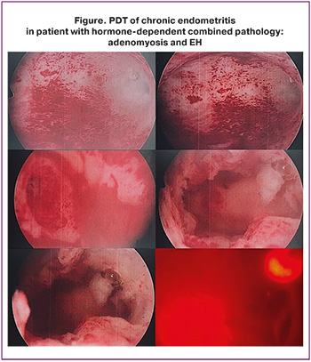
After receiving the results of endometrial biopsy in the outpatient setting and using non-steroidal anti-inflammatory agents or intravenous anesthesia during hysteroscopy (Fig.), the patients with the established diagnosis of chronic endometritis, who had previously signed informed consent, underwent laser-mediated endometrial PDT (the patent for invention RU 2 692 999 C2). For this purpose, photosensitization of the endometrium was performed by intrauterine administration of Radachlorin at a dose of 3.5 mg with exposure time 60–90 minutes. Then uterine walls were irradiated with multi-spot positioning laser at a wave length of 662 nm for 11–13-minutes in non-contact mode until energy density reached 30-40 J/cm2 at radiation power of 0.3–0.4 W using a quartz-polymer light guide connected to the Latus semiconductor laser generator [18].
Assessment of reproductive outcomes included the occurrence of pregnancy and the length of time (months) before getting pregnant after surgery with a maximum follow-up period of 36 months. The effectiveness of treatment was assessed based on clinical data, sonographic findings, and morphological examination of tissue samples obtained by endometrial aspiration biopsy.
Statistical analysis
Statistical data processing was performed using JASP statistics package. The quantitative indicators were represented as the median (Me) and interquartile range [Q1; Q3]. The Mann–Whitney U test was used for intergroup comparisons. The categorical variables were presented as n/N (%); Pearson’s chi-squared test was used for intergroup comparisons. Two-sided Fisher's exact test was used for small numbers of frequencies. For binary outcomes relative risk (RR) and 95% confidence interval (95% CI) were calculated. The differences were considered statistically significant at p<0.05.
Results
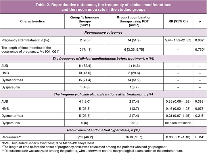
There was no statistically significant difference between the groups in the median age of patients at the beginning of treatment – 38 [35; 42] years in the hormone therapy group and 37 [35; 40,5] years in the combination therapy group, that indicated comparability between the groups by age (p=0.446).
The groups were comparable by the beginning of the menstrual function (p=0.258). The normal age for menarche prevailed in both groups.
No statistically significant difference was found between the groups in the incidence of external genital diseases (p>0.05 for all diseases), that indicates comparability between the groups by somatic status of patients.
Multiparous women, who had given birth to ≥2 babies were most often observed in the hormone therapy group – 10/21 (47.6%), whereas the number of women who had given birth to 1 baby prevailed in the combination therapy group – 17/27 (63%). Reproductive profile was unfavorable in group 2 – spontaneous miscarriages were most common (p=0.008).
No statistically significant differences in the incidence of gynecological and external genital pathologies were found between the groups (p>0,05). In group 2, infertility, external genital endometriosis and low ovarian reserve, as well as burdened surgical history we observed more often (Table 1), that can indicate a more severe baseline profile of patients in the combination therapy group. The intergroup differences in the frequency of the diagnosed chronic endometritis based on morphological examination were comparable between the groups (p=0.390).
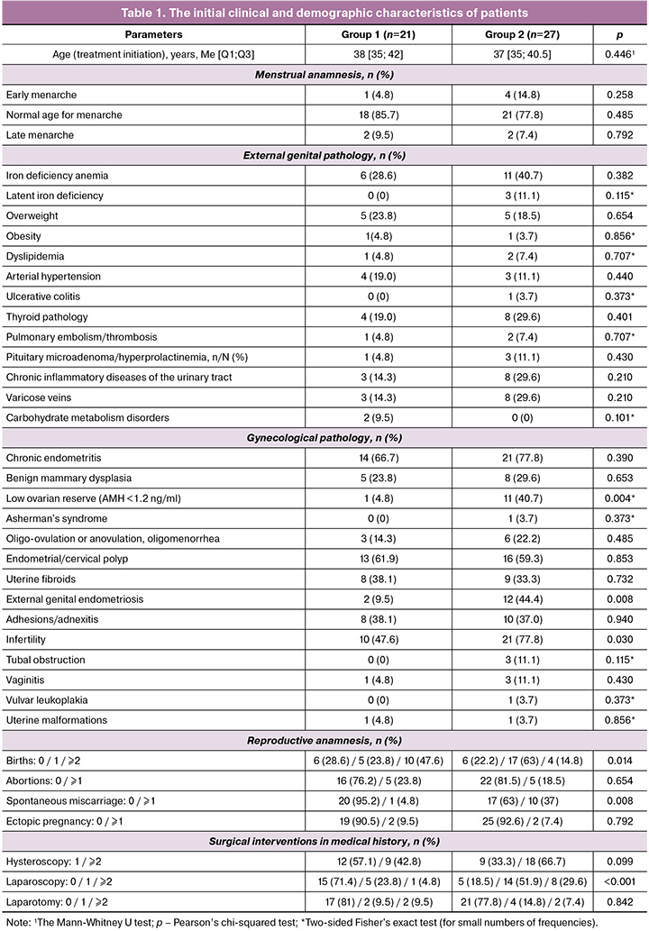
Combination therapy including PDT led to significantly higher pregnancy rate compared with hormone therapy alone. The length of time before the onset of pregnancy among the women who had got pregnant was not significantly different.
The frequency of clinical manifestations after treatment reduced in both groups. Despite lower frequency of AUB, HMB, and dysmenorrhea in the combination therapy group, no statistically significant intergroup differences were found in these parameters. The patients in both groups did not have dyspareunia after treatment.
Reccurrence of EH was less common among the patients who underwent control morphological examination in the combination therapy group. However, no statistically significant differences were found between the groups (Table 2).
Discussion
The patients in both groups were predominantly of late reproductive age (36–45 years) (Table 1). Probably due to a higher percentage of anovulatory cycles and, as a result, relative progesterone deficiency – a key factor in the pathogenesis of EH [19], as well as the diagnostic features at this age, endometrial biopsy is generally performed after the age of 40. In late reproductive age, women experience longer menstrual periods, that means the prolonged exposure to estrogen. Also, adenomyosis is traditionally most commonly diagnosed in women of advanced reproductive age and in multiparous women, as is published in epidemiological reviews and population data [2, 3, 20, 21].
Despite the fact that some authors reported relationship between early menarche and EH or adenomyosis [3, 20–22], in our study the average age of menarche was most common (Table 1). This confirms that the age at menarche is not a mandatory or universal risk factor for adenomyosis or EH. The absence of a shift toward early menarche in these patients can lead to low clinical suspicion and late verification of the disease, and, consequently, the start of therapy. The researchers report that it takes on average up to 10 years from the occurrence of adenomyosis to its diagnosis and treatment initiation [23, 24].
A history of already realized reproductive function is associated with more favorable chances for subsequent pregnancy and live birth, that is confirmed in the studies by other authors [24, 25] and emphasizes low probability of pregnancy in group 2, despite more successful results obtained in our study.
High infertility rate, low ovarian reserve, and external genital endometriosis are comparable with the data reported by other authors in patients with hormone-dependent pathology [7, 26, 27]. In addition, in the studied patient groups, risk factors were identified for the diseases that also contribute to infertility (menstrual irregularities, overweight or obesity, multiple surgeries leading to premature ovarian failure or diminished ovarian reserve due to external genital endometriosis, and chronic endometritis as a result of intrauterine surgical manipulations) (Table 1) [28–31]. A high incidence of chronic endometritis in both groups reflects the clinical characteristics of patients with combined hormone-dependent pathology as a risk factor for the occurrence and recurrence of hyperplastic processes, impaired endometrial receptivity, and reduced implantation potential.
The obtained data demonstrate the advantages of combination therapy using PDT for realization of reproductive function (p=0.002) in the group of patients with EH and adenomyosis. However, the length of time before the occurrence of pregnancy did not significantly differ among the women who got pregnant (p=0.750). Combined hormone-dependent pathology is accompanied by impaired endometrial receptivity, chronic inflammation, altered angiogenesis and local hormonal dysregulation [2, 5–7]. Given the unresolved issues related to the etiology of chronic endometritis, it is suggested that the anti-inflammatory, immunomodulatory, and antibacterial effects of PDT can be a promising, additional pathogenetic therapeutic approach to treatment of this group of patients. In view of the pathogenetic theory of using PDT, it can be suggested that exposure of the endometrium to free oxygen radicals leads to oxidative stress.
After photosensitizer is localized in the uterine cavity, radiation penetrates the tissue and activates mitochondria, that promotes an increase in cellular ATP. In response to oxidative stress, AMP-activated protein kinase (AMPK) is activated, that in turn suppresses mTOR and stimulates autophagy. This can reduce cyclooxygenase 2 levels, since mTOR plays a role in activating inflammatory responses, as has been demonstrated in numerous studies [32–34]. Helping to reduce inflammation, combination of PDT with traditional therapy for adenomyosis, can improve reproductive outcomes in this group of patients and have a better anti-relapse effect due to the impact on chronic endometritis, the main risk factor for the recurrence of EH.
The frequency of clinical manifestations of combined hormone-dependent pathology in both groups reduced after treatment. Despite the absence of statistically significant intergroup differences in the incidence of persistent symptoms after treatment, low incidence of HMB was in the combination therapy group, and the difference in this indicator was borderline significant (p=0.073). This results seems to be clinically significant, since HMB is one of the leading factors associated with reduction in quality of life, development of anemia and reproductive impairments in patients with EH and adenomyosis [28, 29].
Conclusion
In patients with EH and adenomyosis, combination therapy using PDT was associated with higher pregnancy rate compared with hormone therapy alone. The trend was observed in favor of combination therapy with PDT regarding the recurrence rate of EH and persistence of clinical symptoms after treatment. However, no statistically significant intergroup differences were found for these indicators. The obtained results confirm the potential of including PDT in the complex treatment of patients with combined hormone-dependent pathology and justify the need for further research using large samples.
References
- Giudice L.C. Clinical practice. Endometriosis. N. Engl. J. Med. 2010; 362(25): 2389-98. https://dx.doi.org/10.1056/NEJMcp1000274
- Treloar S.A., Bell T.A., Nagle C.M., Purdie D.M., Green A.C. Early menstrual characteristics associated with subsequent diagnosis of endometriosis. Am. J. Obstet. Gynecol. 2009; 202(6): 534.e1-6. https://dx.doi.org/10.1016/j.ajog.2009.10.857
- Nnoaham K.E., Webster P., Kumbang J., Kennedy S.H., Zondervan K.T. Is early age at menarche a risk factor for endometriosis? A systematic review and meta-analysis of case-control studies. Fertil. Steril. 2012; 98(3): 702-712.e6. https://dx.doi.org/10.1016/j.fertnstert.2012.05.035
- Thiruchelvam U., Wingfield M., O’Farrelly C. Natural killer cells: key players in endometriosis. Am. J. Reprod. Immunol. 2015; 74(4): 291-301. https://dx.doi.org/10.1111/AJI.12408
- Гришкина А.А., Чистякова Г.Н., Ремизова И.И., Мелкозерова О.А. Рецептивность эндометрия и экспрессия факторов апоптоза и пролиферации в эндометрии женщин с гиперплазией и бесплодием. Вестник ВолГМУ. 2019; 4(72): 37-9. https://dx.doi.org/10.19163/1994-9480-2019(72)-37-39 [Grishkina A.A., Chistjakova G.N., Remizova I.I., Melkozerova O.A. Endometrial receptivity and expression of apoptosis and proliferation factors in the endometrium of women with hyperplasia and infertility. Journal of Volgograd State Medical University. 2019; 4(72): 37-9 (in Russian). https://dx.doi.org/10.19163/1994-9480-2019(72)-37-39].
- Павловская М.А. Гиперплазия эндометрия у женщин фертильного возраста: клиника, диагностика, патогенез и возможности терапии. Журнал Гродненского государственного медицинского университета. 2015; 2(50): 123-7. [Pavlovskaya M.A. Endometrial hyperplasia in women of reproductive age: clinical features, diagnosis, pathogenesis and treatment. Journal of Grodno State Medical University. 2015; 2(50): 123-7 (in Russian)].
- Оразов М.Р., Радзинский В.Е., Хамошина М.Б., Кавтеладзе Е.В., Шустова В.Б., Цораева Ю.Р., Новгинов Д.С. Бесплодие, ассоциированное c эндометриозом: от легенды к суровой реальности. Трудный пациент. 2019; 17(1-2): 6-12. https://dx.doi.org/10.24411/2074-1995-2019-10001 [Orazov M.R., Radzinsky V.E., Khamoshina M.B., Kavteladze E.V., Shustova V.B., Tsoraeva Yu.R., Novginov D.S. Endometriosis -associated infertility: from myths to harsh reality. Difficult patient. 2019; 17(1-2): 6-12 (in Russian). https://dx.doi.org/10.24411/2074-1995-2019-10001
- Адамян Л.В., Манукян Л.М., Логинова О.Н., Арсланян К.Н., Зайратьянц В.О. Роль матриксных металлопротеиназ в патогенезе эндометриоза (обзор литературы). Проблемы репродукции. 2020; 26(2): 95-103. https://dx.doi.org/10.17116/repro20202602195 [Adamyan L.V., Manukyan L.M., Loginova O.N., Arslanyan K.N., Zayratyants V.O. The role of matrix metalloproteinases in the pathogenesis of endometriosis(literature review). Russian Journal of Human Reproduction. 2020; 26(2): 95‑103 (in Russian). https://dx.doi.org/10.17116/repro20202602195].
- Chodankar R., Critchley H.O.D. Biomarkers in abnormal uterine bleeding. Biol Reprod. 2019; 101(6): 1155-66. https://dx.doi.org/10.1093/biolre/ioy231
- Табеева Г.И., Еловенко А.А., Думановская М.Р., Асатурова А.В., Куземин А.A. Cовременные аспекты патофизиологии, диагностики и лечения хронического эндометрита. Акушерство и гинекология. 2024; 11: 12-20. https://dx.doi.org/10.18565/aig.2024.223 [Tabeeva G.I., Elovenko A.A., Dumanovskaya M.R., Asaturova A.V., Kuzemin A.A. Modern aspects of pathophysiology, diagnosis and treatment of chronic endometritis. Obstetrics and Gynecology. 2024; (11): 12-20 (in Russian). https://dx.doi.org/10.18565/aig.2024.223].
- Сухих Г.Т., Серов В.Н., Адамян Л.В., Баранов И.И., Беженарь В.Ф., Габидуллина Р.И., Дубровина С.О., Козаченко А.В., Подзолкова Н.М., Сметник А.А., Тапильская Н.И., Уварова Е.В., Ших Е.В., Ярмолинская М.И. Алгоритмы ведения пациенток с эндометриозом: согласованная позиция экспертов Российского общества акушеров-гинекологов. Акушерство и гинекология. 2023; 5: 159-76. https://dx.doi.org/10.18565/aig.2023.132 [Sukhikh G.T., Serov V.N., Adamyan L.V., Baranov I.I., Bezhenar V.F., Gabidullina R.I., Dubrovina S.O., Kozachenko A.V., Podzolkova N.M., Smetnik A.A., Tapilskaya N.I., Uvarova E.V., Shikh E.V., Yarmolinskaya M.I. Algorithms for the management of patients with endometriosis: an agreed position of experts from the Russian Society of Obstetricians and Gynecologists. Obstetrics and Gynecology. 2023; (5): 159-76 (in Russian). https://dx.doi.org/10.18565/aig.2023.132].
- Оразов М.Р., Михалева Л.М., Муллина И.А. Рецидивирующая эндометриальная гиперплазия без атипии в репродуктивном возрасте. Прогнозирование и ранняя диагностика. Акушерство и гинекология: новости, мнения, обучение. 2023; 11(3): 30-3. https://dx.doi.org/10.33029/2303-9698-2023-11-3-30-33 [Orazov M.R., Mikhaleva L.M., Mullina I.A. Recurrent endometrial hyperplasia without atypia in reproductive age. Prediction and early diagnosis. Obstetrics and Gynecology: News, Opinions, Training. 2023; 11(3): 30-3 (in Russian). https://dx.doi.org/10.33029/2303-9698-2023-11-3-30-33].
- Kim H., Kim H.J., Ahn H.S. Does endometriosis increase the risks of endometrial hyperplasia and endometrial cancer? Gynecol. Oncol. 2022; 169(10): 147-53. https://dx.doi.org/10.1016/j.ygyno.2022.06.021
- Бахвалова A.A. Риск регенерации слизистой оболочки тела матки после аблации эндометрия у больных с рецидивирующими гиперпластическими процессами в эндометрии. Вестник Российской ассоциации акушеров- гинекологов. 2000; 1: 95-100. [Bakhvalova A.A. Risk of regeneration of the uterine body mucosa after endometrial ablation in patients with recurrent hyperplastic processes in the endometrium. Bulletin of the Russian Association of Obstetricians and Gynecologists. 2000; 1: 95-100 (in Russian)].
- Хачатурян А.Р., Ярмолинская М.И., Папаян Г.В. Отдаленные результаты применения фотодинамической терапии при инфильтративных формах наружного генитального эндометриоза. Research’n Practical Medicine Journal. 2017: 2. [Khachaturyan A.R., Yarmolinskaya M.I., Papayan G.V. Long-term results of application of photodynamic therapy in cases of infiltrative forms of external genital endometriosis. Research’n Practical Medicine Journal. 2017: 2. (in Russian)].
- Арутюнян Н.А., Кацалап С.Н., Акатьева А.С., Хмелевская В.Ф., Алёхин А.И. Фотодинамическая терапия при синдроме «тонкого» эндометрия пациенток с бесплодием. Радиация и риск. 2023; 32(2): 56-65. https://dx.doi.org/10.21870/0131-3878-2023-32-2-56-65 [Arutyunyan N.A., Katsalap S.N., Akatieva A.S., Khmelevskaya V.F., Alekhin A.I. Photodynamic therapy for thin endometrium in patients with infertility. Radiation and Risk. 2023; 32(2): 56-65 (in Russian). https://dx.doi.org/10.21870/0131-3878-2023-32-2-56-65].
- Correia-Barros G., Serambeque B., Carvalho M.J., Marto C.M., Pineiro M., Pinho E Melo T.M.V.D. et al. Applications of photodynamic therapy in endometrial diseases. Bioengineering (Basel). 2022; 9(5): 226. https://dx.doi.org/10.3390/bioengineering9050226
- Никонов С.Д., Пасман Н.М., Безденежных Е.И. Способ лечения хронического эндометрита. Патент № RU 2 692 999 C2. МПК A61N 5/067(2006.01), A61K 38/41(2006.01), A61P 15/00(2006.01), A61M 31/00(2006.01). № 2017136360. 2017: 10 с. [Nikonov S.D., Pasman N.M., Bezdenezhnykh E.I. A method for treating chronic endometritis. Patent No. RU 2 692 999 C2. IPC A61N 5/067(2006.01), A61K 38/41(2006.01), A61P 15/00(2006.01), A61M 31/00(2006.01). No. 2017136360. 2017: 10 p. (in Russian)].
- Galani A., Stavros S., Moustakli E., Potiris A., Zikopoulos A., Anagnostaki I. et al. Endometrial hyperplasia: current insights into pathogenesis and risk factors. Cancers (Basel). 2026; 18(1): 148. https://dx.doi.org/10.3390/cancers18010148
- Upson K., Missmer S.A. Epidemiology of adenomyosis. Semin. Reprod. Med. 2020; 38(2-03): 89-107. https://dx.doi.org/10.1055/s-0040-1718920
- Taran F.A., Stewart E.A., Brucker S. Adenomyosis: epidemiology, risk factors, clinical phenotype and surgical and interventional alternatives to hysterectomy. Geburtshilfe Frauenheilkd. 2013; 73(9): 924-31 https://dx.doi.org/10.1055/s-0033-1350840
- Li Z., Yin J., Liu Y., Zeng F. A risk prediction model for endometrial hyperplasia/endometrial carcinoma in premenopausal women. Sci. Rep. 2025; 15(1): 1019. https://dx.doi.org/10.1038/s41598-024-83568-0
- Breton Z., Gouesbet S., Indersie E., Gabillet M., Tran V., Aflak N. et al. Endometriosis diagnostic delay and its correlates: results from the ComPaRe-Endometriosis cohort. J. Womens Health (Larchmt). 2025; 35(2): 172-88. https://dx.doi.org/10.1177/15409996251380129
- Vercellini P., Bandini V., Viganò P., Ambruoso D., Cetera G.E., Somigliana E. Proposal for targeted, neo-evolutionary-oriented secondary prevention of early-onset endometriosis and adenomyosis. Part II: medical interventions. Hum. Reprod. 2024; 39(1): 18-34. https://dx.doi.org/10.1093/humrep/dead206
- Li Z., Chen W., Sun X., Zhao X., Hu J., Ge L. et al. Nomogram to predict live birth in infertile women with adenomyosis undergoing IVF/ICSI treatment: a retrospective cohort study. Front. Endocrinol. (Lausanne). 2025; 16: 1473923. https://dx.doi.org/10.3389/fendo.2025.1473923
- He W., Chen S., Huang J., Zhang X., Hu L., Xue Z. et al. Association between type of infertility and live birth in couples with a single intrauterine insemination resulting in pregnancy: a propensity score matching cohort study. Front. Endocrinol. 2022; 13: 926183. https://dx.doi.org/10.3389/fendo.2022.926183
- Адамян Л.В., Салимова Д.Ф., Кондратович Л.М. Патогенетические аспекты эндометриоз-ассоциированного бесплодия. Проблемы репродукции. 2015; 21(6): 90-6. https://dx.doi.org/10.17116/repro201521682-88 [Adamyan L.V., Salimova D.F., Kondratovich L.M. Pathogenetic aspects of endometriosis-associated infertility. Russian Journal of Human Reproduction. 2015; 21(6): 90-6 (in Russian). https://dx.doi.org/10.17116/repro201521682-88].
- Song D., Feng X., Zhang Q., Xia E., Xiao Y., Xie W. et al. Prevalence and confounders of chronic endometritis in premenopausal women with abnormal bleeding or reproductive failure. Reprod. Biomed. 2018; 36(1): 78-83. https://dx.doi.org/10.1016/j.rbmo.2017.09.008
- Саламова К.К., Сапрыкина Л.В., Рамазанова А.М., Мильдзихова З.Т., Столярова Е.В. Клинические особенности пациенток с гиперпластическими процессами эндометрия. РМЖ. Мать и дитя. 2021; 4(2): 124-9. https://dx.doi.org/10.32364/2618-8430-2021-4-2-124-129 [Salamova K.K., Saprykina L.V., Ramazanova A.M., Mildzikhova Z.T., Stolyarova E.V. Clinical characteristics of women with endometrial hyperplasia. Russian Journal of Woman and Child Health. 2021; 4(2): 124-9 (in Russian). https://dx.doi.org/10.32364/2618-8430-2021-4-2-124-129].
- Tsikouras P., Kritsotaki N., Nikolettos K., Kotanidou S., Oikonomou E., Bothou A. et al. The impact of adenomyosis on Pregnancy. Biomedicines. 2024; 12(8): 1925. https://dx.doi.org/10.3390/biomedicines12081925
- Lee S.U., Choi S.K., Song H., Kim Y.W. Pregnancy outcomes of endometrial hyperplasia with or without atypia. Sci. Rep. 2025; 15(1): 23231. https://dx.doi.org/10.1038/s41598-025-05906-0
- Chen Z., Liu X., Qin H., Lu Z., Huang S., Liu M. et al. The effect of 630 nm photobiomodulation on the anti-inflammatory effect of human gingival fibroblasts. Photonics. 2021; 8(9): 360. https://dx.doi.org/10.3390/photonics8090360
- Ryu J.H., Park J., Kim B.Y., Kim Y., Kim N.G., Shin Y.I. Photobiomodulation ameliorates inflammatory parameters in fibroblast-like synoviocytes and experimental animal models of rheumatoid arthritis. Front. Immunol. 2023; 14: 1122581. https://dx.doi.org/10.3389/fimmu.2023.1122581
- Shamloo S., Defensor E., Ciari P., Ogawa G., Vidano L., Lin J.S. et al. The anti-inflammatory effects of photobiomodulation are mediated by cytokines: evidence from a mouse model of inflammation. Front. Neurosci. 2023; 17: 1150156. https://dx.doi.org/10.3389/fnins.2023.1150156
Received 10.11.2025
Accepted 30.04.2026
About the Authors
Vladimir N. Serov, Academician of the Russian Academy of Sciences, Dr. Med. Sci., Professor, Chief Researcher, Academician V.I. Kulakov National Medical Research Center for Obstetrics, Gynecology and Perinatology, Ministry of Health of Russia, 117997, Russia, Moscow, Ac. Oparina str., 4, +7(495)438-72-87,https://orcid.org/0000-0001-6640-1235
Natalia M. Pasman, Dr. Med. Sci., Professor, Head of the Department of Obstetrics and Gynecology, Institute of Medicine and Medical Technologies, Novosibirsk National Research State University, 630090, Russia, Novosibirsk, Pirogova str., 2, +7(913)916-25-91, https://orcid.org/0000-0002-6095-1954
Nadezhda A. Selyunina, obstetrician-gynecologist, Professor Pasman Clinic LLC; Teaching Assistant, Department of Obstetrics and Gynecology, Institute of Medicine and Medical Technologies, Novosibirsk National Research State University, 630090, Russia, Novosibirsk, Pirogova str., 2, Researcher ID Web of Science: OAJ-2348-2025,
SPIN: 4273-7582, https://orcid.org/0009-0003-9958-6151
Tatyana V. Veretelnikova, obstetrician-gynecologist of the highest category, Professor Pasman Clinic LLC, Russia, Novosibirsk, https://orcid.org/0009-0007-1022-6985
Elizaveta A. Zhdanova, student at the Department of Obstetrics and Gynecology, Institute of Medicine and Medical Technologies, Novosibirsk National Research State University, 630090, Russia, Novosibirsk, Pirogova str., 2, +7(983)406-08-76, e.zhdanova1@g.nsu.ru, SPIN: 8914-4911, https://orcid.org/0009-0001-9259-4840
Corresponding author: Nadezhda A. Selyunina, nadezhda_silk@mail.ru
Similar Articles

