
Proliferative endometrial diseases: cytokine and macroglobulin spectrum
Sabantsev M.A., Shramko S.V., Zorina V.N.
Objective: To explore serum concentrations of cytokines and macroglobulins in patients with different proliferative endometrial diseases.
Materials and methods: A case-control study included 197 women who were divided into 5 groups based on the results of histological examination of the endometrium: endometrial polyps (EP) – 58 cases, endometrial hyperplasia without atypia (EH) – 52, endometrial atypical hyperplasia (AH) – 24, endometrial cancer (EC) – 42, and the control group (С) – 21 women. Enzyme-linked immunosorbent assay and rocket immunoelectrophoresis were used to determine the concentrations of α₂-macroglobulin (α₂M), pregnancy-associated α₂-glycoprotein (PAG) and their immune complexes with IgG (PAG-IgG, α₂M-IgG), lactoferrin (LF), interleukins (IL-6, IL-8), vascular endothelial growth factor (VEGF), tumor necrosis factor-alpha (TNF-α), and interferon-gamma (IFNγ).
Results: The progressive increase in concentrations of VEGF(11-fold higher in EC compared with the control group), TNF-α (15-fold higher compared with the control group), as well as elevated levels of immune complexes α₂M-IgG, and PAG-IgG, and significantly reduced LF levels were found in the spectrum of diseases EP"EH"AH"EC. EH was associated with significantly high IL-8 levels (65.69±11.17 pg/mL), while AH showed low levels of IFNγ.
Conclusion: Proliferative endometrial diseases exhibit specific serum cytokine and macroglobulin profiles reflecting increased angiogenic activity, enhanced immune complex formation, and depletion of anti-inflammatory capacity with disease progression. The obtained data show the important role of immune and inflammatory mechanisms and open prospects for developing new diagnostic and therapeutic algorithms.
Authors' contributions: Sabantsev M.A., Shramko S.V. – the study concept and designs, material collection and processing; Sabantsev M.A. – statistical data processing, manuscript writing; Shramko S.V., Zorina V.N. – manuscript editing.
Conflicts of interest: The authors confirm that they have no conflicts of interest. The authors declare their authorship in compliance with the ICMJE criteria.
Funding: The study was carried out and the article was published without any sponsorship.
Ethical Approval: The study was approved by the local Ethics Committee of Novokuznetsk State Institute for Further Training of Physicians – Branch Campus of the Russian Medical Academy of Continuous Professional Education, Ministry of Health of Russia (protocol No. 4 of June, 2024).
Patient Consent for Publication: The patients have signed informed consent for participation in the study and publication of their data.
Authors' Data Sharing Statement: The data supporting the findings of this study are available on request from the corresponding author after approval from the principal investigator..
For citation: Sabantsev M.A., Shramko S.V., Zorina V.N. Proliferative endometrial diseases:
cytokine and macroglobulin spectrum.
Akusherstvo i Ginekologiya/Obstetrics and Gynecology. 2026; (4): 121-127 (in Russian)
https://dx.doi.org/10.18565/aig.2025.347
Keywords
endometrial polyps
endometrial hyperplasia
endometrial cancer
cytokine
macroglobulin
immune complexes
endothelial growth factor
tumor necrosis factor
interleukin
interferon-gamma
The endometrium is characterized by highly dynamic physiological differentiation and proliferation during menstrual cycles and specific functions, that is associated with increased sensitivity to biologically active substances, in particular hormones and cytokines. Initially, the role of cytokines was considered mainly in the context of immune responses. Currently it is known that apart from the local effect on the interactions between cells, the role of cytokines includes the ability to exert systemic and immunomodulatory effects, influencing both blastocyst implantation in the endometrium [1], carcinogenesis, development of the cytokine storm and septic shock. In addition, significant activation of cytokines and their receptors occurs during the proliferative activgity of the endometrium [2].
Pathologies of the endometrium include a wide range of diseases. The most common proliferative disorder is endometrial polyp (EP). Its prevalence in perimenopause reaches 34.9%. The risk of malignancy in EP increases in postmenopause. Endometrial cancer (EC) is diagnosed in almost 9% of cases. According to systematic reviews and meta-analyses, the rate of atypical and/or malignant EPs reaches 5.4% [3]. Among gynecological diseases, endometrial hyperplasia (EH) ranks second after infectious diseases, and reaches 70% in perimenopause. Atypical endometrial hyperplasia (AEH) is considered to be a precancerous condition, and according to the latest clinical guidelines, the risk of progression to endometrial cancer reaches 40% [4]. EH has become one of the leading conditions in Russia in terms of annual case growth. From 2004 to 2023, the prevalence of EH increased by more than 75% (from 16,707 to 29,233 cases per year).
The objective of the study was to explore serum concentrations of cytokines and macroglobulins in patients with different proliferative endometrial diseases.
Materials and methods
This case-control study was carried out at the Gynecological Department of Novokuznetsk clinical hospital No. 1 named after G.P. Kurbatov of Novokuznetsk State Institute for Further Training of Physicians – Branch Campus of the Russian Medical Academy of Continuous Professional Education, Ministry of Health of Russia from 2022 to 2024. Inclusion criteria in the study were patients with supposed endometrial pathologies – EP, EH, EC; patients’ informed consent to undergo surgical treatment and participate in the study. The study was approved by the local Ethics Committee of Novokuznetsk State Institute for Further Training of Physicians – Branch Campus of the Russian Medical Academy of Continuous Professional Education, Ministry of Health of Russia (Protocol No. 3 of June 25, 2024). In all cases, informed consent was obtained from patients to participate in the study, undergo surgical treatment and use their biological material in accordance with items 25–32 of WMA Declaration of Helsinki.
The patients underwent elective surgery. Hysteroscopy with curettage of the uterine cavity and cervical canal, and histological examination of scrapings was performed. Before surgery, blood samples were obtained from patients to determine protein concentrations of the alpha-macroglobulin family: α₂-macroglobulin (α₂М) and pregnancy-associated α₂-glycoprotein (PAG), the immune complex of IgG α₂-glycoprotein (PAG-IgG) and the immune complex of IgG with α₂-macroglobulin (α₂M-IgG), lactoferrin (LF), the levels of cytokines: interleukin-6 (IL-6), interleukin-8 (IL-8), as well as vascular endothelial growth factor (VEGF), tumor necrosis factor (TNF-α), and interferon gamma (IFNγ).
Exclusion criteria were pregnancy, postpartum or post-abortion period, lactation, exacerbation of chronic disease, HIV infection, intake of hormone medications, any type of medical intervention, that could influence the results of patient examination.
In cases of histological diagnoses of EC and AEH, the results of repeat histological examination – revision of histologic preparations (glasses) and paraffin blocks by pathomorphologists of clinical oncology dispensary were compared with the results obtained after hysterectomy. At the stage of statistical analysis, the cases with ambiguous histology, precancerous condition, and non-endometrial cancer were excluded from the study.
Serum PAG and α₂М levels were determined by quantitative rocket immunoelectrophoresis using monospecific antisera. IgG immune complexes PAG-IgG and α₂M-IgG were determined by modified enzyme-linked immunosorbent assay (ELISA) [5]. Serum levels of LF, IL-6, IL-8, TNF-α, VEGF and IFNγ were detected by solid-phase ELISA using test-system manufactured by LLC “Vector-Best”, Russia.
The total number of women who completed medical examination was 197. Based on the results of histological examination of the endometrium the following groups were formed: EP – 58 patients, EH – 52; atypical EH – 24; EC – 42, and the control group (C) – 21 cases without endometrial pathology.
Statistical analysis
Statistical data processing was performed using IBM SPSS Statistics 22 and Microsoft Office Excel. The threshold for statistical significance was set at α=0.05. The decision rule for null hypothesis was applied based on the obtained p-values: at p≤α, the null hypothesis was rejected; at p>α, we assumed that the null hypothesis was true. The normality of distribution of the quantitative variables was tested using the Kolmogorov–Smirnov test. Five independent groups were compared using the nonparametric Kruskal–Wallis test. If the differences were significant, post hoc pairwise comparisons of groups were performed using the Mann-Whitney U test with Bonferroni correction for multiple comparisons. The critical level of statistical significance in comparisons after correction was at p≤0.005 (the initial p-value=0.05 divided by 10 comparisons). Analysis of association between two variables was performed using Spearman's rank-order correlation. Descriptive statistics is presented as median (Me) and the quartiles (Q1; Q3), where Q1 is the 25th percentile; Q3 is the 75th percentile.
Results
The study cohort comprised 197 patients. EC group included 42 women (the median age 46 years, the mean age 67.7±1.7 years). The age of women in this group was significantly higher compared with other groups (p<0.001–0.009). EP group included 58 women (the median age 45 years, the mean age 45.3±0.9 years). EH group consisted of 52 patients (the median age 46 years, the mean age 46.6±1.2 years). AEH group consisted of 24 women (the median age 53 years, the mean age 53.8±2.5 years). The age of women in this group was significantly higher compared with EP group (p=0.006).
The level of α2M was significantly low in the groups with EP, EH and EC compared with the control group (p≤0.001). At the same time, the lowest levels of α₂M were found in EH group (Table 1). In contrast, increased levels of immune complexex α₂M-IgG were found in the spectrum of diseases EP→EH→AEH→EC, and reached maximum in EC. Furthermore, α₂M-IgG levels in EC were significantly higher compared with EP, EH groups and control, and were comparable with AEH group.
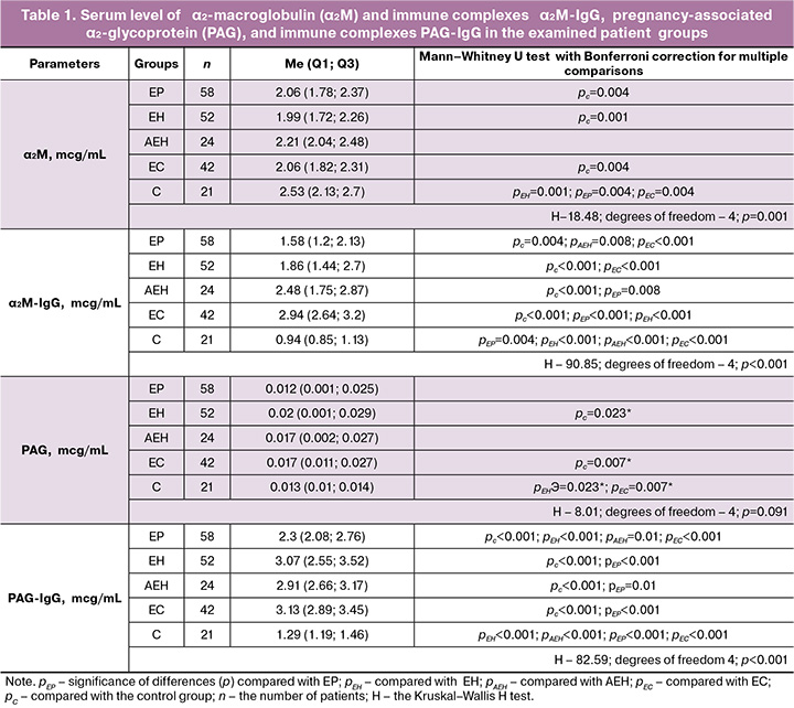
Reduced PAG levels were in EP group compared with the control group, and elevated levels were in other groups. However, the differences between the groups were not statistically significant. Concentrations of PAG-IgG immune complex were significantly higher in all patient groups compared with the control group. At the same time, minimum levels were found in EP, and maximum and comparable levels in EH, AEH, and EC.
Significantly reduced LF levels were found in the spectrum of diseases EP→EH→AEH→EC, and reached statistically significant differences between EP and EC groups. The data are represented in Table 2.
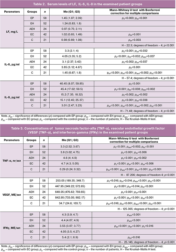
In all patient groups, concentrations of IL-6 and IL-8 were significantly high compared with the control group. Maximum level of IL-8 was found in EH group, and it exceeded the levels of cytokines by 3 times in AEH and EC groups, and by 15 times in the control group.
We found progressive increase in the levels of VEGF and TNF-α in the spectrum of diseases EP→EH→AEH→EC. In patients with EC, VEGF concentration was 24 times higher compared with the control group, and TNF-α concentration was by 16 times higher (Table 3).
Concentrations of IFNγ were significantly low in AEH compared with that in other examined groups. The greatest difference in IFNγ concentration was found in patients with different types of endometrial hyperplasia –without atypia (EH) and atypical endometrial hyperplasia (AEH) (p<0.001).
Correlation analysis showed direct relationship between the levels of proinflammatory cytokines – strong relationship between IL-6 and IL-8 (r=0.662; p<0.001) and moderate between TNF-α and IL-6 (r=0.491; p<0.001), TNF-α and IL-8 (r=0.326; p<0.001).
Discussion
In 2023, the systematic review highlighted the essential role of cytokines in induction of signaling pathways and stimulation of epithelial-mesenchymal transition (EMT). Thereby cytokine-induced EMT can lead to cancer development in the ovaries, cervix, breast, and endometrium [6]. Currently, cytokines are being used as therapeutic targets and diagnostic biomarkers in ovarian and cervical cancer.
The results of our study demonstrate a progressive increase in concentrations of VEGF and TNF-α in the spectrum of proliferative endometrial diseases EP→EH→AEH→EC, that is consistent with general pathogenetic mechanisms [7]. VEGF concentration was 24 times higher compared with the control values, that confirms the importance of neoangiogenesis in malignant transformation. TNF-α concentration was increased 16 times in EC, that is consistent with the role TNF-α in stimulating VEGF expression [8].
Statistically significant difference was found in concentrations of IFNγ – maximum levels in EH, and minimum levels in AEH. This opens prospects for the differential diagnosis of proliferative diseases. According to literature data, IFNγ mediates the cytotoxic effects of lymphocytes and macrophages on neoplastically transformed cells, as well as can inhibit tumor cell proliferation and activate natural killer cells [9].
Significantly reduced levels of α₂М-IgG were in EP, EH and EC compared with the control group. At the same time, the dynamics of levels of immune complexes α₂М-IgG correlated with progression of endometrial pathology in the spectrum of diseases EP→EH→AEH→EC. Our study found maximum high levels of immune complexes PAG-IgG and α₂М-IgG in EC, that is consistent with the concept of the role of immune complexes in the pathogenesis of oncologic diseases. According to literature data, circulating immune complexes promote the formation of an immunosuppressive tumor microenvironment by inhibition of effector cell functions and activating mechanisms of suppression [10]. Thus, accumulation of PAG-IgG and α₂M-IgG can be considered as a laboratory marker of the initiation and progression of the epithelial-mesenchymal transition.
The levels of IL-6 and IL-8 were significantly high in proliferative endometrial diseases. Maximum levels were in EH, and minimum levels in AEH, that indicates the differences in the pathogenesis between two types on endometrial hyperplasia.
The obtained data are consistent with the concept of the role of chronic endometritis in the pathogenesis of endometrial hyperplastic processes [11, 12]. A combination of endometrial hyperplasia and chronic endometritis is diagnosed in every fourth case (25.6%). Immunohistochemical examination of the endometrium in patients of reproductive age with infertility and endometrial hyperplasia determined the diagnostic criteria for chronic endometritis in all cases [13]. Furthermore, inflammatory diseases of the pelvic organs in history increase the recurrence of EH by 2 times [14]. Persistent damage to the endometrium in chronic endometritis leads to impairment of its cyclic transformation and tissue receptivity, as well as changes influencing the potential for pathologic regeneration [15].
Progressively decreased LF levels were in the spectrum of EP→EH→AEH→EC, that can reflect reduction of anti-inflammatory potential in malignancy. The obtained data are consistent with a high prevalence of chronic endometritis in the presence of EP (51.4%) [16]. According to Carvalho F.M. et al., chronic endometritis and EP represent subsequent stages of one common pathological process, which is confirmed by specific vascular changes and molecular data on impairment of signaling pathways, which regulate endometrial proliferation during chronic inflammation [17]. The results of meta-analysis (2024) also confirm a close pathogenetic link between the inflammatory process and EP, that is associated with reproductive disorders, including infertility, miscarriage, and IVF failure [18].
Thus, the results of our study confirm the relationship between the progression of proliferative processes and chronic inflammation in the endometrium.
Conclusions
- Specific changes in concentrations of cytokines and macroglobulins in the blood were determined in different proliferative endometrial diseases.
- Progressive increase in the levels of VEGF and TNF-α and immune complexes α₂М-IgG, PAG-IgG in parallel with reduced anti-inflammatory potential (LF) was found in the spectrum of diseases EP→EH→AEH→EC.
- Close relationship was found between proliferative diseases of the endometrium and accumulation of immune complexes α₂M-IgG and PAG-IgG, that can be considered as a laboratory marker of the initiation and progression of the epithelial-mesenchymal transition.
- The obtained data confirm the importance of chronic inflammation in the pathogenesis of proliferative endometrial diseases. Progressive reduction in LF levels and changes in the cytokine profile are consistent with current concepts of the integration of inflammatory and proliferative processes in endometrial pathology.
- The results of this study open prospects for developing new diagnostic and therapeutic algorithms based on determination of cytokines and macroglobulins, that can improve differential diagnosis and prognosis for the course of proliferative endometrial diseases. EH is characterized by specific inflammatory profile – maximum levels of IL-6 and IL-8, while AEH is characterized by reduced levels of IL-6 and IL-8, and IFNγ. In EC, immunosuppressive profile is formed with maximum activation of angiogenesis.
References
- Seshagiri P.B., Vani V., Madhulika P. Cytokines and blastocyst hatching. Am. J. Reprod. Immunol. 2016; 75(3): 208-17. https://dx.doi.org/10.1111/aji.12464
- Pachomov C.P., Altuchova O.B., Demakova N.A, Krivoshei I.V., Kolesnikov Y.V., Sobyanin F.I. Study of cytokines polymorphous loci connections with rise of endometrium proliferative diseases. Research Journal of Pharmaceutical, Biological and Chemical Sciences. 2014; 5(6): 1473-6.
- Министерство здравоохранения Российской Федерации. Клинические рекомендации. Полипы эндометрия. 2023. [Ministry of Health of the Russian Federation. Clinical guidelines. Endometrial polyps. 2023 (in Russian)].
- Министерство здравоохранения Российской Федерации. Клинические рекомендации. Гиперплазия эндометрия. 2025. [Ministry of Health of the Russian Federation. Clinical guidelines. Endometrial hyperplasia. 2025 (in Russian)].
- Зорина В.Н., Зорина Р.М., Левченко В.Г., Воронина Е.А., Зорин Н.А. Концентрация комплексов макроглобулин-IgG как возможный критерий для прогнозирования гестоза и оценки степени его тяжести. Клиническая и лабораторная диагностика. 2001; 8: 48-9. [Zorina V.N., Zorina R.M., Levchenko V.G., Voronina E.A., Zorin N.A. Macroglobulin-IgG complex con- centration as a criterion for prognosis of gestosis and assessment of its severity. Clinical and Laboratory Diagnostics. 2001; 8: 48-9 (in Russian)].
- Ray I., Michael A., Meira L.B., Ellis P.E. The role of cytokines in epithelial-mesenchymal transition in gynaecological cancers: a systematic review. Cells. 2023; 12(3): 416. https://dx.doi.org/10.3390/cells12030416
- Сабанцев М.А., Шрамко С.В., Левченко В.Г., Волков О.А., Третьякова Т.В. Гиперплазии эндометрия: без атипии и с атипией. Гинекология. 2021; 23(1): 18-24. [Sabantsev M.A., Shramko S.V., Levchenko V.G., Volkov O.A., Tretyakova T.V. Endometrial hyperplasia: without atypia and with atypia. Ginekology. 2021; 23(1): 18-24 (in Russian)]. https://dx.doi.org/10.26442/20795696.2021.1.200666
- Дятлова Л.И., Глухова Т.Н., Понукалина Е.В., Сергеева О.Н., Бизенкова М.Н. О значении дисбаланса цитокинов в патогенезе рака эндометрия. Современные проблемы науки и образования. 2024; 3: 8. [Dyatlova L.I., Glukhova T.N., Ponukalina E.V., Mikhailova Yu.V., Bizenkova M.N. About the significance of cytokine imbalance in the pathogenesis of endometrial cancer. Modern Problems of Science and Education. 2024; 3: 8 (in Russian)]. https://dx.doi.org/10.17513/spno.33412
- Gocher A.M., Workman C.J., Vignali D.A.A. Interferon-γ: teammate or opponent in the tumour microenvironment? Nat. Rev. Immunol. 2022; 22(3): 158-72. https://dx.doi.org/10.1038/s41577-021-00566-3
- Nimmerjahn F., Ravetch J.V. Fcgamma receptors as regulators of immune responses. Nat. Rev. Immunol. 2008; 8(1): 34-47. https://dx.doi.org/10.1038/nri2206
- Кравченко Е.Н., Лаутеншлегер Е.В. Гиперплазия эндометрия и полипы эндометрия: клинико-анамнестическая характеристика женщин. Фундаментальная и клиническая медицина. 2023; 8(4): 16-23. [Kravchenko E.N., Lautenschleger E.V. Endometrial hyperplasia and endometrial polyps: clinical and anamnestic characteristics of women. Fundamental and Clinical Medicine. 2023; 8(4): 16-23 (in Russian)]. https://dx.doi.org/10.23946/2500-0764-2023-8-4-16-23
- Ткаченко Л.В., Свиридова Н.И., Жаркин Н.А., Бурова Н.А., Белан Э.Б. Оценка цитокинового статуса у пациенток с хроническим эндометритом в сочетании с гиперпластическими процессами эндометрия в репродуктивном периоде. Инфекция и иммунитет. 2020; 10(4): 762-8. [Tkachenko L.V., Sviridova N.I., Zharkin N.A., Burova N.A., Belan E.B. Assessing cytokine status of patients with chronic endometritis combined with endometrial hyperplastic processes in reproductive period. Russian Journal of Infection and Immunity. 2020; 10(4): 762-8 (in Russian)]. https://dx.doi.org/10.15789/2220-7619-AOC-1357
- Серебренникова К.Г., Арутюнян Н.А., Кацалап С.Н., Алехин А.И. Фотодинамическая терапия у пациенток с бесплодием при хроническом эндометрите. Российский вестник акушера-гинеколога. 2020; 20(3): 56-61. [Serebrennikova K.G., Arutyunyan N.A., Katsalap S.N., Alekhin A.I. Photodynamic therapy in patients with infertility in chronic endometritis. Russian Bulletin of Obstetrician-Gynecologist. 2020; 20(3): 56-61 (in Russian)]. https://dx.doi.org/10.17116/rosakush20202003156
- Оразов М.Р., Радзинский В.Е., Хамошина М.Б., Михалева Л.М., Муллина И.А., Артеменко Ю.С. Предикторы рецидивирующего течения эндометриальной гиперплазии без атипии у женщин репродуктивного возраста. Клинический разбор в общей медицине. 2023; 4(9): 16-20. [Orazov M.R., Radzinsky V.E., Khamoshina M.B., Mikhaleva L.M., Mullina I.A., Artemenko Yu.S. Predictors of recurrent course of endometrial hyperplasia without atypia in women of reproductive age. Clinical review for general practice. 2023; 4(9): 16-20 (in Russian)]. https://dx.doi.org/10.47407/kr2023.4.9.00304
- Белявская С.А., Дикарева Л.В. Хронический эндометрит как фактор риска пролиферативных заболеваний матки: современные подходы к диагностике. Обзор литературы. Главный врач Юга России. 2024; 4(96): 2-7. [Belyavskaya S.A., Dikareva L.V. Chronic endometritis as a risk factor for proliferative diseases of the uterus: modern approaches to diagnosis. Literature review. Glavnyj vraсh Yuga Rossii. 2024; 4(96): 2-7 (in Russian)].
- Moreno I., Cicinelli E., Garcia-Grau I., Gonzalez-Monfort M., Bau D., Vilella F. et al. The diagnosis of chronic endometritis in infertile asymptomatic women: a comparative study of histology, microbial cultures, hysteroscopy, and molecular microbiology. Am. J. Obstet. Gynecol. 2018; 218(6): 602.e1-e16. https://dx.doi.org/10.1016/j.ajog.2018.02.012
- Carvalho F.M., Aguiar F.N., Tomioka R., de Oliveira R.M., Frantz N., Ueno J. Functional endometrial polyps in infertile asymptomatic patients: a possible evolution of vascular changes secondary to endometritis. Eur. J. Obstet. Gynecol. Reprod. Biol. 2013; 170(1): 152-6. https://dx.doi.org/10.1016/j.ejogrb.2013.05.012
- Оразов М.Р., Михалева Л.М., Пойманова О.Ф. Гистероскопическая диагностика полипов эндометрия, ассоциированных с хроническим эндометритом. Акушерство и гинекология: новости, мнения, обучение. 2024; 12(Спецвып.): 141-7. [Orazov M.R., Mikhaleva L.M., Poimanova O.F. Hysteroscopic diagnosis of endometrial polyps associated with chronic endometritis. Obstetrics and Gynecology: News, Opinions, Training. 2024; 12(S): 141-7 (in Russian)]. https://dx.doi.org/10.33029/2303-9698-2024-12-suppl-141-147
Received 01.12.2025
Accepted 09.04.2026
About the Authors
Maksim A. Sabantsev, PhD student at the Department of Obstetrics and Gynecology, Novokuznetsk State Institute for Further Training of Physicians – Branch Campus of the Russian Medical Academy of Continuous Professional Education, Ministry of Health of the Russian Federation, 654005, Russia, Novokuznetsk, Stroiteley Ave., 5,+7(913)333-23-22, Dr.Sabantsev@ya.ru, https://orcid.org/0000-0001-7104-1852
Svetlana V. Shramko, Dr. Med. Sci., Associate Professor, Professor at the Department of Obstetrics and Gynecology, Novokuznetsk State Institute for Further Training of Physicians – Branch Campus of the Russian Medical Academy of Continuous Professional Education, Ministry of Health of the Russian Federation, 654005, Russia, Novokuznetsk, Stroiteley Ave., 5, +7(961)714-00-13, shramko_08@mail.ru, https://orcid.org/0000-0003-1299-165X
Veronika N. Zorina, Dr. Bio. Sci., Leading Researcher at the Laboratory of Applied Toxicology and Pharmacology of Toxicology Division, Golikov Research Center of Toxicology, 192019, Russia, St. Petersburg, Bekhtereva str., 1 lit A, +7(812)372-51-23, nilimmun@yandex.ru, https://orcid.org/0000-0001-9183-7663
Corresponding author: Maksim A. Sabantsev, Dr.Sabantsev@ya.ru
Similar Articles

