
Small-for-gestational-age fetuses: a heterogeneous group requiring reassessment of clinical outcomes
Sidelnikova M.D., Kan N.E., Tyutyunnik V.L., Soldatova E.E., Domanskaya M.A.
Objective. To investigate the association between antenatal small-for-gestational-age (SGA) status and adverse perinatal outcomes in term neonates.
Materials and methods. This retrospective cohort study included 200 pregnant women who delivered at the
V.I. Kulakov National Medical Research Center for Obstetrics, Gynecology, and Perinatology (2020–2025). The study group (n=100) comprised women with singleton term pregnancies complicated by an SGA fetus, while the comparison group (n=100) included women with normally grown fetuses. Clinical and anamnestic characteristics, pregnancy and delivery outcomes, and early neonatal complications were also evaluated. Statistical analyses were performed using JASP software (Student's t-test, Mann–Whitney U test, chi-square test, Fisher's exact test, univariable and multivariable logistic regression; p<0.05).
Results. SGA status was significantly associated with an increased risk of adverse perinatal outcomes. The incidence of the composite adverse outcome was significantly higher in the SGA group (24%) than in the control group (8%) (p=0.002). Neonates with SGA demonstrated a marked trend toward higher rates of cephalohematoma formation, central nervous system (CNS) disorders, and infectious-inflammatory conditions. In the univariable logistic regression analysis, SGA increased the risk of adverse outcomes by 3.6-fold (odds ratio [OR]=3.63; 95% confidence interval [CI]: 1.54–8.55; p=0.003). Multivariable analysis adjusted for maternal age confirmed that SGA remained an independent predictor of adverse outcomes (adjusted OR=3.62; 95% CI: 1.52–8.62; p=0.004).
Conclusion. SGA is an independent risk factor for adverse perinatal outcomes and warrants reconsideration of traditional views regarding its clinical significance due to the heterogeneity of the population and challenges associated with its diagnosis.
Authors' contributions. Sidelnikova M.D., Kan N.E., Tyutyunnik V.L., Soldatova E.E., Domanskaya M.A. – the study conception and design, data acquisition, literature review, data processing and analysis, statistical analysis, drafting of the manuscript, editing of the manuscript.
Conflicts of interest. The authors have no conflicts of interest to declare.
Funding. There was no funding for this study.
Ethical Approval. The study was reviewed and approved by the Research Ethics Committee of the V.I. Kulakov NMRC for OG&P, Ministry of Health of Russia.
Generative Artificial Intelligence. No artificial intelligence tools were used in the preparation of this manuscript.
Patient Consent for Publication. All patients provided informed consent for the publication of their data.
Authors' Data Sharing Statement. The data supporting the findings of this study are available upon request from the corresponding author after approval from the principal investigator.
For citation: Sidelnikova M.D., Kan N.E., Tyutyunnik V.L., Soldatova E.E., Domanskaya M.A.
Small-for-gestational-age fetuses: a heterogeneous group requiring reassessment of clinical outcomes.
Akusherstvo i Ginekologiya/Obstetrics and Gynecology. 2026; (6): 92-100 (in Russian)
https://dx.doi.org/10.18565/aig.2026.23
Keywords
small-for-gestational-age fetus
fetal growth restriction
perinatal outcomes
According to the current clinical guidelines issued by the Ministry of Health of the Russian Federation, the term "small-for-gestational-age fetus" (SGA) is considered synonymous with a constitutionally small fetus, for whom favorable perinatal outcomes are generally expected [1]. However, emerging evidence has begun to challenge this interpretation, indicating that the SGA population is heterogeneous and includes subgroups at an increased risk of perinatal complications [2]. Furthermore, national guidelines in several countries now classify fetuses with an estimated fetal weight below the 10th percentile as being at potential risk for adverse outcomes, reflecting the accumulating evidence of the heterogeneity within this group [3]. This issue is compounded by the limitations of antenatal diagnostics, which often struggle to distinguish true fetal growth restriction, which is associated with increased perinatal morbidity and mortality, from constitutionally small fetuses, for whom such outcomes are less likely. This study examines this emerging research paradigm and explores the associations between antenatally diagnosed SGA status and a spectrum of adverse perinatal outcomes.
This study aimed to investigate the association between antenatal small-for-gestational-age status and adverse perinatal outcomes in term neonates.
Materials and methods
This retrospective cohort study included 200 pregnant women who delivered at the V.I. Kulakov National Medical Research Center for Obstetrics, Gynecology, and Perinatology, Ministry of Health of the Russian Federation between 2020 and 2025. Participants were divided into two groups: the study group (n=100), consisting of women with a diagnosis of a small-for-gestational-age fetus at term according to the definition provided in the Russian clinical guidelines [1], and the control group (n=100), consisting of women with normally grown fetuses who were consecutively selected from deliveries occurring during the same period and who met the eligibility criteria. Individual matching by maternal age, parity, or other characteristics was not conducted.
The inclusion criteria were singleton pregnancies and gestational age at delivery between 37+0 and 41+6 weeks. The exclusion criteria included confirmed fetal chromosomal abnormalities and/or congenital malformations, multiple pregnancies, surrogate pregnancies, and preterm deliveries before 37 completed weeks of gestation.
The following data were analyzed: baseline clinical and anamnestic characteristics, pregnancy and delivery features, and outcomes during the early neonatal period. Clinical information regarding pregnancy and neonatal outcomes was obtained from the archived maternity and neonatal medical records.
For this study, adverse perinatal outcomes were defined as the presence of at least one of the following conditions:
- Perinatal mortality, a critically important clinical endpoint and key indicator of the quality of obstetric and neonatal care.
- Suboptimal birth outcomes (confirmed birth asphyxia or admission to the neonatal intensive care unit [NICU]) reflect severe impairment of neonatal adaptation and an increased risk of neurological complications. These conditions may have long-term developmental effects.
- Neonatal morbidity, including:
- Respiratory disorders (respiratory failure, transient tachypnea of the newborn, and neonatal apnea), which may require respiratory support and are associated with an increased risk of long-term respiratory morbidity, hypoxia, and brain injury.
- Gastrointestinal disorders (necrotizing enterocolitis, gastric hemorrhage), which pose serious threats to neonatal survival, may require urgent surgical intervention, and can result in sepsis, intestinal perforation, or death;
- Central nervous system (CNS) disorders (intraventricular hemorrhage, CNS depression syndrome), which may lead to neurological impairment, cerebral palsy, cognitive deficits, and long-term developmental consequences;
- Cephalohematoma formation, a clinically significant condition that requires prolonged medical observation and treatment when necessary. It may result in complications such as anemia, jaundice, infection, and, rarely, neurological sequelae;
- Hematologic disorders (congenital anemia, disseminated intravascular coagulation syndrome, neonatal jaundice, neonatal thrombocytopenia), which may lead to hypoxia, hemorrhage, and other serious complications. Severe cases may require blood transfusion and intensive care;
- Cardiovascular disorders (persistent fetal circulation, atrial septal defect, neonatal cardiac arrhythmia, ventricular septal defect, and circulatory insufficiency), which may result in hypoxia and heart failure. Severe forms may require surgical intervention;
- Infectious and inflammatory diseases (infection specific to the neonatal period, urinary tract infection, other specified neonatal respiratory conditions, congenital pneumonia, neonatal sepsis), which are among the leading causes of neonatal morbidity and mortality, may progress to multiorgan failure and death.
- Prolonged neonatal hospitalization for medical indications (>5 days) indicates significant health problems requiring extended treatment and monitoring. Even in the absence of overtly severe disease, prolonged hospitalization may reflect underlying health concerns and potential long-term consequences.
A composite adverse neonatal outcome was used because of the heterogeneous nature of the SGA population. This approach enabled the aggregation of multiple clinically relevant complications associated with low fetal weight, thereby increasing the statistical power given the relatively low frequency of individual outcomes. It also provides a more comprehensive assessment of the overall risk, enhancing the clinical and prognostic relevance of the findings.
Statistical analysis
Statistical analysis was performed using JASP version 0.9.1 (Netherlands). The normality of the data distribution was assessed using the Shapiro–Wilk test. Parametric data are presented as mean and standard deviation, and nonparametric data are reported as median and interquartile range. Categorical variables are presented as absolute frequencies and percentages of the total.
Continuous variables were compared between groups using the Student's t-test or the Mann–Whitney U test, depending on the distribution of the data. Categorical variables were compared using the chi-square or Fisher's exact test, as appropriate. Multivariable logistic regression analysis was used to evaluate the independent association between exposure and outcome while accounting for the influence of other variables. The results are presented as adjusted odds ratio (OR) with 95% confidence interval (95% CI). Statistical significance was set at p<0.05.
Results
Comparative characteristics of the study groups are presented in detail in Table 1. The groups were comparable in terms of key anthropometric and demographic characteristics, as well as the prevalence of extragenital and most gynecological conditions. However, nulliparous women and pregnancies conceived via assisted reproductive technologies (ART) were significantly more common in the SGA group. Conversely, a history of cesarean delivery was more frequent in the control group.
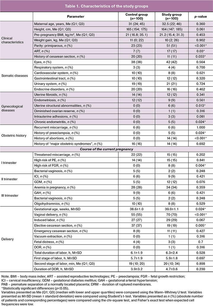
Women carrying an SGA fetus had a significantly higher incidence of uterine anomalies, chronic endometritis, a history of induced abortion, and previous preeclampsia. Analysis of the current pregnancy revealed only one statistically significant difference: a high risk of fetal growth restriction (FGR) according to first-trimester prenatal screening was more common in the SGA group. Other aspects of pregnancy course, including the incidence of early pregnancy nausea and vomiting, threatened miscarriage, threatened preterm birth, maternal anemia, cervical insufficiency, gestational diabetes mellitus, and hypertensive disorders, were comparable between groups (Table 1).
Delivery characteristics showed that gestational age at birth was significantly greater in the study group (Table 1). The rate of vaginal delivery was also higher in this group, while elective operative delivery occurred significantly more frequently in the control group. No significant differences were observed between groups in the rates of emergency operative delivery, vacuum-assisted delivery, labor induction, fetal distress, or premature placental abruption.
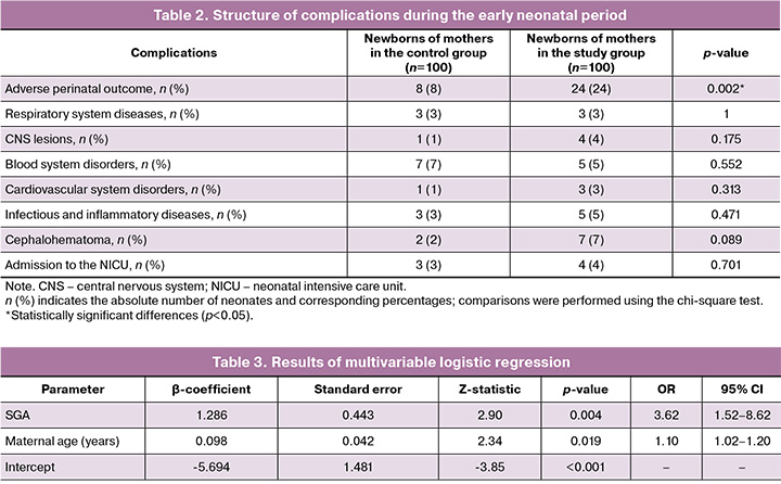
Assessment of perinatal outcomes demonstrated that neonates in the study group experienced adverse outcomes substantially more often. The incidence of the composite adverse perinatal outcome was significantly higher in this group than among neonates born to mothers with an appropriately grown fetus [24/100 (24%) vs. 8/100 (8%), p<0.05].
Analysis of the individual components of the composite adverse perinatal outcome (Table 2) revealed a trend toward a higher frequency of cephalohematoma, CNS injury, and infectious-inflammatory conditions among neonates in the study group. A tendency toward more frequent admission to the NICU was also observed in the study group, although these differences did not reach statistical significance. In contrast, hematologic disorders were predominantly observed among neonates with normal birth weight.
Logistic regression was applied for further analysis, as the dependent variable was a composite adverse perinatal outcome. Univariate analysis demonstrated that SGA was significantly associated with adverse perinatal outcomes (p<0.05). The logistic regression equation for the univariate analysis was:
log(p/(1-p)) = -2.44 + 1.29×SGA,
where p represents the probability of an adverse perinatal outcome. Neonates born to mothers in the study group (SGA) had a 3.6-fold higher risk of adverse outcomes compared with neonates in the control group (OR=3.63; 95% CI: 1.54–8.55; p=0.003).
To determine the independent association between SGA and adverse perinatal outcomes while controlling for potential confounding factors, multivariable logistic regression was performed. Variables demonstrating a statistically significant association with the outcome in univariate analysis (p<0.05), along with the clinically relevant factor of maternal age, were included in the model. The variable "High risk of FGR according to first-trimester prenatal screening" was excluded a priori from the multivariable model due to its pathogenetic relationship with the dependent variable (SGA); simultaneous inclusion of both variables would have introduced conceptual multicollinearity and underestimated the independent contribution of SGA. Independent variables were assessed for collinearity using variance inflation factors (VIFs); all values ranged from 1.01 to 1.14, indicating the absence of meaningful multicollinearity. Final variable selection was performed using backward stepwise elimination. The logistic regression equation for the multivariable analysis was:
log(p/(1-p)) = -5.69 + 1.29 × SGA + 0.098 × Maternal age,
where p represents the probability of an adverse perinatal outcome.
The results of the multivariable analysis are presented in Table 3.
The overall model was statistically significant (likelihood-ratio test: χ²=15.5; df=2; p<0.001). Nagelkerke's pseudo-R² was 0.128, indicating moderate explanatory power, which is typical of medical studies investigating outcomes with multifactorial etiologies. The remaining proportion of outcome variance (87.2%) was likely attributable to factors for which the groups were not initially comparable (e.g., parity and use of ART). This limitation highlights a potential direction for future prospective studies aimed at validating the observed associations. External validation in an independent cohort was not performed.
Multivariable analysis demonstrated that SGA remained an independent predictor of adverse outcomes after adjustment for maternal age, with an adjusted OR of 3.62 (95% CI: 1.52–8.62; p=0.004). Maternal age was also identified as an independent predictor, with each additional year increasing the odds of an adverse outcome by approximately 10% (OR=1.10; 95% CI: 1.02–1.20; p=0.019).
Discussion
The present study demonstrated that fetal SGA, even at term, is associated with an increased risk of adverse perinatal outcomes. This association persisted after adjusting for several clinical factors, underscoring the independent role of low fetal weight as a predictor of perinatal complications.
In contrast to some previously published studies [4, 5], most maternal demographic and anthropometric characteristics (age, BMI, and gestational weight gain) were not significantly associated with the development of fetal SGA in the present cohort. Although maternal height formally differed between groups (median 165 cm vs. 164 cm; p=0.061), the substantial overlap in distribution ranges precludes meaningful clinical interpretation. This finding is unlikely to represent a biologically relevant risk factor. The absence of such differences may indicate the predominance of other etiological factors underlying SGA in our population or suggest that these parameters are not major predictors of the condition, which is consistent with the growing recognition of the heterogeneity of SGA.
However, several important differences were identified, including a higher proportion of primigravid women in the SGA group. Yang L. et al. [6] suggested that this association may be related to incomplete maternal vascular adaptation to pregnancy, differences in trophoblast invasion, and the absence of established immunological tolerance to fetal tissues, which is characteristic of subsequent pregnancies. The higher prevalence of previous cesarean delivery among women with appropriately grown fetuses in our study was attributable to the greater proportion of multiparous women in this group. Another notable finding was the higher frequency of pregnancies conceived using ART in the SGA group. This observation is consistent with studies reporting an increased risk of impaired fetal development following ART conception [7].
The prevalence of extragenital diseases was comparable between the groups, suggesting that SGA was not directly associated with the presence of common maternal somatic disorders. Similarly, the prevalence of gynecological conditions, including endometriosis, uterine fibroids, ovarian cysts, and infertility, did not differ significantly between the groups.
Nevertheless, uterine structural abnormalities and chronic endometritis were significantly more prevalent in the SGA group. These conditions may directly affect implantation and placental development and, consequently, fetal growth, thereby increasing the risk of SGA.
Analysis of obstetric history revealed no differences in the rates of spontaneous miscarriage, missed miscarriage or recurrent pregnancy loss. However, a history of induced abortion was more common among women in the SGA group, consistent with previous reports [8]. A history of preeclampsia is a well-established risk factor for fetal growth abnormalities [1], and this association was confirmed in our study.
The only statistically significant difference identified during early pregnancy was a higher predicted risk of Fetal Growth Restriction (FGR) according to first-trimester prenatal screening in the SGA group. This finding highlights the importance of early identification and close monitoring in high-risk patients [9]. In contrast, pregnancy complications such as early pregnancy nausea and vomiting, threatened miscarriage, threatened preterm birth, maternal anemia, cervical insufficiency, gestational diabetes mellitus, and hypertensive disorders did not differ significantly between the groups.
Analysis of the delivery characteristics revealed several statistically significant differences. The higher gestational age at delivery and greater frequency of vaginal birth in the SGA group may reflect management strategies aimed at maximizing gestational prolongation in such pregnancies. Conversely, the higher proportion of planned operative deliveries in the control group was attributable to the greater number of multiparous women, among whom 20% had a uterine scar following a previous cesarean section. The absence of differences in the rates of emergency operative delivery, vacuum extraction, labor induction, fetal distress, and placental abruption may indicate that these acute complications were either not predominant concerns in this population of SGA fetuses or were successfully managed in this population.
These findings demonstrate that, despite the absence of statistically significant differences in several demographic and somatic characteristics, the SGA group differed from the control group in terms of the proportion of primigravid women, frequency of ART-conceived pregnancies, uterine structural abnormalities, chronic endometritis, history of induced abortion, history of preeclampsia, and increased risk of FGR identified during prenatal screening. These factors likely contribute to the elevated risk of adverse perinatal outcomes in SGA pregnancies.
Our study confirmed that fetal SGA, even at term, is a significant risk factor for adverse perinatal outcome. The incidence of the composite adverse outcome was significantly higher among neonates in the SGA group (24%) than among those in the control group (8%), consistent with numerous studies evaluating the impact of low birth weight on perinatal health [2, 10, 11].
Analysis of the individual components of the composite outcome revealed complication patterns characteristic of the SGA group. The predominance of cephalohematomas, central nervous system (CNS) injuries, and infectious-inflammatory conditions in this group may be related to intrauterine hypoxia and inflammation, which frequently accompany fetal growth abnormalities [12]. Chronic hypoxia may exert neurotoxic effects, contribute to CNS injury, and increase susceptibility to infections. The higher incidence of cephalohematoma may reflect increased birth trauma in the setting of pre-existing fetal distress. Although not statistically significant, the trend toward a higher rate of NICU admission among SGA neonates further supports the presence of unfavorable outcomes in this population.
Interestingly, hematologic disorders were more common in neonates with normal fetal weights. This finding suggests that adverse outcomes in the two groups arise through different etiological mechanisms. It is possible that, among appropriately grown fetuses, specific factors unrelated to hypoxia or infection affect hematopoietic function.
Univariable logistic regression analysis demonstrated that SGA was associated with a 3.6-fold increase in the risk of adverse perinatal outcomes (OR 3.63; 95% CI 1.54–8.55; p=0.003). Multivariable logistic regression was used to evaluate the independent effect of SGA after adjusting for maternal age. Even after adjustment, SGA remained an independent predictor of adverse outcomes, with an adjusted odds ratio of 3.62 (95% CI 1.52–8.62; p=0.004). These findings indicate that low fetal weight itself, independent of other analyzed factors, represents a substantial risk factor for newborns.
Limitations and interpretation of findings. Several aspects of the present study warrant further investigation. First, the predominance of cephalohematomas in the SGA group, together with the higher rate of vaginal delivery, raises the question of the contribution of labor biomechanics to the composite outcomes. However, the persistence of a significant association between SGA and adverse outcomes after adjustment for maternal age in the multivariable model suggests that SGA fetuses possess increased biological vulnerability rather than being affected solely by differences in obstetric management practices. Nevertheless, delivery management strategies for SGA pregnancies may also contribute to outcomes and should be evaluated in prospective studies.
Second, the heterogeneity of the SGA group, as emphasized in the study title, was reflected in our data by the coexistence of constitutionally small fetuses (without evidence of placental insufficiency or abnormal Doppler findings) and fetuses with an antenatally identified risk of growth restriction detected during first-trimester screening. The latter subgroup was observed exclusively in the SGA cohort (8/100 vs. 0/100), although its independent prognostic contribution in the multivariable model was largely subsumed by SGA status itself. The stratification of SGA cases according to specific subtypes remains an important objective for future research.
Third, the control group consisted of patients from the same institution with appropriately grown fetuses delivered at term, which may partly explain the relatively low prevalence of certain adverse historical factors, including induced abortions, preeclampsia, and uterine structural abnormalities. Therefore, selection bias cannot be completely excluded, and the estimated odds ratios may be somewhat inflated. External validation of the model in an independent cohort represents an important direction for future research.
In summary
- SGA as an independent risk factor: The study findings demonstrate that fetal SGA is an independent risk factor for adverse perinatal outcomes.
- Higher prevalence of adverse outcomes in the SGA group: The incidence of composite adverse perinatal outcomes was significantly higher among neonates in the SGA group than among neonates born to mothers carrying appropriately grown fetuses.
- Clinical relevance of the composite outcome: The use of a composite adverse outcome enabled a more comprehensive assessment of the overall risk, considering the heterogeneity of the SGA population and the relatively low frequency of individual complications.
- Independence of SGA as a predictor: Multivariable logistic regression adjusted for maternal age confirmed that SGA remains an independent risk factor for adverse perinatal outcomes (adjusted OR 3.62; 95% CI 1.52–8.62; p=0.004), supporting its independent prognostic significance.
Conclusion
The present findings confirm that SGA represents a significant clinical problem associated with an increased risk of adverse perinatal outcomes, even in term pregnancies. The heterogeneous nature of the SGA population, challenges of antenatal diagnosis, and relatively low frequency of individual complications support the use of a composite approach to risk assessment. Regardless of its underlying etiology, low fetal weight may be considered an independent predictor that requires close clinical attention. Future research should focus on developing more accurate prenatal diagnostic methods capable of distinguishing risk subgroups within the SGA population and optimizing management strategies for these pregnancies to minimize adverse perinatal outcomes.
References
- Министерство здравоохранения Российской Федерации. Клинические рекомендации. Недостаточный рост плода, требующий предоставления медицинской помощи матери (задержка роста плода). М.; 2025. 73 c. [Ministry of Health of the Russian Federation. Clinical guidelines. Insufficient fetal growth requiring medical care for the mother (fetal growth retardation). Moscow; 2025. 73 p. (in Russian)].
- Ruiz-Martinez S., Delgado J.L., Paules C., Cavallaro A., De Paco C., Villar J. et al. Clinical phenotypes for risk stratification in small-for-gestational-age fetuses. Ultrasound Obstet. Gynecol. 2022; 59(4): 490-6. https://dx.doi.org/10.1002/uog.23765
- Giouleka S., Tsakiridis I., Mamopoulos A., Kalogiannidis I., Athanasiadis A., Dagklis T. Fetal growth restriction: a comprehensive review of major guidelines. Obstet. Gynecol. Surv. 2023; 78(11): 690-708. https://dx.doi.org/10.1097/OGX.0000000000001203
- Devaguru A., Gada S., Potpalle D., Dinesh Eshwar M., Purwar D. The prevalence of low birth weight among newborn babies and its associated maternal risk factors: a hospital-based cross-sectional study. Cureus. 2023; 15(5): e38587. https://dx.doi.org/10.7759/cureus.38587
- Saccone G., Gragnano E., Ilardi B., Marrone V., Strina I., Venturella R. et al. Maternal and perinatal complications according to maternal age: a systematic review and meta-analysis. Int. J. Gynaecol. Obstet. 2022; 159(1): 43-55. https://dx.doi.org/10.1002/ijgo.14100
- Yang L., Feng L., Huang L., Li X., Qiu W., Yang K. et al. Maternal factors for intrauterine growth retardation: systematic review and meta-analysis of observational studies. Reprod. Sci. 2023; 30(6): 1737-45. https://dx.doi.org/10.1007/s43032-021-00756-3
- Larsen M.L., Krebs L., Hoei-Hansen C.E., Kumar S. Assessment of fetal growth trajectory identifies infants at high risk of perinatal mortality. Ultrasound Obstet. Gynecol. 2024; 63(6): 764-771. https://dx.doi.org/10.1002/uog.27610
- Mohammad N., Sohaila A., Rabbani U., Ahmed S., Ahmed S., Ali S.R. Maternal predictors of intrauterine growth retardation. J. Coll. Physicians Surg. Pak. 2018; 28(9): 681-5. https://dx.doi.org/10.29271/jcpsp.2018.09.681
- Relph S., Coxon K., Vieira M.C., Copas A., Healey A., Alagna A. et al. Effect of the growth assessment protocol on the DEtection of small for GestatioNal age fetus: process evaluation from the DESiGN cluster randomised trial. Implement. Sci. 2022; 17(1): 60. https://dx.doi.org/10.1186/s13012-022-01228-1
- Mendoza M., Hurtado I., Bonacina E., Garcia-Manau P., Serrano B., Tur H. et al. Individual risk assessment for prenatal counseling in early-onset growth-restricted and small-for-gestational-age fetuses. Acta Obstet. Gynecol. Scand. 2021; 100(3): 504-12. https://dx.doi.org/10.1111/aogs.14032
- Гасымова Ш.Р., Тютюнник В.Л., Кан Н.Е., Волочаева М.В., Донников А.Е. Клинико-анамнестические факторы риска и прогностические модели развития задержки роста плода. Акушерство и гинекология. 2024; 10: 52-9. https://dx.doi.org/10.18565/aig.2024.166 [Gasimova Sh.R., Tyutyunnik V.L., Kan N.E., Volochaeva M.V., Donnikov A.E. Clinical and anamnestic risk factors and prognostic models for the development of fetal growth retardation. Obstetrics and Gynecology. 2024; (10): 52-9 (in Russian). https://dx.doi.org/10.18565/aig.2024.166].
- Song P. Re: Prognostic value of angiogenic markers in pregnancy with fetal growth restriction. Ultrasound Obstet. Gynecol. 2024; 63(6): 840. https://dx.doi.org/10.1002/uog.27674
Received 26.01.2026
Accepted 20.05.2026
About the Authors
Maria D. Sidelnikova, obstetrician-gynecologist at the 1st Obstetric Physiological Department, Academician V.I. Kulakov National Medical Research Center for Obstetrics, Gynecology and Perinatology, Ministry of Health of Russia, 117997, Russia, Moscow, Ac. Oparina str., 4, +7(903)220-45-13, m_sidelnikova@oparina4.ru,https://orcid.org/0000-0002-0805-4703
Natalia E. Kan, Professor, Dr. Med. Sci., Honored Scientist of the Russian Federation, Deputy Director General for Research – Director of the Institute of Obstetrics, Academician V.I. Kulakov National Medical Research Center for Obstetrics, Gynecology and Perinatology, Ministry of Health of Russia, 117997, Russia, Moscow,
Ac. Oparina str., 4, kan-med@mail.ru, Researcher ID: B-2370-2015, SPIN: 5378-8437, Authors ID: 624900, Scopus Author ID: 57008835600,
https://orcid.org/0000-0001-5087-5946
Victor L. Tyutyunnik, Professor, Dr. Med. Sci., Honored Doctor of the Russian Federation, Leading Researcher at the Center of Scientific and Clinical Researches,
Academician V.I. Kulakov National Medical Research Center for Obstetrics, Gynecology and Perinatology, Ministry of Health of Russia, 117997, Russia, Moscow,
Ac. Oparina str., 4, tioutiounnik@mail.ru, Researcher ID: B-2364-2015, SPIN: 1963-1359, Authors ID: 213217, Scopus Author ID: 56190621500,
https://orcid.org/0000-0002-5830-5099
Ekaterina E. Soldatova, PhD, Researcher at the Institute of Obstetrics, Academician V.I. Kulakov National Medical Research Center for Obstetrics, Gynecology and Perinatology, Ministry of Health of Russia, 117997, Russia, Moscow, Ac. Oparina str., 4, katerina.soldatova95@bk.ru, https://orcid.org/0000-0001-6463-3403
Maria A. Domanskaya, clinical resident, Academician V.I. Kulakov National Medical Research Center for Obstetrics, Gynecology and Perinatology, Ministry of Health of Russia, 117997, Russia, Moscow, Ac. Oparina str., 4, mdomanskaya@yandex.ru, https://orcid.org/0009-0007-8882-5603
Corresponding author: Maria D. Sidelnikova, m_sidelnikova@oparina4.ru
Similar Articles

