
Wick effect Wick effect: A comparative study of its potential for development using polypropylene and titanium mesh implant models
Ivanov O.A., Bezhenar V.F., Palastin P.M.
Background. Implant-associated infections are primarily driven by bacterial biofilm formation. While the physicochemical properties of the material surface are recognized as key factors in adhesion, the role of implant macromorphology, particularly its wicking effect that facilitates microbial spread, remains understudied. A comparative analysis of Staphylococcus aureus adhesion, a primary pathogen in such infections, on widely used polypropylene and titanium meshes in surgery is of significant interest for assessing long-term risks.
Objective. To conduct a comparative analysis of Staphylococcus aureus adhesion on titanium and polypropylene implants, focusing on the distribution pattern of microorganisms and assessing the potential biofilm formation risk associated with the wicking properties of the materials.
Materials and methods. This in vitro study used commercial mesh implants: polypropylene (Gynemesh PS, Johnson & Johnson, USA) and titanium (Titanium Silk, Elastic Titanium Implants LLC, Russia). Samples were incubated overnight with S. aureus VT209 at 37°C for 24 h. The number of adherent bacteria was quantitatively assessed using a culture-based method with colony-forming unit (CFU) counting. Scanning electron microscopy (SEM) and energy-dispersive X-ray spectroscopy (EDS) were used to analyze the bacterial distribution and surface characteristics.
Results. The quantitative adhesion levels of S. aureus to titanium (Me=4.30×10² CFU [4.25×10²; 4.40×10²]) and polypropylene (Me=4.45×10² CFU [4.40×10²; 4.50×10²]) implants were comparable (p>0.05). However, a fundamental difference in the distribution patterns was observed. On titanium implants, adhesion was strictly localized to the edge areas with increased roughness and micro-defects, as confirmed by SEM and EDS. In contrast, the polypropylene implants showed a uniform distribution of bacteria over the entire surface, indicating the pronounced wicking properties of the material that facilitate passive spread and colonization of the entire available area.
Conclusion. Despite similar adhesion levels, the identified differences in microbial distribution indicate a higher potential risk of forming extensive biofilms that are difficult to eradicate with antibiotics and the immune system when using polypropylene implants owing to their pronounced wicking effect. Conversely, the localized nature of adhesion on titanium potentially simplifies the identification and surgical debridement of the infection focus. These findings challenge the safety of polypropylene from a long-term infectious risk perspective and highlight the critical need to develop implants with suppressed wicking properties or apply antimicrobial coatings over their entire surface.
Authors' contributions. Ivanov O.A. – conception and design of the study, data collection and analysis, drafting of the manuscript; Bezhenar V.F. –approval of the final version to be submitted; Palastin P.M. – data collection and analysis, editing of the manuscript.
Conflicts of interest. The authors have no conflicts of interest to declare.
Funding. There was no funding for this study.
Ethical Approval. Ethical approval was not required because the study involved only standard bacterial reference strains and non‑biological materials and did not include human or animal subjects.
Generative Artificial Intelligence. No artificial intelligence tools were used in the preparation of this manuscript.
Patient Consent for Publication. All patients provided informed consent for the publication of their data.
Authors' Data Sharing Statement. The data supporting the findings of this study are available upon request from the corresponding author after approval from the principal investigator.
For citation: Ivanov O.A., Bezhenar V.F., Palastin P.M. Wick effect: A comparative study of
its potential for development using polypropylene and titanium mesh implant models.
Akusherstvo i Ginekologiya/Obstetrics and Gynecology. 2026; (6): 135-143 (in Russian)
https://dx.doi.org/10.18565/aig.2025.336
Keywords
mesh implant
polypropylene
titanium
adhesion
Staphylococcus aureus
wicking effect
biofilm formation
Pelvic organ prolapse (POP) is a highly prevalent clinical condition that substantially impairs the quality of life of patients [1]. Surgical correction using mesh implants has become firmly established in contemporary gynecological practice, demonstrating high efficacy in restoring pelvic floor anatomy and function [2]. However, this approach carries the risk of serious complications, collectively referred to as mesh-associated morbidity [3–5]. Among these, infectious complications occupy a particularly important position, and the difficulty of their management, sometimes necessitating complete implant removal, is largely attributable to the capacity of bacteria to adhere to implant surfaces and form biofilms [6, 7].
Staphylococcus aureus is one of the most virulent pathogens responsible for implant-associated infections. Its pronounced adhesive capacity and propensity to form biofilms resistant to both antibiotics and host immune defenses are the principal drivers of persistent infection [8, 9]. The adhesion process is governed by complex interactions between the physicochemical properties of the implant surface (roughness, hydrophobicity, and surface charge) and those of the microorganism [10, 11]. The choice of implant material is, therefore, of critical importance.
In current surgical practice, polypropylene implants are the most widely used for POP repair, with titanium implants gaining increasing adoption. Existing comparative studies have largely focused on parameters such as biocomcompatibility, mechanical strength, and elasticity [12, 13]. However, for the assessment of long-term infectious risk, the spatial distribution of bacteria across the implant surface is at least as important as the total number of adherent bacteria.
A notable gap exists in the literature regarding the comparative analysis of the spatial distribution patterns of bacteria on these two materials. While some studies have identified edge adhesion on metallic implants as a consequence of mechanical processing [14], another equally important phenomenon has received insufficient attention: the wicking properties of polypropylene. As a synthetic polymer, polypropylene can actively absorb and redistribute fluids – and the microorganisms suspended within them – throughout its bulk macroporous structure via capillary forces [15, 16]. This fundamental material property may predetermine a diffuse, uniform pattern of surface colonization rather than a localized one, creating optimal conditions for the formation of extensive and treatment-refractory biofilms.
Accordingly, conventional approaches that only assess quantitative adhesion parameters may substantially underestimate the infectious risks associated with polymeric mesh implants. A localized infection at the edge of a titanium implant may potentially be amenable to targeted debridement, whereas a biofilm distributed uniformly across the entire surface of a polypropylene prosthesis almost invariably necessitates complete implant removal.
This study aimed to conduct a comparative analysis of the quantity of adherent Staphylococcus aureus and the spatial distribution pattern of bacterial adhesion on polypropylene and titanium implant surfaces.
Materials and methods
Study design
An experimental comparative in vitro study was conducted to analyze the adhesion and spatial distribution of Staphylococcus aureus on two types of mesh implants. Thirty samples were examined and divided into two groups: polypropylene (n=15) and titanium (n=15). The study group included samples of the Gynemesh PS polypropylene implant, whereas the comparison group included samples of the silk-titanium implant.
Study setting and ethics
All experiments were conducted in the first quarter of 2025 in the laboratories of the Department of Microbiology, Pavlov First Saint Petersburg State Medical University (St. Petersburg, Russia). Microbiological procedures were performed in a Class II biological safety cabinet in accordance with standard microbiological protocols. Ethical approval was not required for this study, as it involved standard bacterial strains and non-biological materials.
Implant samples
Two commercially available mesh implants used in gynecological practice were selected for the study: the Gynemesh PS polypropylene implant (Johnson & Johnson, USA) and Titanium Silk titanium implant (Elastic Titanium Implants LLC, Russia). Under sterile conditions, 15 standardized fragments measuring 1×1 cm were cut from each implant using Cooper’s scissors. This procedure simulated the intraoperative implant preparation.
Preparation
A 24-hour culture of the reference strain Staphylococcus aureus VT209 from the Department of Microbiology was used. The culture was grown on Columbia blood agar base (Thermo Fisher Scientific, USA) at 37°C. Bacterial cells were suspended in sterile 0.9% sodium chloride solution, and the suspension concentration was adjusted to 1×10⁵ CFU/mL using the McFarland turbidity standard, followed by spectrophotometric verification (optical density at a wavelength of 600 nm).
Modeling of bacterial adhesion and assessment of the wicking effect
To evaluate the adhesive properties and bacterial distribution patterns, each implant fragment was placed in an individual sterile Petri dish (9 cm in diameter) and immersed in 20 mL of bacterial suspension (1×10⁵ CFU/mL). The dishes were incubated at 37°C for 24 h to allow bacterial adhesion. Following incubation, the fragments were washed thrice in sterile 0.9% sodium chloride solution to remove non-adherent (loosely attached) bacterial cells. Each washing step was performed for 1 min in a fresh portion of the solution with gentle agitation on a shaker (50 rpm) to standardize the procedure.
Cultivation, visualization, and quantitative analysis of bacterial distribution
Washed mesh fragments were aseptically transferred onto the surface of meat-peptone agar (State Research Center for Applied Microbiology and Biotechnology, Russia) in Petri dishes. To immobilize the implants and facilitate subsequent visualization of bacterial adhesion zones, the fragments were overlaid with a layer of semi-solid agar (0.7%), which fixed the mesh within its thickness after it solidified. The dishes were then incubated at 37°C for 24 h. After incubation, colony-forming units (CFU) that grew on the agar surface directly beneath and around the implant were counted. Colonies originating from bacteria that remained adhered to the mesh and were not detached during agar overlaying were considered indicators of strong adhesion. The primary analytical parameter was not only the total number of CFU but also the visual assessment of their distribution pattern, which served as an indicator of the wicking properties of the material.
The adhesion index (AI) was calculated using the following formula:
AI = lg(N) / lg(N₀),
where N is the number of adherent bacteria (CFU/sample), and N₀ is the number of bacteria in the initial suspension (CFU/mL).
Analysis of surface microstructure and chemical composition
To explain the observed differences in bacterial distribution, the implant surfaces were also analyzed.
Scanning electron microscopy (SEM) was used. The surface microstructures of the central and peripheral regions of the implants were examined using a Tescan Mira 3 scanning electron microscope (TESCAN, Czech Republic). Analyses were performed at multiple magnifications to assess the surface topography and the presence of microdefects.
Energy-dispersive X-ray spectroscopy (EDS). The chemical compositions of the same regions were analyzed using an energy-dispersive spectrometer integrated into a Tescan Mira 3 microscope (TESCAN, Czech Republic). The qualitative and quantitative elemental compositions were determined.
Statistical analysis
Statistical analysis was performed using GraphPad Prism 9.0 software (GraphPad Software, USA). The normality of the distribution of continuous adhesion parameters (CFU/sample) in the polypropylene and titanium groups was assessed using the Shapiro–Wilk test. As the distributions in both groups deviated significantly from normality (p< 0.05), comparisons between the two independent groups were performed using the nonparametric Mann–Whitney U test. Results are presented as median (Me) and interquartile range (25th and 75th percentiles), reported as Me (Q1; Q3). Differences were considered statistically significant at p<0.05.
Results
Quantitative assessment of Staphylococcus aureus adhesion
Quantitative analysis of colony-forming unit (CFU) counts of Staphylococcus aureus firmly adhered to the surface of the implants revealed no statistically significant difference between groups. On polypropylene implants, the number of adherent bacteria was 4.45×10² [4.40×10²; 4.50×10²] CFU/sample. The corresponding value on titanium implants was comparable, at 4.30×10² [4.25×10²; 4.40×10²] CFU/sample (p>0.05). Accordingly, the adhesion index (AI) was identical for both materials, at 0.53.
These data indicate that the overall capacity of the S. aureus strain for primary adhesion to polypropylene and titanium surfaces is statistically equivalent.
Qualitative analysis of bacterial distribution: identification of a wicking effect
Despite the similarity in quantitative parameters, visual analysis revealed a fundamental and clinically relevant difference in the spatial distribution of bacterial colonies on the two materials.
On polypropylene implants, colonies were distributed uniformly and diffusely across the entire surface of the fragment (Fig. 1b). Bacteria adhered to the mesh filaments and the pores between them without forming distinct macroconglomerates. This pattern is consistent with the passive spread of the bacterial suspension across the material through capillary forces – a wicking effect – resulting in colonization of the entire available surface.
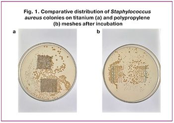
On titanium implants, by contrast, distribution was markedly localized. The majority of colonies were concentrated almost exclusively along the perimeter of the fragment, while the central region remained largely free of microbial colonization (Fig. 1a). This pattern suggests that adhesion to titanium is not an intrinsic bulk property of the material but rather a consequence of local surface alterations at the edges, introduced during mechanical cutting.
Preparation for scanning electron microscopy and X-ray spectral analysis
Analysis was performed on sterile implant specimens. Fragments measuring 1×1 cm were mounted on aluminum stubs using double-sided conductive carbon tape. For scanning electron microscopy (SEM), specimens were sputter-coated with a thin conductive gold layer (~10 nm) using a Quorum Q150R S magnetron sputtering system (Quorum Technologies, United Kingdom) to prevent surface charge accumulation and improve image quality.
Because energy-dispersive X-ray spectroscopy (EDS) requires minimal spectral background from the coating layer, a separate set of identical specimens was carbon-coated using the same equipment.
Surface morphology (SEM) and elemental composition (EDS) were assessed using a Tescan Mira 3 scanning electron microscope (TESCAN, Czech Republic) equipped with an energy-dispersive spectrometer.
Scanning electron microscopy
SEM examination of the titanium implant surface revealed clear microstructural differences between the central and edge regions. The central regions (Fig. 2a–c) were characterized by a relatively smooth, homogeneous surface with minimal microdefects. This indicates that these areas were subjected to less mechanical stress during preparation and use, a factor that may contribute to their structural stability and reduced susceptibility to bacterial adhesion.
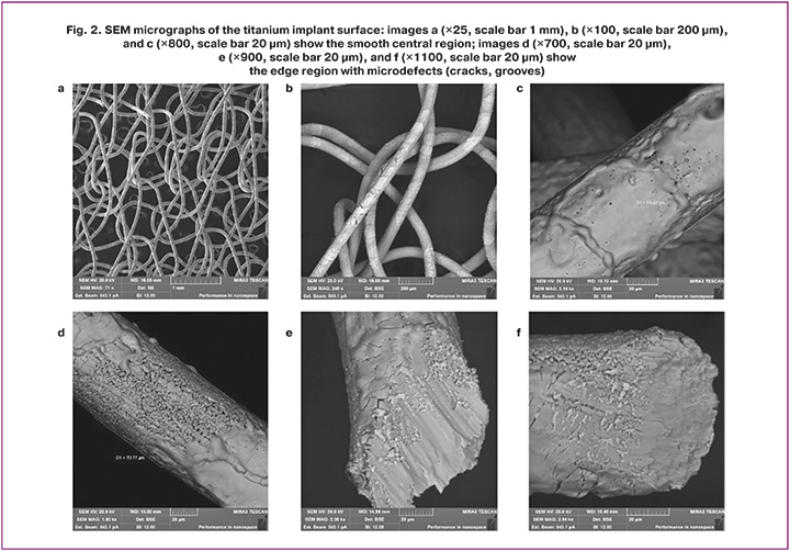
The edge regions (Fig. 2d–f), in contrast, exhibited numerous cracks, grooves, and surface irregularities, likely resulting from mechanical processing, such as cutting the implant from bulk material during surgical preparation. These defects create zones of increased roughness capable of altering the adhesive properties of the surface. The cracks and grooves at the edges may generate local microniches favorable for bacterial attachment, rendering these regions more susceptible to microbial colonization.
X-ray spectral microanalysis
EDS analysis of the central and edge regions revealed variation in chemical composition between the two zones. In the central region, titanium was, as expected, the predominant element, with content ranging from 66.4% to 96.34% by weight. This predominance confirms the chemical stability of this zone, which likely contributes to its relatively low susceptibility to contamination and mechanical damage. However, other elements were detected at low concentrations, including carbon (2.05–3.84%), oxygen (4.52–29.42%), aluminum, iron, and calcium (Figure 3). These elements most likely reached the surface through interaction with the environment or oxidative processes occurring during implant processing or use.
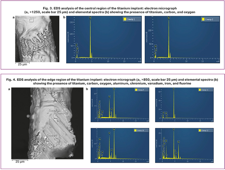
A more complex elemental distribution was observed in the edge regions. In addition to titanium, whose content decreased markedly to 17.10% in certain zones, elevated levels of carbon (up to 17.26%) and oxygen (up to 11.61%) were detected. These changes may reflect active oxidative processes and accumulation of organic contaminants at the implant edges. Additional elements identified included fluorine (up to 2.42%), aluminum, silicon, vanadium, chromium, iron (up to 58.86%), and copper (Fig. 4a–c). The presence of these elements at the edge region may be attributable to mechanical processing and the use of surgical instruments during implant preparation for placement.
Discussion
This comparative in vitro study identified a fundamental difference in the mode of interaction between Staphylococcus aureus and the surfaces of titanium versus polypropylene implants, a distinction that cannot be captured by conventional colony-forming unit (CFU) enumeration alone. The principal finding of our study is that polypropylene, by virtue of its wicking properties, promotes uniform bacterial distribution across the entire implant surface, thereby creating conditions conducive to extensive biofilm formation, whereas adhesion to titanium is strictly localized.
At first glance, the absence of a statistically significant difference in the number of adherent bacteria (4.48×10² vs. 4.31×10² CFU/sample) might suggest a comparable infectious risk between the two materials. However, our analysis demonstrates that this interpretation is fundamentally insufficient for long-term risk assessment. The clinical significance of implant-associated infections is determined not by the initial number of adherent microorganisms but by their spatial distribution and potential for mature biofilm development [17].
The localized pattern of adhesion on titanium implants, confined to zones of mechanical damage at the sample edges, is consistent with previously published data [18]. Scanning electron microscopy (SEM) and atomic force microscopy confirmed that these zones are characterized by increased surface roughness, microdefects, and chemical contamination (Fe, Cr, and Al) introduced by surgical instrumentation, all of which are well-established factors that promote bacterial adhesion [19]. However, this problem is focal in nature and potentially manageable. Intraoperative avoidance of forceful trimming, use of laser cutting, or prefabrication of implants to the required dimensions could substantially mitigate this risk [20].
In contrast, the uniform bacterial distribution observed across the polypropylene surfaces was not a consequence of localized surface defects but rather an intrinsic material property: its pronounced wicking effect. As a hydrophobic polymer with a well-developed macroporous architecture, polypropylene actively absorbs and redistributes liquid media containing suspended microorganisms throughout its bulk structure using capillary forces. The result is not focal but diffuse, volumetric colonization, in which the entire implant surface becomes a potential substrate for biofilm formation.
These adhesion patterns have direct and clinically significant implications. Localized infection on titanium – a process confined to the marginal zone – is theoretically amenable to targeted intervention, including surgical débridement or local application of antimicrobial agents, without requiring complete implant removal [21]. In contrast, diffuse infection on polypropylene implies that even minor intraoperative contamination may initiate the formation of an extensive, poorly vascularized biofilm integrated throughout the mesh structure. Such biofilms are largely inaccessible to antibiotics and immune effector cells [22]. In most cases, the only effective treatment is complete implant removal, a technically demanding procedure associated with substantial risk to the patient.
Our findings support the hypothesis that polypropylene may carry a higher long-term risk of severe infectious complications than titanium, precisely because of its capacity to promote diffuse rather than localized pathogen distribution.
Limitations. The present study has several limitations. The 24-hour in vitro model adequately captured only the initial phase of bacterial adhesion and did not reflect the dynamics of mature biofilm formation. The use of a single S. aureus strain limits the generalizability of the findings to other microorganisms. Furthermore, the model does not account for the potential effects of biological fluids (serum, wound exudate) or host immune factors, which may modulate the wicking behavior of the material used.
In contrast to the conventional paradigm, which focuses on the quantity of adherent bacteria, this study reframes the spatial distribution of microbial cells as a key determinant of infectious risk. The wicking effect of polypropylene represents a concealed hazard, facilitating diffuse colonization and creating conditions for the formation of extensive and treatment-resistant biofilms. Titanium implants, despite the issue of edge adhesion, exhibit a localized infection pattern that is potentially more amenable to targeted clinical management. These findings highlight the need to reassess current approaches for implant material safety evaluation and develop novel strategies aimed at suppressing the wicking properties of polymeric meshes.
Conclusion
This comparative in vitro study of Staphylococcus aureus adhesion to titanium and polypropylene implants supports the following principal conclusions:
Quantitative CFU analysis of firmly adherent bacteria revealed no statistically significant differences between the polypropylene and titanium implants. The key distinction between the two materials lies not in the total number of adherent microorganisms but in their spatial distribution on the implant surface.
Titanium implants exhibited strictly localized bacterial adhesion confined to zones of mechanical damage and chemical contamination at the sample edges. Polypropylene implants, by contrast, showed uniform, diffuse bacterial distribution across the entire surface – a direct consequence of the material's pronounced wicking effect.
The localized nature of the infection on titanium potentially permits the use of targeted therapeutic strategies (débridement, marginal resection) without necessitating complete implant removal. The uniform bacterial distribution on polypropylene creates optimal conditions for the formation of an extensive biofilm that is highly resistant to both antibiotics and immune clearance, which in most cases requires complete implant removal and is associated with substantial risks to the patient.
In light of the long-term risks of biofilm formation, polypropylene implants may carry a higher potential for serious infectious complications than titanium implants, whose edge adhesion problem is technically addressable. When selecting an implant, clinicians should consider not only the mechanical characteristics but also the fundamental material properties that may facilitate infection propagation. The present findings question the unconditional safety of polypropylene meshes and underscore the need for a thorough reassessment of their risk profile in view of the wicking effect identified here.
References
- Беженарь В.Ф., Богатырева Е.В., Цыпурдеева А.А., Цуладзе Л.К., Русина Е.И., Гусева Е.С. Новые возможности хирургической коррекции тазового пролапса с использованием синтетических имплантов: пути профилактики послеоперационных осложнений. Акушерство, гинекология и репродукция. 2012; 6(2): 6-13. [Bezhenar V.F., Bogatyreva E.V., Tsypurdeeva A.A., Tsuladze L.K., Rusina E.I., Guseva E.S. New possibilities for surgical correction of pelvic prolapse using synthetic implants: ways to prevent postoperative complications. Obstetrics, Gynecology and Reproduction. 2012; 6(2): 6-13 (in Russian)].
- Попов А.А., Бабаева С.А., Федоров А.А., Тюрина С.С., Идашкин А.Д., Клюшников И.Д., Ефремова Е.С. Изучение клинической эффективности и безопасности методов хирургической коррекции апикального пролапса гениталий. Российский вестник акушера-гинеколога. 2023; 23(4): 58‑64. https://dx.doi.org/10.17116/rosakush20232304158 [Popov A.A., Babaeva S.A., Fedorov A.A., Tyurina S.S., Idashkin A.D., Klyushnikov I.D., Efremova E.S. Study of the clinical efficacy and safety of surgical methods for correction of apical genital prolapse. Russian Bulletin of Obstetrician-Gynecologist. 2023; 23(4): 58-64 (in Russian). https://dx.doi.org/10.17116/rosakush20232304158].
- Беженарь В.Ф., Паластин П.М., Толибова Г.Х. Эрозии влагалища в отдаленные сроки после постановки синтетических имплантатов при гинекологических операциях. РМЖ. Медицинское обозрение. 2018; 10: 17-21. [Bezhenar V.F., Palastin P.M., Tolibova G.Kh. Vaginal erosion at a long time after synthetic implants insertion during gynecological surgery. RMJ. Medical Review. 2018; 10: 17-21 (in Russian)].
- Seifalian A, Basma Z, Digesu A, Khullar V. Polypropylene pelvic mesh: what went wrong and what will be of the future? Biomedicines. 2023; 11(3): 741. https://dx.doi.org/10.3390/biomedicines11030741
- da Silveira S.D.R.B., Auge A.P., Jarmy-Dibella Z.I., Margarido P.F., Carramao S., Alves Rodrigues C. et al. A multicenter, randomized trial comparing pelvic organ prolapse surgical treatment with native tissue and synthetic mesh: a 5-year follow-up study. Neurourol. Urodyn, 2020; 39(3): 1002-11. https://dx.doi.org/10.1002/nau.24323
- Chapple C.R., Cruz F., Deffieux X., Milani A.L., Arlandis S., Artibani W. et al. Consensus statement of the european urology association and the European urogynaecological association on the use of implanted materials for treating pelvic organ prolapse and stress urinary incontinence. Eur. Urol. 2017; 72(3): 424-31. https://dx.doi.org/10.1016/j.eururo.2017.03.048
- Галлямов Э.А., Бусырев Ю.Б., Гвоздев А.А., Шалыгин А.Б., Федоров А.В. Эндоскопическое стентирование как метод выбора в лечении послеоперационной стриктуры гепатикоеюноанастомоза. Эндоскопическая хирургия. 2023; 29(6): 18-24. https://dx.doi.org/10.17116/endoskopi20231218 [Gallyamov E.A., Busyrev Yu.B., Gvozdev A.A., Shalygin A.B., Fedorov A.V. Endoscopic stenting as a method of choice in the treatment of postoperative stricture of hepaticojejunal anastomosis. Endoscopic Surgery. 2023; 29(6): 18-24 (in Russian). https://dx.doi.org/10.17116/endoskopi20231218].
- Sharma D., Misba L., Khan A.U. Antibiotics versus biofilm: an emerging battleground in microbial communities. Antimicrob. Resist. Infect. Control. 2019; 8: 76. https://dx.doi.org/10.1186/s13756-019-0533-3
- Борисова М.И., Лазакович Д.Н., Сидорова Н.А., Савушкин А.И. Биопленкообразующая активность и феномен персистенции микроорганизмов. Journal of Biomedical Technologies. 2015; 2: 28-35. https://dx.doi.org/10.15393/j6.art.2015.3382 [Borisova M.I., Lazakovich D.N., Sidorova N.A., Savushkin A.I. Biofilm-forming activity and the phenomenon of microbial persistence. Journal of Biomedical Technologies. 2015; 2: 28-35 (in Russian). https://dx.doi.org/10.15393/j6.art.2015.3382].
- Wassmann T., Kreis S., Behr M., Buergers R. The influence of surface texture and wettability on initial bacterial adhesion on titanium and zirconium oxide dental implants. Int. J. Implant. Dent. 2017; 3(1): 32. https://dx.doi.org/10.1186/s40729-017-0093-3
- Гветадзе Р.Ш., Дмитриева Н.А., Воронин А.Н. Особенности адгезии микроорганизмов к стоматологическим материалам, используемым для формирования контура десны при протезировании с опорой на дентальные имплантаты. Стоматология. 2019; 98(5): 118-23. https://dx.doi.org/10.17116/stomat201998051118 [Gvetadze R.Sh., Dmitrieva N.A., Voronin A.N. Adhesion of microorganisms to various dental materials used to form a gum contour in implant-retained restorations. Stomatology. 2019; 98(5): 118-23 (in Russian). https://dx.doi.org/10.17116/stomat201998051118].
- Камоева С.В., Маковская Д.С., Доброхотова Ю.Э. Хирургическая коррекция пролапса тазовых органов у женщин с использованием сетчатых эндопротезов на основе титанового «шелка»: результаты 5-летнего наблюдения. РМЖ. Мать и дитя. 2024; 7(3): 227-235. https://dx.doi.org/10.32364/2618-8430-2024-7-3-6 [Kamoeva S.V., Makovskaya D.S., Dobrokhotova Yu.E. Surgical management of female pelvic organ prolapse using titanium "silk" mesh replacement: 5-year results. Russian Journal of Woman and Child Health. 2024; 7(3): 227-35 (in Russian). https://dx.doi.org/10.32364/2618-8430-2024-7-3-6].
- Heinonen P., Ala-Nissilä S., Aaltonen R., Kiilholma P. Trocar-guided polypropylene mesh for pelvic organ prolapse surgery-perioperative morbidity and short-term outcome of the first 100 patients. Gynecol. Surg. 2011; 8: 165-70. https://dx.doi.org/10.1007/s10397-010-0628-6
- Villegas M., Bayat F., Kramer T., Schwarz E., Wilson D., Hosseinidoust Z. et al. Emerging strategies to prevent bacterial infections on titanium-based implants. Small. 2024; 20(46): e2404351. https://dx.doi.org/10.1002/smll.202404351
- Zacher A.T., Schwarz F., Dailey S.K., Rupp M., Alt V., Schmitz P. et al. Biofilm formation of Staphylococcus aureus on various implants used for surgical treatment of destructive spondylodiscitis. Sci. Rep. 2024; 14: 19364. https://dx.doi.org/10.1038/s41598-024-70244-6
- Паластин П.М., Беженарь В.Ф., Петров А.В. Реакция местного иммунитета на введение полипропиленового синтетического имплантата (экспериментальное исследование). Российский вестник акушера-гинеколога. 2019; 19(2): 45-9. https://dx.doi.org/10.17116/rosakush20191902147 [Palastin P.M., Bezhenar V.F., Petrov A.V. The reaction of local immunity to the introduction of a polypropylene synthetic implant (an experimental study). Russian Bulletin of Obstetrician-Gynecologist. 2019; 19(2): 45-9 (in Russian). https://dx.doi.org/10.17116/rosakush20191902147].
- Sanchez C.J., Mende K., Beckius M.L., Akers K.S., Romano D.R., Wenke J.C. et al. Biofilm formation by clinical isolates and the implications in chronic infections. BMC Infect. Dis. 2013; 13: 47. https://dx.doi.org/10.1186/1471-2334-13-47
- Swalsky A., Noumbissi S.S., Wiedemann T.G. The systemic and local interactions related to titanium implant corrosion and hypersensitivity reactions: a narrative review of the literature. Int. J. Implant. Dent. 2024; 10: 58. https://dx.doi.org/10.1186/s40729-024-00578-3
- Rezaei N.M., Hasegawa M., Ishijima M., Nakhaei K., Okubo T., Taniyama T. et al. Biological and osseointegration capabilities of hierarchically (meso-/micro-/nano-scale) roughened zirconia. Int. J. Nanomedicine. 2018; 8(13): 3381-95. https://dx.doi.org/10.2147/IJN.S159955
- Valente F., Scarano A., Murmura G., Varvara G., Sinjari B., Mandelli F. et al. Collagen fibres orientation in the bone matrix around dental implants: does the implant’s thread design play a role? Int. J. Mol. Sci. 2021; 22(15): 7860. https://dx.doi.org/10.3390/ijms22157860
- Черкасов М.Ф., Черкасов Д.М., Меликова С.Г., Старцев Ю.М., Галашокян К.М., Ендоренко К.В. Стратегия сохранения сетчатых имплантатов при инфицированных послеоперационных ранах передней брюшной стенки. Хирургия. Журнал им. Н.И. Пирогова. 2022; 11: 49-54. https://dx.doi.org/10.17116/hirurgia202211149 [Cherkasov M.F., Cherkasov D.M., Melikova S.G., Startsev Yu.M., Galashokyan K.M., Endorenko K.V. Strategy for maintaining mesh implants in infected postoperative wounds of anterior abdominal wall. Pirogov Russian Journal of Surgery. 2022; (11): 49-54 (in Russian). https://dx.doi.org/10.17116/hirurgia202211149].
- Артюшков Е.Л., Лызиков А.А., Каплан М.Л., Дорошко Е.Ю., Осипов Б.Б. Бактериальная биопленка как фактор риска инфекционных осложнений при применении полипропиленовых эндопротезов в хирургии грыж передней брюшной стенки. Проблемы здоровья и экологии. 2025; 22(1): 15-22. https://dx.doi.org/10.51523/2708-6011.2025-22-1-02 [Artyushkov E.L., Lyzikov А.A., Kaplan M.L., Doroshko Y.Y., Osipov B.B. Bacterial biofilm as a risk factor for infectious complications in the use of polypropylene endoprostheses in surgery of anterior wall hernias. Health and Ecology Issues. (in Russian). https://dx.doi.org/10.51523/2708-6011.2025-22-1-02].
Received 19.11.2025
Accepted 08.06.2026
About the Authors
Oleg A. Ivanov, PhD Student, Department of Obstetrics, Gynecology and Neonatology, Senior Laboratory Assistant, Department of Obstetrics, Gynecology and Neonatology, Pavlov First Saint Petersburg State Medical University, Ministry of Health of Russia, 197022, Russia, St. Petersburg, Leo Tolstoy str., 6-8, ivanoffmd@gmail.com, eLibrary SPIN: 8620-9749, https://orcid.org/0000-0002-6596-4105Vitaly F. Bezhenar, Dr. Med. Sci., Professor, Head of the Departments of Obstetrics, Gynecology and Neonatology/Reproductology, Head of the Clinic of Obstetrics and Gynecology, Pavlov First Saint Petersburg State Medical University, Ministry of Health of Russia, 197022, Russia, St. Petersburg, Leo Tolstoy str., 6-8; Main Supernumerary Specialist Obstetrician-Gynecologist of the Health Committee of St. Petersburg, eLibrary SPIN: 8626-7555, https://orcid.org/0000-0002-7807-4929
Peter M. Palastin, PhD, Associate Professor at the Department of Obstetrics, Gynecology and Neonatology, Pavlov First Saint Petersburg State Medical University, Ministry of Health of Russia, 197022, Russia, St. Petersburg, Leo Tolstoy str., 6-8, eLibrary SPIN: 8008-8723, https://orcid.org/0000-0003-3502-2499
Similar Articles

