
Possibilities of ultrasonic shear wave elastography in the diagnosis of cervical cancer (first experience)
Objective. To determine the diagnostic value of two-dimensional shear wave elastography in patients with cervical cancer. Materials and methods. Two-dimensional shear wave elastography was performed in the control group (in 20 women) and in 18 patients with confirmed cervical cancer. The study was performed using the Aixplorer ultrasound system (SSI, France). Results. There was a statistically significant (p<0.001) increase in the stiffness (Me=66.6 kPa (51; 88.7)) of cervical tissues affected by cancer, compared to the control group (Me=21.1 kPa (20; 23.5). ROC analysis showed that when using a 32 kPa cut-off value, the sensitivity, specificity, and prognostic value of the positive and negative results were 96%, 89%, 86%, and 95%, respectively. AUROC for this threshold reaches 0.97. Conclusion. Two-dimensional share wave elastography may be a new objective method for diagnosing cervical cancer.Zykin B.I., Ogryzkova V.L., Ionova E.A.
Keywords
cervical cancer
ultrasound two-dimensional shear wave elastography
Early detection of cervical cancer has recently become a particularly relevant issue. Despite the fact that mortality due to this pathology has significantly decreased owing to modern diagnostic methods, the dynamics of morbidity indicators remains hazardous; the increase in morbidity was 23.83% per 100,000 population of Russia in 2004–2014 [1]. At the same time, there is a negative trend towards an increase in the number of cases of the disease at a young age.
Ultrasound examinations, as the most simple and informative, have proven to be effective in the diagnosis of cervical cancer. However, the value of ultrasound elastography in the examination of such patients still remains understudied. Moreover, some doctors have not developed a complete understanding of the fact that in addition to ultrasound compression elastography, there is shear elastography [2].
Compression elastography has already been known and used for the diagnosis of cervical cancer [3]. Unfortunately, this largely qualitative method is lacking objective digital parameters. Its results are largely determined by the subjective perception of the doctor. Even the use of the elasticity score suffers from instability [4], since the stiffness of the cervical neoplasm is compared to the extremely uncertain stiffness of the non-standardized reference tissue.
At the same time, ultrasonic shear wave elastography is distinguished by the ability to evaluate the studied tissues using objective parameters, as shear wave velocity is measured in kPa, or in m/s. The experience of using ultrasonic shear wave elastography for the diagnosis of cervical cancer is still insignificant [5, 6].
The objective of the study was to evaluate the diagnostic capabilities of the method of ultrasonic shear wave elastography using a transvaginal sensor in the examination of patients with cervical cancer.
Materials and Methods
The study included the results of shear wave elastography and elastometry of the cervix of 18 patients aged 30 to 62 years (Me=44.5 years (38; 52) at different stages of malignancy confirmed during subsequent surgical treatment (tissue in situ (Tis) – one patient, T1 stage – eleven patients, T2 stage – five patients, and T3 stage – one patient). The control group included 20 reproductive-aged women (Me = 24.5 years (23; 25.5) with a normal echographic image of the cervix (according to the ultrasound assessment conducted for determining the position of the intrauterine contraceptive device); the women had no clinical manifestations of any gynecological diseases.
 Ultrasound examination including echography and shear wave elastography of the cervix was performed using the Aixplorer™ ultrasound system (SSI, France) with a transvaginal broadband (3–12 MHz) microconvex transducer. The shape, size, and internal structure of the cervix were evaluated during echography. During the elastographic study it was necessary to exclude the pressure of the sensor on the cervix in order to rule out possible artifacts. The settings of the elastography window made it possible to get an image of elasticity of the entire cervix. The color scale with an upper limit of 150 kPa was chosen and did not change from study to study. Measuring windows of 5–10 mm were used for elastometry. During the study, the cervical elasticity was measured three times.
Statistical analysis
Retrospective processing of ultrasound elastometry results was performed using the MedCalc v.7.4 program using nonparametric statistics (median (Me) and quartiles Q1 and Q3 (Q1; Q3). The difference in results was evaluated using the Mann–Whitney U-test and considered statistically significant at p<0.05. In order to determine the threshold values of elasticity of cervical cancer foci, ROC analysis was performed to identify the sensitivity, specificity, prognostic value of positive and negative results, as well as the area under the curve (AUROC).
Results
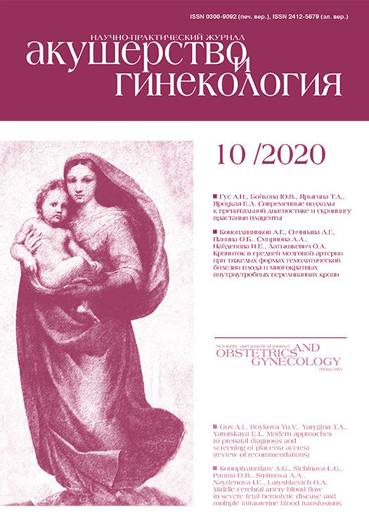
When performing shear wave elastography in the control group, the cervical tissue was steadily stained with a homogeneous blue color (Fig. 1). The cervix elasticity was Me=21.1 kPa (20; 23.5) in women of the control group.
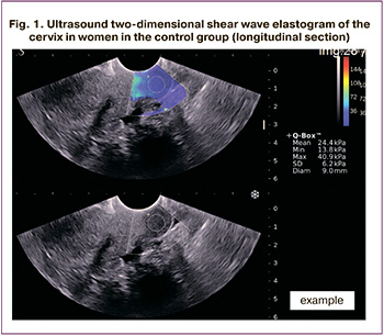
The patients with cancer had a tumor stained in an intense yellow-red color (Fig. 2).
According to elastometry, the median elasticity of the tumor in patients with T1 and T2 stages increased significantly compared to the controls and reached Me=65.7 kPa (56.1; 101.2) and Me= 67.6 kPa (45.3; 80.6), respectively. Two women with Tis and T3 stage showed a clear tendency to increase stiffness up to 38.1 kPa and 138.9 kPa, respectively, but these patients were singular and did not allow us to make a final conclusion. The group distribution of elastometry data is shown in Table 1 and Figure 3.
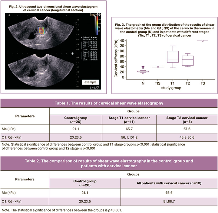
 In the combined group of patients with cervical cancer, the parameter Me (Q1; Q3) of tumor elasticity was 66.6 kPa (51; 88.7). The comparison showed a statistically significant increase in the elasticity of cervical tissues affected by cancer (p<0.001) compared with the control group (Table 2 and Fig. 4).
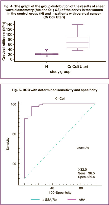
The ROC-analysis made it possible to establish that when the threshold of 32 kPa was applied, the sensitivity, specificity, and prognostic value of a positive and negative result reached 96%, 89%, 86%, and 95%, respectively, and AUROC reached 0.97 (Fig. 5).
Discussion
The search for early signs of cervical cancer is an obvious challenge for modern ultrasound diagnostics. Today, ultrasound Doppler imaging can reach rather high accuracy (89.5% sensitivity and 93.5% specificity) in the assessment of cervical cancer [7]. However, it should be noted that space-occupying lesions that are already visible on the screen are discussed in such publications. Our research suggests that shear wave elastography is a new approach to solving the problem of early instrumental diagnosis of cervical cancer. There is no doubt the results presented by us are based on a small amount of material. However, the obtained data are consistent with the published results [5, 6], which showed that the shear wave velocity in the tissues of the unaltered cervix is significantly lower than in those affected by the malignant process.
Conclusion
Therefore, there is every reason to believe that ultrasound shear wave elastography is a promising objective method for assessing the condition of the cervix.
References
- Статистические данные и рак шейки матки. Available at: https://www.oncoforum.ru/o-rake/statistika-raka/rak-sheyki-matki-statistika-i-prognozy-vyzhivaemosti.html [Statistics and cervical cancer. (in Russian)].
- Зыкин Б.И., Постнова Н.А., Медведев В.Е. Эластография: анатомия метода. Променева дiагностика, променева терапiя. 2012; 2-3: 107-13. [Zykin B.I., Postnova N.A., Medvedev V.E. Elastography: anatomy of the method. Promeneva diagnostics, promeneva therapy/Diagnostic radiology and radiotherapy. 2012; 2-3: 107-13. (in Russian)].
- Головко Т.С., Бакай О.А. Возможности эластографии в диагностике новообразований шейки матки. Здоровье женщины. 2014; 8: 112-7. [Golovko T.S., Bakay O.A. Possibilities of elastography in the diagnosis of cervical neoplasms. Women's health. 2014; 8 (94): 112-7. (in Russian)].
- Lu R., Xiao Y., Liu M., Shi D. Ultrasound elastography in the Ddfferential diagnosis of benign and malignant cervical lesions. J. Ultrasound Med. 2014; 33(4): 667-71. https://dx.doi.org/10.7863/ultra.33.4.667.
- Su Y., Du L., Wu Y., Zhang J., Zhang X., Jia X. et al. Evaluation of cervical cancer detection with acoustic radiation force impulse ultrasound imaging. Exp. Ther. Med. 2013; 5(6): 1715-9. https://dx.doi.org/10.3892/etm.2013.1057.
- Liu C., Li T., Hu Z., Li Y., Cheng X., Zhu Y, Lu M. Transvaginal real-time shear wave elastography in the diagnosis of cervical disease. J. Ultrasound Med. 2019; 38(12): 3173-81. https://dx.doi.org/10.1002/jum.15018.
- Dodampahala S.H., Jayakody S.N., Gunathilake W.C.C., Rahubaddha A.N., Dodampahala S.K. Transvaginal color Doppler in the assessment of cervical carcinoma and pre-cancer: evidence from a case control study using colour Doppler ultrasonography pulsatility index of uterine vasculature. Advanc. Reprod. Sci. 2016; 4: 93-9.
Received 15.05.2020
Accepted 13.09.2020
About the Authors
Boris I. Zykin, MD, professor of Department of Diagnostic radiology, Medical-Biological University of Innovation and Continuing Education of I.I. Burnazyan Federal Medical Biological Center, FMBA of the Russian Federation. Tel.: +7(499)190-96-92. E-mail: b.zykin@yandex.ru. 123182, Russia, Moscow, Zhivopisnaya str., 46.Vera L. Ogryzkova, Ph.D., Leading Researcher of the National Center of Oncology of the Reproductive Organs (branch of the Scientific Medical Research Center for Radiology of the named after P.A. Herzen), Ministry of Health of Russian Federation. Tel.: +7(495)150-11-22. E-mail: verogr@yandex.ru.
119121, Russia, Moscow, Pogodinskaya str., 6, bld. 1.
Elena A. Ionova, MD, Head of the Department of Diagnostic radiology, Medical-Biological University of Innovation and Continuing Education of I.I. Burnazyan Federal Medical Biological Center, FMBA of the Russian Federation. Tel.: +7(964)762-99-49. E-mail: ionela60@mail.ru.
For citation: Zykin B.I., Ogryzkova V.L, Ionova E. A. Possibilities of ultrasonic shear wave elastography in the diagnosis of cervical cancer (first experience).
Akusherstvo i Ginekologiya / Obstetrics and gynecology. 2020; 10: 113-117 (in Russian)
https://dx.doi.org/10.18565/aig.2020.10.113-117
Similar Articles

