Частота встречаемости аномалий мюллеровых протоков (АМП) в общей популяции невелика, варьирует от 2 до 7% [1]. Однако у пациенток с бесплодием, привычным невынашиванием беременности, с эктопической беременностью процент АМП достигает 18–20% и зачастую только АМП является единственной причиной [1–3]. Несвоевременная диагностика, неправильная интерпретация порока могут стать причиной такого грозного осложнения, как гибель плода при разрыве замкнутого функционирующего рога, спонтанного прерывания беременности, преждевременных родов, фетоплацентарной недостаточности, преждевременной отслойки аномально расположенной плаценты, перфорации матки во время внутриматочной хирургии. На протяжении последних 15 лет магнитно-резонансная томография (МРТ) является единственным достоверным методом диагностики АМП, однако повсеместное использование метода ограничено [4]. В последние два десятилетия при помощи двухмерного ультразвукового исследования и ультразвуковой эхогистерографии и гистеросальпингографии можно было судить о факте наличия АМП, но окончательно верифицировать его, с учетом современной классификации и клинических требований, не представлялось возможным. Помимо отсутствия четких критериев, долгое время отсутствовала единая классификация АМП. В 2013 г. сообществами ЕSHRE/ESGE была предложена классификация CONUTA (Congenital Uterine Anomalies), объединяющая группы по эмбриологии и патогенезу, клиническим проявлениям и необходимости хирургической коррекции: отдельно выделены пороки развития тела матки, шейки матки и влагалища, которые могут сочетаться друг с другом или нет, в отдельный параграф вынесены сочетанные экстрагенитальные аномалии [5, 6]. Однако предложенные для оценки процентильные значения вместо численных показателей оказались неудачными и привели к гипердиагностике внутриматочной перегородки, что инициировало увеличение частоты хирургических вмешательств [7].
Материалы и методы
Для определения возможностей различных режимов трехмерной эхографии и эхогистерографии (3D-УЗИ/3D-ГСГ) нами были обследованы 450 пациенток репродуктивного возраста (от 15 до 45 лет) с различными АМП. Результаты эхографии сопоставляли с данными МРТ в 70% наблюдений, с данными гистероскопии – в 92%, с данными лапароскопии(томии) – в 23% и 8,5% наблюдений соответственно. С учетом вышеперечисленных исследований нами были выявлены: седловидная форма полости матки (класс U0) у 60 пациенток; Т-форма полости матки и ACUM (класс U1) у 4 и 2 пациенток соответственно, неполная и полная внутриматочные перегородки (класс U2a/b) у 78 и 85 обследованных, двурогая матка (класс U3a) – у 92; удвоение тела матки (класс U3b) – у 91, перегородчатая форма (класс U3с) – у 7; однорогая матка (класс U4a/b) – у 31. Критериями включения пациенток в исследование стали: подозрение на наличие АМП по данным предшествующих УЗИ, аномальные маточные кровотечения (АМК), бесплодие и привычное невынашивание беременности. Следует отметить, что у 23% обследованных выявленный порок не сопровождался никакими клиническими проявлениями, был находкой рутинного УЗИ при диспансеризации. Анализируя менструальную функцию пациенток, мы выяснили: возраст наступления менархе от 11 до 16 лет, у 28% были выявлены различные нарушения менструального цикла и у 70% – жалобы на АМК (пациентки с АМП класса U1–U3), у 8% (пациентки класса U3 и U4) были указания на альгодисменорею, лишающую трудоспособности и у 9% из них (класс U4) требующую госпитализации и проведения оперативной лапароскопии по поводу формирования гематосальпинкса больших размеров. Синдром хронических тазовых болей имел место у 11% (пациентки класса U1, U3 и U4). Бесплодие различной продолжительности диагностировано во всех группах у 80%, в анамнезе у 1% обследованных имеется указание на полное самопроизвольное прерывание беременности на ранних сроках, у 47% – на неполное. Привычное невынашивание беременности диагностировано у 41% (равноценно встречалось во всех группах наблюдения). У 2 пациенток 25 и 28 лет группы U4a произошел разрыв не диагностированного вовремя добавочного рога на 19-й и 21-й неделе гестации, сопровождавшийся внутрибрюшным кровотечением и геморрагическим шоком 3 степени. Учитывая неясную интраоперационную картину и отсутствие условий для проведения реконструктивного объема, потребовалось проведение повторной хирургической операции. Ранние преждевременные роды зафиксированы в 2% наблюдений на 22–28-й неделях беременности с перинатальными потерями (группа U2/U3), преждевременная отслойка аномально расположенной на перегородке плаценты произошла у 1% пациенток группы U2a в сроках гестации 28–32 недели. Самопроизвольные неосложненные роды произошли у 26% пациенток (группа U3/U4).
3D-УЗИ/3D-ГСГ выполняли на аппарате Canon Aplio MX с использованием трехмерного трансвагинального датчика (6,5 МГц). УЗИ проводилось в день первичного обращения, а также повторно на 5–7-й день менструального цикла. Для получения информативных эхограмм использовали различные имеющиеся режимы трехмерной эхографии: режим мультипланарной реконструкции (МПР), Omni-view, инверсии изображения, сосудистый режим Glass body, TUI. 3D-ГСГ выполнялась по нашей стандартной методике с использованием баллонных катетеров, стерильного физиологического раствора, подача которого осуществлялась автоматически помпой Hamou Endomat (KarlStorz, Германия). Построение и интерпретация трехмерной эхограммы порока не являются рутинными и требуют от специалиста достаточной квалификации, должны выполняться согласно регламенту и анализироваться с учетом нижеизложенных задач. Последовательно должны быть оценены такие параметры матки и зоны параметрия, как форма тела матки и ее размер, наружный контур миометрия (НКМ), форма полости матки, ее объем, дополнительные анатомические структуры в виде двух отражений от эндометрия или/и от эндоцервикса, дополнительные образования в проекции параметрия, имеющие связь с телом матки. Основным операторнезависимым преимуществом 3D-УЗИ является получение фронтального среза (ФС) органов малого таза, позволяющего параллельно интерпретировать основные критерии АМП. 3DУЗИ предоставило не только возможность параллельной оценки (НКМ) и внутриполостной архитектоники, аналогично МРТ или сочетанной гистеро-/лапароскопии, но и получать эхограммы на новом, до сих пор не существующем виртуальном уровне.
Результаты
Параметрами анатомической нормы тела матки (класс U0) во ФС являлись: НКМ в виде подковы, полость матки в виде треугольника. Качественным критерием тела матки стали вариабельные ее формы, такие как подковообразный контур, наличие инвагинации различной степени выраженности или полигональная форма. Количественным критерием – глубина инвагинации НКМ, определяемая согласно правилу лучевой диагностики R.N. Troiano и S.M. McCarthy (2004) как расстояние от интеркорнуальной линии до самой отдаленной точки миометрия [8]. Качественный критерий полости – вариабельность ее формы в виде треугольной, дугообразной, V/Y-образной различной степени выраженности, Г-образной или зеркальной Г-образной. Количественным критерием диагностики полости стала оценка степени пролабирования миометрия в полость, определяемая по формуле R.N. Troiano и S.M. McCarthy (2004) расстоянием от интеростиальной линии до самого отдаленного ориентира полости, а также протяженность интеростиальной оси [8]. Параллельная оценка качественных и количественных критериев, а также детальная оценка изменений шейки матки и цервикального канала и области параметрия позволили определить паттерн каждой АМП. Седловидная форма полости – вариант нормы (класс U0) – зачастую не имеет клинических проявлений, однако нехарактерная ультразвуковая картина заставляет проводить диагностику с АМП. Критерием является визуализация неизмененного НКМ в сочетании с дугообразной или V-образной формой полости. Схожая ультразвуковая картина наталкивает на проведение дифференциальной диагностики с U2a. Диагностическим критерием является расстояние от интеростиальной линии до наиболее удаленной точки полости, определяемой во ФС. При выявлении внутриполостной инвагинации протяженностью более 14 мм диагностировали внутриматочную перегородку. При 3D-ГСГ критерии были идентичными, более четко оценивалась внутриполостная архитектоника и проходимость маточных труб.
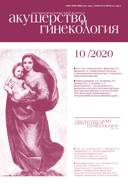
 К классу UI относят редчайшую Т-форму полости матки, повышающую риск привычного невынашивания беременности или бесплодия из-за уменьшенного объема полости матки. 3D-критериями стали: неизмененный НКМ, уменьшение поперечного размера матки, Т-форма полости матки с уменьшением ее объема в 3–4 раза в сравнении с нормой. Диагностируется тол ко при отсутствии изменений миометрия, инициирующих сужение полости (рис. 1). Двум пациенткам с привычным невынашиванием произведена резекция латеральных стенок полости петлей биполярного гистерорезектоскопа с последующей терапией, направленной на профилактику образования синехий, а также гормональная терапия КОК сроком 6 месяцев, после чего обе пациентки самостоятельно забеременели, обе беременности завершились самопроизвольными неосложненными срочными родами.
Также к классу UI можно отнести дискутабельную ACUM (accessory and cavitated uterine mass), диагностируемую у пациенток до 30 лет с хроническими тазовыми болями и АМК, патогенетически связанную с дублированием ткани мюллерова протока в критической области круглой связки. Гистологическое заключение исключает аденомиоз, однако патологические находки соответствуют полости, выстланной эндометриальным эпителием, представленным железами и стромой, заполненной шоколадным содержимым. 3D-критериями стали: интактная форма полости, измененный НКМ по латеральной или верхнелатеральной поверхности за счет выбухания в брюшную полость, инициированного образованием, расположенным в миометрии гипоэхогенной структуры с крупнодисперсной аваскулярной взвесью и отсутствием дополнительного источника кровоснабжения в виде «сосудистой ножки». Также, в отличие от миоматозного узла, отсутствует капсула, ACUM имеет нечеткие границы, дезорганизующие мышечные волокна (рис. 2). Учитывая юный возраст пациенток, стоит вопрос не только в постановке диагноза, но и в выборе адекватной органосохраняющей хирургической тактики, сопряженной с высокими хирургическими рисками. Одной пациентке мы произвели лапароскопию, иссечение образования, гистологическое исследование подтвердило нашу находку. Вторая пациентка 15 лет на данный момент получает препараты АГнРГ, ввиду отказа от хирургического лечения.

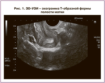
У пациенток с неполной внутриматочной перегородкой (класс U2a) наличие перегородки практически не нарушало целостность НКМ, сочетаясь с дуго-V-образной формой полости матки различной степени выраженности за счет внутриполостной мышечной инвагинации более 14 мм. В режиме Glass Body мы диагностировали выраженное оскудение сосудистого рисунка перегородки вплоть до его тотального исчезновения по направлению к ее дистальным отделам, что в сочетании с отягощенным акушерско-гинекологическим анамнезом может являться абсолютным показанием к иссечению перегородки (рис. 3).
Интраоперационное 3D-УЗИ позволит хирургу провести завершенную резекцию перегородки, сформировать адекватную треугольную полость и избежать неполной резекции и формирования седловидной формы полости матки. Объемные виртуальные эхограммы позволят наглядно презентовать пациентке произведенную резекцию и акцентировать ее внимание на объеме и форме вновь сформированной полости матки (рис. 4).
 При проведении 3D-ГСГ определялась сепарированная полость за счет внутриполостной структуры клювообразной формы и более четко в условиях гидрометры оценивалась толщина перегородки. Также на фоне контраста стали более четко определяться тонкие синехии, не диагностированные ранее.
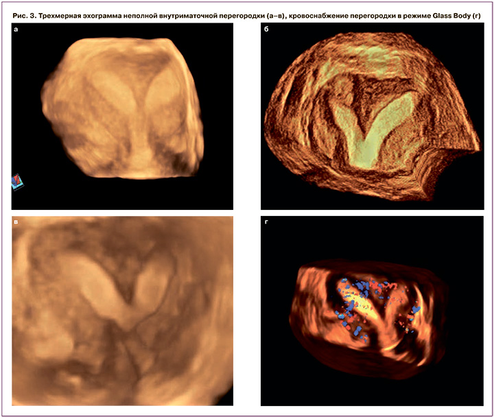
Трехмерными критериями полной внутриматочной перегородки (класс U2b) стали: интактный НКМ, V/Y-образная форма полости, сформированная внутриполостной структурой, разделяющей ее на две гемиполости, доходящая до внутреннего зева шейки матки. В ФС мы могли оценить симметричность обеих гемиполостей и объем каждой из них, что может быть решающей информацией перед выполнением вспомогательных репродуктивных технологий и переноса эмбриона. Режим TUI позволил более четко определить вариабельность толщины перегородки (рис. 5), которая в нашем исследовании составила максимально от 15 до 27 мм и минимально от 0,3 до 2,2 мм. При проведении 3D-ГСГ катетер устанавливался в проекции внутреннего зева шейки матки для адекватной параллельной инсуффляции обеих гемиполостей, при невозможности – катетер попеременно вводился в каждую гемиполость. У 43% обследованных с отягощенным акушерско-гинекологическим анамнезом или отсутствием (выраженным оскудением) кровотока перегородки мы произвели ее резекцию биполярной петлей с обязательным введением противоспаечного барьера и назначением комбинированных оральных контрацептивов на 3–6 месяцев, у 22% из них в течение последующих 2 лет беременность закончилась самопроизвольными срочными родами.
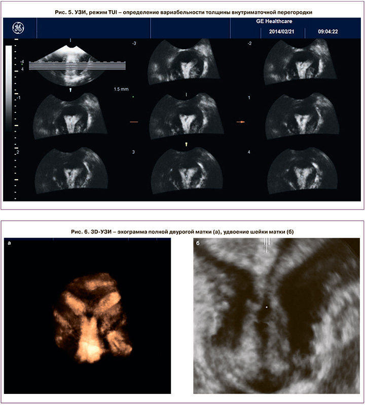
Трехмерными паттернами двурогой матки (класс U3a) стали: измененная форма тела матки за счет инвагинации НКМ глубиной от 2,5 до 16 мм, V/Y-образная форма полости. При наличии Y/V-образной формы полости и незначительном изменении НКМ измеряли расстояние от интеркорнуальной линии до НКМ, если оно было менее 4 мм, ставился диагноз «двурогая матка», если больше 4 мм – «внутриматочная перегородка». В зависимости от протяженности перегородки выделяют полную и неполную формы, однако, вне зависимости от ее формы, коррекция порока при помощи гистерорезектоскопии сопряжена с высочайшим риском перфорации матки. При неправильной интерпретации порока и диагностировании перегородки эндоскопический ориентир – устья маточных труб – будет ошибочным и произойдет непреднамеренная перфорация в проекции инвагинации НКМ, которая находится на линии, расположенной ниже устьев маточных труб. Такой редкий порок, как полная двурогая матка с удвоением шейки, диагностирован у 2 пациенток (рис. 6).
Режим Glass body позволил нам получить характерную архитектонику кровотока в виде M-образной формы, что разительно отличает порок от удвоения матки (рис. 7).
Ультразвуковые паттерны 3D-ГСГ аналогичны, дополнительная информация получена за счет более четкого измерения толщины перегородки на фоне гидрометры и в определении проходимости маточных труб у пациенток с бесплодием.
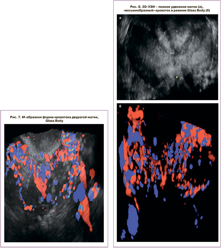
Удвоение тела матки (класс U3b) характеризуется прерывающимся и вновь восстанавливающимся НКМ, разделенным инвагинацией различной протяженности и глубины (от 5 до 7 см), зеркальными Г-образными формами гемиполостей. Glass Body позволил нам диагностировать «восьмиобразный» тип кровотока, свидетельствующий о полном равнозначном удвоении (рис. 8).
При визуализации данного порока делается акцент на состояние шейки, области перешейка и направлении гемиполостей, эта информация будет решающей при планировании и в ходе выполнения любой внутриматочной манипуляции, особенно при наличии одной шейки и выраженном изменении области перешейка с его укорочением и истончением (рис. 9). ФС шейки матки также облегчают постановку диагноза относительно ее удвоения (рис. 10).

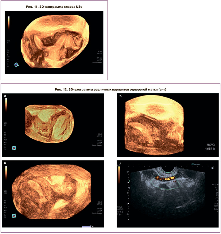
Ультразвуковые паттерны класса U3с позволяют наглядно оценить возможность гистероскопической коррекции порока для формирования адекватной полости, но только под интраоперационной ультразвуковой навигацией (рис. 11).
Самый большой процент диагностических и тактических ошибок касается класса U4, объединяющего различные однорогие матки. 3D-критериями в ФС стали: уменьшенный поперечный размер матки (от 20 до 24 мм), полигональная форма НКМ, «бананообразная»/Г-образная форма полости, асимметричное расположение яичников. Важной является панорамная оценка зоны параметрия для идентификации дополнительного рога в виде структуры, расположенной справа или слева от основного рога, контактирующего с ним и имеющего различную форму – от полигональной до линейной, объемом, не превышающим 1/3 объема основного рога. Добавочный рог, помимо полигональной формы, может иметь различную структуру: от однородной средней эхогенности без активного кровоснабжения до гиперваскулярной гетерогенной структуры. Важной является идентификация функциональной способности рога за счет обнаружения полости с эндометрием, что ни в коем случае не должно быть пропущено, поскольку данная аномалия колоссально повышает риск эктопической беременности с возможными грозными осложнениями и снижает возможность ее диагностики, а также повышает риск заболеваний эндометрия или формирования гематосальпинкса (рис. 12). Только проведение 3D-ГСГ позволит определить наличие сообщения между основным и добавочным рогом в виде затекания контраста из основной полости в рудиментарную. Всем пациенткам данного класса в незамедлительном порядке мы рекомендовали проведение лапароскопии, удаление рога с маточной трубой.
Заключение
Таким образом, трехмерная эхография предлагает качественно новый метод ультразвуковой диагностики АМП, аналогичный МРТ, позволяющий получать эхограмму в виде виртуального отображения реальной анатомии. Специфичные трехмерные эхографические паттерны позволят с легкостью классифицировать порок. Комбинация различных режимов позволит получить дополнительную информацию относительно сосудистой архитектоники, спрогнозировать возможные осложнения беременности, сформировать четкие показания для коррекции порока, осуществить навигацию во время внутриматочной хирургии. Трехмерная эхогистерография – дополнительный высокоспецифичный метод, применяемый для дифференциальной диагностики или в случае трудно интерпретируемой измененной анатомии, сопряженной с другими заболеваниями эндометрия и миометрия.