Рак молочной железы (РМЖ) занимает первое место среди всех онкологических заболеваний у женщин, и, несмотря на успехи в его диагностике и лечении, имеет место рост заболеваемости и смертности от РМЖ. Прослеживается тенденция к увеличению числа женщин молодого возраста с РМЖ. В 2018 г. взяты на учет 64 544 больных с впервые выявленным злокачественным новообразованием молочной железы [1]. Объективная оценка предиктивных и прогностических факторов является ключевой задачей при обследовании и лечении больных онкологическими заболеваниями. Современный стандарт диагностики РМЖ включает гистологическое и иммуногистохимическое (ИГХ) исследования РМЖ, включающие оценку в опухоли уровня экспрессии рецепторов эстрогенов и прогестерона, HER2/neu и Ki-67, необходимую для планирования адекватной терапии [2, 3]. Одним из важнейших прогностических факторов при РМЖ является маркер пролиферации Ki-67, выявляемый ИГХ на биопсийном и операционном материале [4, 5]. Метод позволяет определить процент позитивно окрашенных ядер опухолевых клеток, т.е. дать характеристику пролиферативной активности опухоли. В настоящее время указанный показатель оценивают визуально, результаты исследования характеризуются высокой вариабельностью и нередко зависят от опыта врача-патоморфолога [6–8], в связи с этим объективизация оценки экспрессии Ki-67 является актуальной диагностической задачей [2, 9, 10]. Количественная оценка экспрессии маркера Ki-67 при РМЖ различных гистологических типов и степеней злокачественности представляется необходимой для изучения исследования фундаментальных аспектов онкоморфологии РМЖ [11, 12].
Цель исследования – сравнительный анализ уровня экспрессии маркера пролиферации Ki-67 в РМЖ неспецифического типа, выявленного с помощью традиционной визуальной оценки и при применении метода цифровой микроскопии с использованием программного обеспечения PatternQuant (QuantCenter 3DHISTECH).
Материалы и методы
Количественное ретроспективное морфологическое исследование экспрессии Ki-67 проведено с использованием метода цифровой микроскопии и программного обеспечения PatternQuant (QuantCenter 3DHISTECH) на парафиновых срезах операционного материала толщиной 4 мкм у 50 пациенток с инвазивной карциномой молочной железы неспецифического типа в Калининградской области (архив лаборатории иммуногистохимической и патологоанатомической диагностики КДЦ БФУ им. И. Канта). Методы исследования – клинико-морфологический анализ, гистологическое исследование с использованием критериев Ноттингемской классификации, ИГХ-окрашивание на иммуногистостейнере BondMax с использованием антител Ki67 (Clone MIB 1, Dako), Progesteron Receptor (16), Estrogen Receptor (6F11), статистическая обработка результатов с помощью программ Statistica 10.0 и Exсel 2010. Показатели экспрессии Ki-67, полученные врачом при визуальном количественном исследовании препаратов РМЖ (3–5 полей зрения, подсчет экспрессии в 100 опухолевых клетках в участках опухоли с максимально выраженной пролиферацией), сравнивали с результатами автоматического подсчета цифровых изображений опухоли на идентичных по площади участках опухоли (3 поля зрения, ув. ×400) [6, 7, 13, 14]. Учитывали позитивное ядерное окрашивание опухолевых клеток (%): 1) при относительно однородном распределении Ki-67+ опухолевых клеток количественную оценку проводили в трех случайно выбранных полях зрения; 2) при гетерогенном распределении позитивно окрашенных опухолевых клеток исследовали краевые участки опухоли/«горячие точки» – области с максимально выраженной экспрессией Ki-67 [10, 15, 16]
Результаты и обсуждение
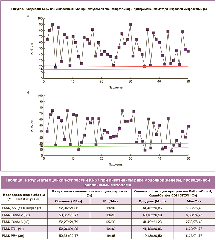
В основной исследованной выборке (50 случаев) инвазивного РМЖ неспецифического типа средний возраст пациенток составил 61,2±12,8 года. Распределение РМЖ по степеням злокачественности было следующим: РМЖ Grade2 выявлен у 36 пациенток, Grade3 – у 13 больных, Grade1 – у 1 женщины, что в целом соответствует описанным ранее данным [17]. Положительная экспрессия рецепторов эстрогенов в опухолях имела место у 41 женщины, прогестерона – у 39. Результаты оценки показателей экспрессии Ki-67 традиционным способом при визуальной количественной оценке врачом и при автоматизированном подсчете процента позитивно окрашенных клеток опухоли с помощью программы PatternQuant представлены в таблице. Установили, что средние показатели экспрессии Ki-67 в изученных выборках (общая выборка, опухоли Grade2) при оценке врачом и с помощью программы PatternQuant продемонстрировали сходные тенденции. Тем не менее при оценке врачом они были несколько выше по сравнению с автоматизированным подсчетом. Это может быть обусловлено выбором различных полей зрения для исследования при ретроспективном анализе архивных препаратов с помощью программы PatternQuant с целью последующего сравнения с ранее изученными препаратами при комплексной гистологической и ИГХ-диагностике РМЖ. В пользу данного утверждения свидетельствуют различия в величине минимальных и максимальных показателей экспрессии Ki-67 при оценке двумя использованными методами (рисунок), что является особенно значимым при определении порогового уровня экспрессии Ki-67 (14%) в каждом индивидуальном случае для определения молекулярного субтипа РМЖ на основе оценки суррогатных ИГХ-маркеров (люминальный тип А или люминальный тип В).
Для оценки степени соответствия результатов подсчета средних показателей экспрессии Ki-67 при визуальной количественной оценке и при использовании цифровых изображений опухоли оценкой с помощью программы PatternQuant выполнен корреляционный анализ данных основной выборки (50 случаев). Выявлена линейная корреляционная зависимость высокой величины, r=0,72, p<0,05.
Таким образом, по-видимому, в настоящее время для объективной оценки уровня экспрессии важного прогностического маркера пролиферации Ki-67, входящего в стандартную панель ИГХ-исследования опухоли у пациенток с РМЖ, может быть рекомендовано применение визуальной количественной оценки опытным специалистом-патологом. В этом случае возможны адекватный учет гистологических особенностей строения опухоли и степени ее злокачественности, проведение оценки пролиферативной активности в фокусах с ее максимальной выраженностью. Автоматизированный подсчет процента позитивно окрашенных клеток опухоли с помощью программы PatternQuant (QuantCenter 3DHISTECH) на единице площади среза дает удовлетворительные результаты при условии правильного выбора участков опухоли для последующего анализа их цифровых изображений. С учетом полученных данных для объективизации количественной оценки экспрессии Ki-67 может быть рекомендовано дальнейшее одновременное унифицированное исследование показателей пролиферации в максимально идентичных участках опухоли в расчете на одинаковое число опухолевых клеток, что особенно значимо в случаях выраженной гетерогенности опухолей.
Заключение
Средние показатели экспрессии Ki-67 при РМЖ неспецифического типа при традиционной количественной визуальной оценке врачом-патологом и с помощью программы PatternQuant продемонстрировали сходные тенденции как в общей выборке РМЖ неспецифического типа, так и в подгруппах, разделенных по степени злокачественности и экспрессии рецепторов эстрогенов и прогестерона в опухоли.
Выявлены различия в определении максимальных и минимальных показателей пролиферации при РМЖ. При их оценке врачом-патологом результаты были несколько выше по сравнению с автоматизированным подсчетом, что может быть особенно значимым при определении порогового уровня экспрессии Ki-67 (14%) в каждом индивидуальном случае для определения молекулярного субтипа опухоли – люминального типа А или люминального типа В.
Несмотря на достаточно высокую эффективность количественной оценки экспрессии Ki-67 при РМЖ с помощью метода цифровой микроскопии и программного обеспечения QuantCenter 3DHISTECH, для объективизации результатов рекомендуется унифицированное исследование показателей пролиферации в максимально идентичных участках опухоли в расчете на одинаковое число опухолевых клеток, что особенно значимо в случаях выраженной гетерогенности опухолей.