
Amino acid composition of the endometrium in patients with diminished ovarian reserve
Dubinskaya E.D., Mglinets A.P., Gasparov A.S., Alyoshkina E.V.
Objective. To determine the characteristics of the endometrial amino acid profile in patients with diminished ovarian reserve (DOR) compared with patients with normal ovarian reserve in early and middle reproductive age.
Materials and methods. An observational cross-sectional comparative study was conducted in 110 reproductive-age patients (20–35 years) undergoing preparation for assisted reproductive technology (ART) programs. Patients were divided into two groups: group 1 comprised 80 patients with DOR (AMH<1.2 ng/mL, AFC<5–7 in each ovary); group 2 (control) comprised 30 patients with normal ovarian reserve. Endometrial samples were obtained by pipelle biopsy during the mid-secretory phase of the menstrual cycle. Twenty-three amino acids and their metabolites were analyzed using UPLC-MS/MS). Statistical analyses were performed using the MetaboAnalyst v5.0 platform and included univariate analysis (Mann–Whitney U test, FDR) and multivariate statistical analysis (PCA, PLS-DA), as well as metabolic pathway analysis.
Results. Statistically significant differences were identified for 11 amino acids (q<0.05). Patients with DOR showed marked reductions in sarcosine concentration (7.91-fold; FC=0.13), tryptophan (FC=0.68), pipecolic acid (FC=0.76), and leucine (FC=0.89). At the same time, increased levels of alanine (FC=1.64), γ-aminobutyric acid (FC=1.61), asparagine (FC=1.56), glutamine (FC=1.55), proline (FC=1.44), hydroxyproline (FC=1.17), and aspartic acid (FC=1.15) were observed. Multivariate PLS-DA demonstrated clear separation of the groups based on their metabolic profiles. The most pronounced changes involved the alanine, aspartate and glutamate, and arginine and proline metabolic pathways.
Conclusion. DOR in patients of early and middle reproductive age is associated with the development of a local metabolic phenotype of the endometrium characterized by alterations in amino acid composition. These changes may be associated with the functional state of the endometrium and its implantation potential.
Authors' contributions. Dubinskaya E.D. – conception and design of the study, scientific supervision, editing of the manuscript; Mglinets A.P. – data collection and analysis, statistical analysis, literature review, drafting of the manuscript; Gasparov A.S. – consultation, editing of the manuscript; Alyoshkina E.V. – data processing and drafting of the manuscript.
Conflicts of interest. The authors have no conflicts of interest to declare.
Funding. There was no funding for this study.
Ethical Approval: The study was reviewed and approved by the Research Ethics Committee of the Patrice Lumumba Peoples' Friendship University of Russia (Ref. No: 4).
Generative Artificial Intelligence. No artificial intelligence tools were used in the preparation of this manuscript.
Patient Consent for Publication. All patients provided informed consent for the publication of their data.
Authors' Data Sharing Statement. The data supporting the findings of this study are available upon request from the corresponding author after approval from the principal investigator.
For citation: Dubinskaya E.D., Mglinets A.P., Gasparov A.S., Alyoshkina E.V. Amino acid composition
of the endometrium in patients with diminished ovarian reserve.
Akusherstvo i Ginekologiya/Obstetrics and Gynecology. 2026; (10): 89-99 (in Russian)
https://dx.doi.org/10.18565/aig.2026.293
Keywords
endometrium
endometrial receptivity
amino acid composition
diminished ovarian reserve
metabolomics
According to various authors, the prevalence of infertility among patients with diminished ovarian reserve (DOR) in early and middle reproductive age ranges from 19 to 38.4% [1, 2]. This cohort of patients clearly represents a complex clinical challenge, not only in terms of the quality of embryos obtained but also with regard to the implantation potential of the endometrium.
The clinical pregnancy rate achieved with assisted reproductive technology (ART) programs is substantially lower in this group of patients than in women with normal ovarian reserve [3, 4]. However, the literature regarding the relationship between ovarian reserve parameters and embryo quality remains contradictory. Some studies have demonstrated an association between anti-Müllerian hormone (AMH) levels and embryo euploidy rates [5], whereas others have found no independent association between AMH levels and euploidy [6]. According to our previously published data, the rate of recurrent implantation failure in patients with DOR is 61% following the transfer of good-quality embryos [7].
However, successful embryo implantation is known to depend not only on embryo morphological quality and chromosomal status but also on the condition of the endometrium, which forms the so-called “window of implantation” [8, 9]. The effect of DOR on endometrial receptivity, however, remains unexplored.
To date, efforts to assess the implantation properties of the endometrium as a whole are ongoing. In recent years, investigation of the endometrial metabolomic profile has emerged as a promising approach for characterizing the overall spectrum of low-molecular-weight metabolites in biological tissues and fluids [10, 11]. The metabolomic profile reflects the ultimate result of interactions among genetic, hormonal, and environmental factors and may serve as a more sensitive indicator of tissue functional status than morphological or isolated hormonal parameters [12].
According to the available literature, the amino acid profile most objectively reflects the state of cellular metabolism and tissue homeostasis [13]. Unlike hormonal parameters, which predominantly characterize systemic regulation of reproductive function, analysis of the endometrial amino acid profile makes it possible to assess local features of its functional state [14].
The scientific hypothesis underlying the present study was that the endometrium of patients with DOR differs from that of patients with normal ovarian reserve in amino acid composition, reflecting a local metabolic phenotype and potentially affecting its implantation properties.
In view of the above, the aim of the present study was to determine the characteristics of the endometrial amino acid profile in patients with DOR compared with patients with normal ovarian reserve in early and middle reproductive age.
Materials and methods
Study design
This was an observational, cross-sectional, comparative study involving 110 reproductive-age women (20–35 years) undergoing assisted reproductive technology (ART) at the «Ya Zdorova» clinic, affiliated with the Department of Obstetrics and Gynecology with a Course in Perinatology, Patrice Lumumba Peoples' Friendship University of Russia. The study was conducted between 2024 and 2026.
The study employed an exploratory metabolomic design to identify potential biomarkers of endometrial receptivity in women with DOR. Consequently, no a priori sample size calculation was performed. The sample size was determined by the availability of eligible patients meeting the inclusion criteria during the specified study period. The findings are intended to serve as a baseline for subsequent validation studies in independent cohorts.
Study group allocation
Patients were assigned to two groups based on ovarian reserve status: Group 1 (study group) comprised 80 patients with DOR, and Group 2 (control group) comprised 30 patients with normal ovarian reserve.
DOR was defined as AMH concentration <1.2 ng/mL and an antral follicle count (AFC) <5–7 in each ovary. These criteria were based on current approaches to ovarian reserve assessment [15, 16]. Follicle-stimulating hormone (FSH) and estradiol were not used as independent grouping criteria but were considered when comparing the clinical and laboratory characteristics of patients.
Inclusion and exclusion criteria
Inclusion criteria: age 20–35 years, tubal-peritoneal infertility, absence of pregnancy after at least one year of regular unprotected intercourse, preparation for an ART program, and written informed consent to participate in the study.
Exclusion criteria: chronic endometritis (N71.1), endometrial polyp (N84.0), endometrial hyperplasia (N85.0–N85.1), intrauterine adhesions (N85.6), submucosal uterine leiomyoma (D25.0), premature ovarian insufficiency (E28.3), polycystic ovary syndrome (E28.2), ovarian endometriomas (N80.1), benign ovarian neoplasms (D27), history of unilateral oophorectomy, use of hormonal medications, dietary supplements, or vitamin-mineral complexes, and other contraindications to ART according to Order No. 803n of the Ministry of Health of the Russian Federation dated July 31, 2020.
Sample collection and preparation
The research material comprised endometrial samples obtained via pipelle biopsy during the mid-secretory phase of the menstrual cycle (days 20–23 of a 28-day cycle, or on day 7 post-ovulation confirmed by folliculometry for cycles of different duration).
Following collection, samples were immediately frozen and stored at -80°C until analysis. Repeated freeze-thaw cycles were avoided. Laboratory analyses were performed at the "Archimed" laboratory in compliance with established laboratory protocols. Prior to analysis, samples were thawed, spiked with isotopically labeled internal standards, subjected to protein precipitation using an organic solvent, centrifuged, and derivatized with butanol.
Amino acid composition analysis
Quantitative analysis of 23 amino acids and their derivatives was conducted using ultra-performance liquid chromatography-tandem mass spectrometry (UPLC-MS/MS) on a Sciex QTrap 4500 system coupled with a Waters Acquity UPLC chromatograph. Detection was carried out in multiple reaction monitoring (MRM) mode, and quantification was performed using the internal standard method.
Statistical analysis
Primary analysis included the evaluation of quantitative variable distributions using the Shapiro–Wilk test. Quantitative data are presented as mean ± standard deviation or median (Me) and interquartile range [Q1; Q3]. Between-group comparisons of quantitative variables were performed using Student's t-test or the Mann–Whitney U test, depending on the distribution of the data. Categorical variables were compared using Pearson's χ² test or Fisher's exact test. Statistical analysis was performed using StatTech software, version 4.11.0 (StatTech LLC, Russia).
Downstream data processing was conducted using the MetaboAnalyst platform, version 5.0. During preprocessing, metabolites with >20% missing values were excluded from the analysis, and remaining missing values were imputed. Potential outliers were evaluated using the interquartile range (IQR) method; values corresponding to technical artifacts were excluded. Prior to statistical analysis, data were subjected to median normalization, log10 transformation, and autoscaling.
To assess relative changes in parameters, fold change (FC) was calculated for the comparison "diminished ovarian reserve/normal ovarian reserve". Statistical evaluation of differences between groups in the metabolomic analysis was performed using the non-parametric Wilcoxon rank-sum test (Mann–Whitney U test). Correction for multiple comparisons was executed using the Benjamini–Hochberg method. Post-correction results are presented as false discovery rate (FDR, q-values).
Principal component analysis (PCA) was used to explore the overall data structure, while partial least squares-discriminant analysis (PLS-DA) was applied to evaluate intergroup differences. Metabolic pathways were identified using the MetaboAnalyst platform based on the KEGG and HMDB databases.
Ethical considerations
The study was conducted in accordance with the principles of the Declaration of Helsinki. All patients provided written informed consent prior to participation in the study. The study was reviewed and approved by the Research Ethics Committee of the Patrice Lumumba Peoples' Friendship University of Russia (Ref. No: 4).
Results
Clinical and anamnestic characteristics of the study participants
The main characteristics of the patients across the study groups are presented in Table 1.
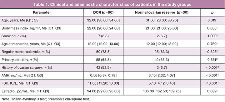
Analysis of clinical and anamnestic features demonstrated that patients in the study groups were comparable in age, body mass index, age at menarche, menstrual cycle characteristics, and smoking status (p>0.05), indicating sample homogeneity and minimizing the potential confounding effect of these factors on the study results. However, previous ovarian surgery was significantly more prevalent in patients with decreased ovarian reserve (DOR) compared with the control group (42/80 (52.5%) vs. 2/30 (6.7%); p<0.001), which aligns with current understanding regarding the impact of surgical treatment on ovarian reserve depletion.
Statistically significant differences were identified in parameters evaluating ovarian reserve. Compared with controls, patients with diminished reserve exhibited elevated FSH levels, reduced AMH levels, and decreased estradiol concentrations (p<0.05). These findings reflect typical hormonal changes associated with reduced ovarian reserve, thereby confirming the appropriateness of the study group allocation.
Overall, the analysis demonstrated group comparability regarding key clinical and baseline demographic characteristics alongside expected differences in hormonal markers of ovarian reserve, confirming the validity of the sample design.
Comparative analysis of endometrial amino acid composition
Univariate analysis results are presented in Table 2.
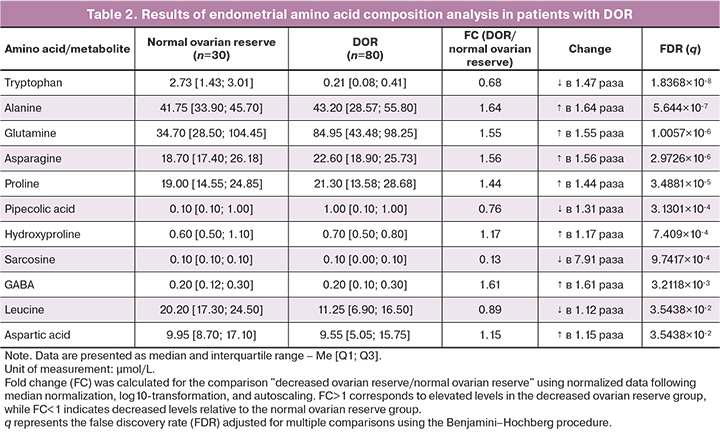
Following Benjamini–Hochberg adjustment for multiple comparisons, statistically significant differences were observed for 11 metabolites (q<0.05). The most prominent reduction in concentration was recorded for sarcosine (FC=0.13), which was 7.91-fold lower compared with the control group. Furthermore, decreased levels of tryptophan (FC=0.68), pipecolic acid (FC=0.76), and leucine (FC=0.89) were noted. Concurrently, increased concentrations were observed for alanine (F =1.64), γ-aminobutyric acid (GABA) (FC=1.61), asparagine (FC=1.56), glutamine (FC=1.55), proline (FC=1.44), hydroxyproline (FC=1.17), and aspartic acid (FC=1.15). Differences for all listed metabolites retained statistical significance after multiple comparison adjustment.
Visualization of differentially altered metabolites
To perform an integrative evaluation of the identified changes, a volcano plot (Figure 1) was constructed, combining the magnitude of concentration fold changes with levels of statistical significance.
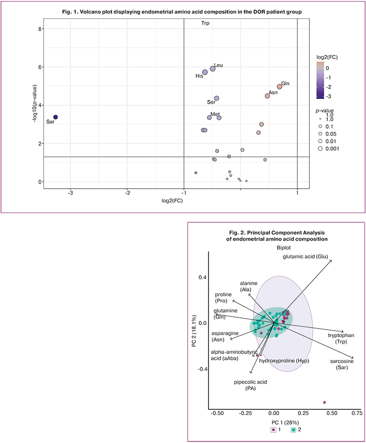
The most substantial contributions to group separation were made by sarcosine, tryptophan, alanine, GABA, asparagine, glutamine, and proline, which simultaneously exhibited high statistical significance and large effect sizes. The remaining differentially expressed amino acids also reached statistical significance, albeit with less pronounced concentration shifts.
Multivariate analysis
Principal component analysis (PCA) revealed partial segregation between the study groups based on the overall metabolite profile (Fig. 2).
Despite an area of overlap, the distribution of samples demonstrated distinct differences in the endometrial amino acid profile between patients with diminished and normal ovarian reserve. At the same time, the overlapping cluster region reflected biological heterogeneity within the study sample, emphasizing the necessity for supervised multivariate analytical methods.
Partial least squares-discriminant analysis (PLS-DA) achieved clearer separation between the study groups compared with PCA. Cross-validation identified a two-component model as optimal, yielding an explained variance (R2) of 0.54, a predictive ability parameter (Q2) of 0.41, and a classification accuracy of 0.89. For the single-component model, R2 and Q2 were 0.43 and 0.36, respectively. The resulting R2 and Q2 values indicated satisfactory explanatory and predictive performance of the model, whereas permutation testing revealed no evidence of random separation (empirical p=0.01; 0 out of 100 permutations), demonstrating that the model significantly differed from random permutation models (Fig. 3).
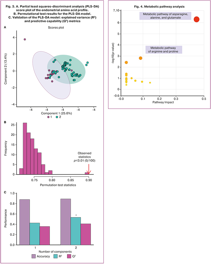
The derived PLS-DA performance metrics should not be construed as definitive clinical validation of the classification model. Given the exploratory nature of the study and the absence of an independent validation cohort, this multivariate analysis results are interpreted as exploratory and warrant further verification in independent cohorts.
Pathway analysis
To investigate the biological relevance of the identified differences, pathway analysis was performed using the MetaboAnalyst platform integrated with the KEGG and HMDB databases (Fig. 4).
Pathway analysis demonstrated that the most pronounced and statistically significant alterations involved alanine, aspartate, and glutamate metabolism (FDR q=1.41×10⁻⁵, Pathway Impact=0.41; identified metabolites: L-alanine, L-asparagine, L-aspartate, L-glutamine, and GABA) and arginine and proline metabolism (FDR q=2.05×10⁻², Pathway Impact=0.09; identified metabolites: L-proline and 4-hydroxyproline). The high Pathway Impact values, combined with statistical significance after multiple testing corrections, indicate substantial alterations in interconnected pathways of amino acid metabolism in endometrial tissue associated with DOR. These findings are consistent with univariate analysis results and support the involvement of energy metabolism, nitrogen balance, and protein synthesis in driving the altered endometrial metabolic profile.
In conclusion, the combination of univariate analysis, multivariate statistical methods, and metabolic pathway analysis demonstrated robust differences in the endometrial amino acid profile between patients with diminished and normal ovarian reserve in early and middle reproductive age. These findings indicate interconnected local metabolic alterations within endometrial tissue in DOR that affect multiple pathways of amino acid metabolism, supporting the utility of the endometrial amino acid profile as a source of potential biomarkers for assessing endometrial functional status [10, 14, 16].
Discussion
The present study offers the first comprehensive assessment of the amino acid profile of endometrial tissue in patients of early and middle reproductive age with diminished ovarian reserve (DOR) during the putative window of implantation. Multivariate analysis (PLS-DA) and metabolic pathway analysis revealed significant differences in the amino acid composition of the endometrium between patients with DOR and women with preserved ovarian function. Of the 23 amino acids and their derivatives examined, 11 compounds showed statistically significant changes after correction for multiple comparisons (q<0.05).
Current concepts of endometrial receptivity characterize it as a dynamically changing, metabolically active tissue whose functional readiness is determined not only by hormonal stimulation but also by local energy adaptation [17], epigenetic programming [18], immune regulation [19], and structural remodeling of the extracellular matrix [20]. Recent metabolomic studies have demonstrated that changes in the concentrations of low-molecular-weight compounds can reflect the functional state of the endometrium and potentially identify novel biomarkers of receptivity [21, 22].
One of the most significant findings of this study was the identification of disturbances in the alanine, aspartate, and glutamate metabolic pathway, which contributed most significantly to the differences between the groups. Patients with DOR exhibited elevated levels of alanine (FC=1.64; q<0.001), glutamine (FC=1.55; q<0.001), and asparagine (FC=1.56; q<0.001). The amino acids of this metabolic axis play a central role in maintaining cellular energy metabolism. Glutamine is an important source of carbon skeletons for the tricarboxylic acid cycle, participates in redox balance regulation, and supports biosynthetic processes under conditions of high cellular metabolic activity [23]. During decidualization, endometrial stromal cells undergo substantial functional reorganization, accompanied by changes in energy requirements and mitochondrial metabolism. Therefore, the observed accumulation of glutamine, alanine, and asparagine may reflect impaired metabolic adaptation of the endometrium in DOR, including altered processes of their transport, interconversion, or utilization.
The elevated asparagine level may also be considered in the context of the cellular response to altered availability of nutrient substrates. It is known that amino acid imbalance can activate adaptive mechanisms of the amino acid response (AAR), primarily regulated through the ATF4 pathway, which helps maintain cellular homeostasis under metabolic stress [24].
Of particular note are the changes in proline and hydroxyproline content. The study revealed elevated concentrations of proline (FC=1.44; q<0.001) and hydroxyproline (FC=1.17; q<0.001), which are the principal structural components of collagen and important markers of extracellular matrix metabolism. The formation of a receptive endometrium requires strictly regulated remodeling of the extracellular matrix, which ensures changes in stromal structure, cell migration, and subsequent embryo implantation. Disruption of the balance between synthesis and degradation of matrix components may lead to altered properties of the tissue microenvironment and potentially affect decidualization processes [17, 20]. The observed increase in proline and hydroxyproline content may indicate altered dynamics of collagen turnover in DOR and suggests a possible association with extracellular matrix remodeling processes. Confirmation of this hypothesis requires further studies assessing matrix metalloproteinase expression and structural characteristics of the endometrium.
The most pronounced difference between the groups was identified for sarcosine: its concentration in endometrial tissue of patients with DOR was reduced by nearly 8-fold (FC=0.13; q<0.001). Sarcosine is an intermediate metabolite of one-carbon metabolism and is associated with the regulation of the S-adenosylmethionine pool – the principal methyl group donor in DNA and protein methylation processes [25].
One-carbon metabolism plays an important role in cellular differentiation and epigenetic regulation. The formation of a receptive endometrium is accompanied by changes in the expression of a large number of genes regulating adhesion, immune response, and decidual transformation [26]. Therefore, reduced sarcosine levels may indicate altered metabolic support of epigenetic plasticity in endometrial tissue and suggest a possible change in one-carbon metabolism.
In the present study, reduced levels of the essential amino acids leucine (FC=0.89; q=0.035) and tryptophan (FC=0.68; q<0.001), as well as the lysine metabolite pipecolic acid (FC=0.76; q<0.05), were identified. These compounds participate in the regulation of cellular metabolism and immune homeostasis of the endometrium. Leucine is a key nutritional signal regulating the activity of the mTORC1 complex through the amino acid sensing system [27]. Altered leucine availability may influence the intensity of anabolic processes and cellular adaptation, which is potentially relevant to the functional maturation of the endometrium.
Tryptophan participates in the regulation of the immune microenvironment through the kynurenine pathway of its catabolism, including the activity of indoleamine 2,3-dioxygenase 1 (IDO1) [28]. This metabolic mechanism is considered one of the factors contributing to the establishment of immunological tolerance at the maternal–embryonic interface. However, a reduced tryptophan concentration in endometrial tissue of patients with DOR cannot be unequivocally regarded as a sign of effective immune adaptation. The obtained data may reflect either increased tryptophan consumption upon activation of the kynurenine pathway or altered regulation of this metabolic node, resulting in a reduced available pool of the amino acid.
Pipecolic acid is a physiological modulator of GABAergic transmission and neuroimmune signaling pathways [29]. Since the local GABAergic system of the endometrium participates in the regulation of decidual transformation, trophism, and immune tolerance [30], the reduced pipecolic acid level, in combination with decreased leucine content, may indicate altered metabolic signals potentially associated with the functional state of the endometrium.
Thus, the obtained results indicate that DOR is associated not only with impaired ovarian reproductive potential but also with the formation of a specific metabolic phenotype of endometrial tissue. The identified changes in the amino acid profile involve key pathways related to cellular energy supply, extracellular matrix remodeling, one-carbon metabolism, and immunometabolic regulation.
It should be noted that the identified changes in metabolite concentrations do not permit direct determination of the activity of the corresponding signaling and functional pathways. Therefore, the considered relationships between individual metabolites and the processes of one-carbon metabolism, mTORC1-dependent regulation, the kynurenine pathway, and extracellular matrix remodeling should be regarded as putative mechanisms requiring further experimental confirmation.
Given our previously obtained data on the low implantation rate following transfer of good-quality embryos in patients with DOR [7], from a clinical perspective, the results of the present study allow the endometrium of patients with DOR to be considered as a potentially functionally altered tissue whose state may influence the effectiveness of implantation processes in ART programs. The obtained data create a basis for the development of metabolomic markers for assessing endometrial readiness for implantation and for further identification of personalized approaches to endometrial preparation in patients with DOR.
At the same time, the identified associations require further confirmation in studies with larger sample sizes, the inclusion of functional methods for assessing endometrial receptivity, and analysis of the molecular mechanisms regulating the identified metabolic changes.
Limitations of the study
Interpretation of the obtained results should consider several methodological and clinical limitations of the present work.
- Study design and causal relationships: within the framework of this observational, cross-sectional comparative study, the assessment of the metabolomic profile of the endometrium was performed once (in a cross-sectional format during the window of implantation). The single-time nature of tissue sampling allows pronounced metabolic associations to be identified and a specific endometrial phenotype in DOR to be described; however, it does not permit an unambiguous determination of the causal relationship between changes in the levels of specific metabolites and reduced receptivity.
- Sample size and power calculation: the limited size of the study groups and the absence of an a priori calculation of statistical power for individual subgroups with rare metabolic shifts require caution when extrapolating the data. Despite the application of stringent control for false-positive results (q<0.05), the identified patterns require independent validation in larger cohorts.
- Single-center design: the study was conducted within a single clinical center, which may limit the geographic and population representativeness of the obtained sample.
- Potential influence of confounders (surgical interventions): a substantial proportion of patients in the study group (52.5%) had a history of ovarian surgical interventions. Chronic surgical trauma, local inflammation, and changes in pelvic hemodynamics may act as additional confounders capable of exerting an independent effect on the metabolic profile of endometrial tissue.
- Absence of correlation with clinical ART outcomes: in this study, assessment of the endometrial metabolome was performed in isolation during the putative "window of implantation" without comparison with direct clinical outcomes of ART programs (implantation rate, achievement of ongoing pregnancy, live birth) in the current or subsequent embryo transfer cycles.
- Absence of reference intervals: to date, standardized reference ranges for tissue amino acid content in the endometrium are lacking in clinical practice, which complicates direct comparison of absolute values with general population indicators.
- Absence of independent validation: the study was exploratory in nature and aimed at the primary identification of features of the endometrial amino acid profile in diminished ovarian reserve. Accordingly, independent validation of the analytical method employed and separate assessment of its characteristics, including the limits of detection and quantification (LOD and LOQ), were not performed within the framework of the present study. Furthermore, the protocol did not provide for special procedures for batch-effect correction, randomization of sample analysis order, or operator blinding. The obtained results should be regarded as exploratory; their confirmation requires subsequent studies with independent validation of the analytical method and in independent samples.
Conclusion
The present study demonstrated that the endometrium of patients of early and middle reproductive age with DOR is characterized by an altered local metabolic phenotype. Patients with DOR showed statistically significant changes in the levels of amino acids and their metabolites involving several interconnected metabolic pathways. These findings indicate differences in the metabolic profile of the endometrium in DOR and suggest that it may represent a promising source of potential metabolomic markers of tissue functional status.
Future research should focus on confirming these associations in independent cohorts, correlating metabolic changes with functional characteristics of the endometrium, and assessing their association with clinical outcomes of ART programs (achievement of intrauterine pregnancy).
References
- Choi R., Park W., Chun G., Lee S.G., Lee E.H. Investigation of the prevalence of diminished ovarian reserve in Korean women of reproductive age. J. Clin. Med. 2023; 12(15): 5099. https://dx.doi.org/10.3390/jcm12155099
- Carson S.A., Kallen A.N. Diagnosis and management of infertility: a review. JAMA. 2021; 326(1): 65-76. https://dx.doi.org/10.1001/jama.2021.4788
- Liu F.X., Ming H.X., Huang K.L., Yi S.J., Liang X.F., Luo W.W. et al. Clinical pregnancy outcomes in young women with diminished ovarian reserve undergoing frozen embryo transfer: a comprehensive analysis with exploratory insights into endometrial aging. Front. Endocrinol. (Lausanne). 2025; 16: 1608200. https://dx.doi.org/10.3389/fendo.2025.1608200
- Zhu S., Jiang W., Liao X., Sun Y., Chen X., Zheng B. Effect of diminished ovarian reserve on the outcome of fresh embryo transfer in IVF/ICSI cycles among young women: a retrospective cohort study. BMC Womens Health. 2024; 24(1): 230. https://dx.doi.org/10.1186/s12905-024-03039-6
- Li H.J., Seifer D.B., Tal R. AMH independently predicts aneuploidy but not live birth per transfer in IVF PGT-A cycles. Reprod. Biol. Endocrinol. 2023; 21(1): 19. https://dx.doi.org/10.1186/s12958-023-01066-w
- Eubanks A., Flannagan K., Le K., Namath A., Rambhatla A., Reckhow J. et al. Beyond numbers: antimüllerian hormone and antral follicle count are not independent predictors of embryo euploidy in in vitro fertilization with preimplantation genetic testing for aneuploidy. Fertil. Steril. 2026; 125(2): 237-46. https://dx.doi.org/10.1016/j.fertnstert.2025.08.013
- Дубинская Е.Д., Гаспаров А.С., Мглинец А.П., Лукьянова Я.С.,Мацкевич Е.Н. Клинико-анамнестические предикторы неудач имплантации при низком овариальном резерве: ретроспективное исследование. Акушерство и гинекология. 2026; 5: 140-7. https://dx.doi.org/10.18565/aig.2026.58 [Dubinskaya E.D., Gasparov A.S., Mglinets A.P., Lukyanova Ya.S., Matskevich E.N. Clinical and anamnestic predictors of implantation failures in women with diminished ovarian reserve: retrospective study. Obstetrics and Gynecology. 2026; (5): 140-7 (in Russian). https://dx.doi.org/10.18565/aig.2026.58].
- Радзинский В.Е., Михалёва Л.М., Оразов М.Р., Силантьева Е.С., Камилова Д.П., Мидибер К.Ю., Орехов Р.Е. Рецептивность эндометрия пациенток с повторными неудачами имплантации. Доктор.Ру. 2022; 21(1): 27-33. https://dx.doi.org/10.31550/1727-2378-2022-21-1-27-33 [Radzinsky V.E., Mikhaleva L.M., OrazovM.R., Silantieva E.S., Kamilova D.P., Midiber K.Yu., Orekhov R.E. Endometrium receptivity in patients with repeated implant failures. Doctor.ru. 2022; 21(1): 27-33 (in Russian). https://dx.doi.org/10.31550/1727-2378-2022-21-1-27-33].
- Hiraoka T., Osuga Y., Hirota Y. Current perspectives on endometrial receptivity: a comprehensive overview of etiology and treatment. J. Obstet. Gynaecol. Res. 2023; 49(10): 2397-409. https://dx.doi.org/10.1111/jog.15759
- Molina N.M., Jurado-Fasoli L., Sola-Leyva A., Sevilla-Lorente R., Canha-Gouveia A., Ruiz-Durán S. et al. Endometrial whole metabolome profile at the receptive phase: influence of Mediterranean Diet and infertility. Front. Endocrinol. (Lausanne). 2023; 14: 1120988. https://dx.doi.org/10.3389/fendo.2023.1120988
- Gao J., Xiao Y. Metabolomics and its applications in assisted reproductive technology. IET Nanobiotechnol. 2023; 17(5): 399-405. https://dx.doi.org/10.1049/nbt2.12141
- Wang J., Zenere A., Wang X., Bergström G., Edfors F., Uhlén M. et al. Longitudinal analysis of genetic and environmental interplay in human metabolic profiles and the implication for metabolic health. Genome Med. 2025; 17: 68. https://dx.doi.org/10.1186/s13073-025-01492-y
- Ling Z.N., Jiang Y.F., Ru J.N., Lu J.H., Ding B., Wu J. Amino acid metabolism in health and disease. Signal Transduct. Target Ther. 2023; 8(1): 345. https://dx.doi.org/10.1038/s41392-023-01569-3
- Shu Q., Zhao Y., Wang A., Li W., Xu H., Dong W. et al. [Analysis of the difference in metabolites and gene expressions between pre-receptive and receptive endometria]. Zhonghua Yi Xue Yi Chuan Xue Za Zhi. 2023; 40(12): 1496-503. https://dx.doi.org/10.3760/cma.j.cn511374-20221108-00769
- Ferraretti A.P., La Marca A., Fauser B.C.J.M., Tarlatzis B., Nargund G., Gianaroli L. ESHRE consensus on the definition of “poor response” to ovarian stimulation for in vitro fertilization: the Bologna criteria. Hum. Reprod. 2011; 26(7): 1616-24. https://dx.doi.org/10.1093/humrep/der092
- Wang M.J., Lin M.H., Yang J.H., Lee R.K.-K., Lee K.S. Low antimüllerian hormone (<1.2 ng/ml) does not impact oocyte quality and IVF/ICSI outcomes in women ≤40 years old. Taiwan. J. Obstet. Gynecol. 2025; 64(2): 248-52. https://dx.doi.org/10.1016/j.tjog.2024.02.007
- Harden S.L., Zhou J., Gharanei S., Diniz-da-Costa M., Lucas E.S., Cui L. et al. Exometabolomic analysis of decidualizing human endometrial stromal and perivascular cells. Front. Cell. Dev. Biol. 2021; 9: 626619. https://dx.doi.org/10.3389/fcell.2021.626619
- Adamczyk M., Wender-Ozegowska E., Kedzia M. Epigenetic factors in eutopic endometrium in women with endometriosis and infertility. Int. J. Mol. Sci. 2022; 23(7): 3804. https://dx.doi.org/10.3390/ijms23073804
- Panagodimou E., Terzopoulou I., Triantafyllidou O., Markantes G., Georgopoulos N., Vlahos N. et al. The role of immunologic factors in endometrial receptivity: an embryo-endometrium dialogue. Int. J. Mol. Sci. 2026; 27(10): 4588. https://dx.doi.org/10.3390/ijms27104588
- Gebril M., Mulder S., Das R., Nallasamy S. Extracellular matrix reorganization during endometrial decidualization. Histochem. Cell. Biol. 2025; 163(1): 84. https://dx.doi.org/10.1007/s00418-025-02411-3
- Jia Z., Wei Y., Zhang Y., Song K., Yuan J. Metabolic reprogramming and heterogeneity during the decidualization process of endometrial stromal cells. Cell. Commun. Signal. 2024; 22(1): 385. https://dx.doi.org/10.1186/s12964-024-01763-y
- Liu X., Xu S-j. Research progress in metabolomics for assessing endometrial receptivity in women with repeated implantation failure. Journal of International Obstetrics and Gynecology. 2026; 53(2): 165-9. https://dx.doi.org/10.12280/gjfckx.20251189
- Tang L., Xu X.H., Xu S., Liu Z., He Q., Li W. et al. Dysregulated Gln-Glu-α-ketoglutarate axis impairs maternal decidualization and increases the risk of recurrent spontaneous miscarriage. Cell. Rep. Med. 2023; 4(5): 101026. https://dx.doi.org/10.1016/j.xcrm.2023.101026
- Neill G., Masson G.R. A stay of execution: ATF4 regulation and potential outcomes for the integrated stress response. Front. Mol. Neurosci. 2023; 16: 1112253. https://dx.doi.org/10.3389/fnmol.2023.1112253
- Ducker G.S., Rabinowitz J.D. One-carbon metabolism in health and disease. Cell. Metab. 2017; 25(1): 27-42. https://dx.doi.org/10.1016/j.cmet.2016.08.009
- Kong S., Zhou C., Bao H., Ni Z., Liu M., He B. et al. Epigenetic control of embryo-uterine crosstalk at peri-implantation. Cell. Mol. Life Sci. 2019; 76(24): 4813-28. https://dx.doi.org/10.1007/s00018-019-03245-8
- Yue S., Li G., He S., Li T. The Central role of mTORC1 in amino acid sensing. Cancer Res. 2022; 82(17): 2964-74. https://dx.doi.org/10.1158/0008-5472.CAN-21-4403
- Seo S.K., Kwon B. Immune regulation through tryptophan metabolism. Exp. Mol. Med. 2023; 55(7): 1371-9. https://dx.doi.org/10.1038/s12276-023-01028-7
- Kovaříková V., Špirková A., Šefčíková Z., Pisko J., Kalatová L., Koppel J. et al. Gamma-aminobutyric acid (GABA) can affect physiological processes in preimplantation embryos via GABAA and GABAB receptors. Reprod. Med. Biol. 2023; 22(1): e12528. https://dx.doi.org/10.1002/rmb2.12528
- Chen W., Zhang Q., Wang H., Tan D., Tan Y. Unique and independent role of the GABAB1 subunit in embryo implantation and uterine decidualization in mice. Genes. Dis. 2019; 8(1): 79-86. https://dx.doi.org/10.1016/j.gendis.2019.06.005
Received 01.09.2026
Accepted 14.09.2026
About the Authors
Ekaterina D. Dubinskaya, Dr. Med. Sci., Professor at the Department of Obstetrics, Gynecology with a Course in Perinatology, Medical Institute, Patrice Lumumba Peoples’ Friendship University of Russia, 8 Miklukho-Maklaya str., Moscow, 117198, Russia, +7(903)117-55-58, eka-dubinskaya@yandex.ru, https://orcid.org/0000-0002-8311-0381Anna P. Mglinets, PhD Student, Department of Obstetrics, Gynecology with a Course in Perinatology, Medical Institute, Patrice Lumumba Peoples’ Friendship University
of Russia, 8 Miklukho-Maklaya str., Moscow, 117198, Russia, +7(906)056-49-98, mg.anna@mail.ru, https://orcid.org/0009-0001-3985-3954
Alexander S. Gasparov, Dr. Med. Sci., Professor, Professor at the Department of Obstetrics, Gynecology with a Course in Perinatology, Medical Institute, Patrice Lumumba Peoples’ Friendship University of Russia, 8 Miklukho-Maklaya str., Moscow, 117198, Russia, +7(985)776-77-78, 13513520@mail.ru, https://orcid.org/0000-0001-6301-1880
Elizaveta V. Alyoshkina, Teaching Assistant at the Department of Obstetrics, Gynecology with a Course in Perinatology, Medical Institute, Patrice Lumumba Peoples’ Friendship University of Russia, 8 Miklukho-Maklaya str., Moscow, 117198, Russia, +7(926)768-44-27, alyoshkina.ev@yandex.ru, https://orcid.org/0000-0001-5339-1285
Similar Articles