
Organ-sparing surgery with vacuum-assisted laparostomy for advanced obstetric peritonitis: a series of 58 consecutive cases
Churina D.E., Makarenko T.A., Cherdantsev D.V.
Background. Obstetric peritonitis is a severe purulent-septic complication following cesarean section, with an incidence ranging from 0.1% to 1.5%. Mortality from postpartum sepsis accounts for 37–40% of maternal deaths. The primary challenge in surgical management lies in balancing the need for radical source control with the desire to preserve the uterus in young women of reproductive age.
Objective. To improve surgical outcomes in patients with advanced obstetric peritonitis by presenting clinical experience with a staged surgical approach using vacuum-assisted laparostomy in a consecutive series of patients after abdominal delivery.
Materials and methods. We conducted a retrospective analysis of a consecutive case series of 58 patients with obstetric peritonitis, which developed after cesarean section, treated at the Krasnoyarsk Regional Clinical Hospital between 2018 and 2025. The study was conducted in accordance with the PROCESS 2023 guideline. All procedures utilized vacuum-assisted laparostomy, performed according to an original technique (Russian Federation Patent No. 2816058). Statistical analyses were descriptive. Continuous variables are summarized as medians with first and third quartiles, and categorical variables as counts and percentages.
Results. The uterus was preserved in 49 (84.5%) of the 58 patients. Vacuum-assisted laparostomy was employed in 56 women (96.6%), and hysterectomy was avoided in 49 of these patients, representing 87.5% of those who underwent laparostomy. No deaths occurred in the case series (0%). Two patients became pregnant again after discharge and subsequently had term deliveries. No intraoperative injuries to adjacent organs were observed. Complications, characterized by failure of vacuum-assisted laparostomy (including progression of sepsis, absence of favorable laboratory trends, and total myometrial necrosis), occurred in 7 patients and necessitated hysterectomy.
Conclusion. In this case series, a staged surgical approach using vacuum-assisted laparostomy in patients with obstetric peritonitis was associated with a high rate of uterine preservation (84.5%) and zero mortality. These findings suggest the potential for preserving reproductive function in some patients experiencing this complication.
Authors' contributions. Churina D.E. – conception of the study, clinical data collection and analysis, statistical analysis, drafting of the manuscript; Makarenko T.A. – study design, editing of the manuscript, clinical supervision; Cherdantsev D.V. – development of the vacuum-assisted laparostomy method, performance of surgical interventions, drafting of the manuscript.
Conflicts of interest. The authors have no conflicts of interest to declare.
Funding. The study was conducted within the framework of the state R&D assignment (EGISU No. 122040600142-6) titled "New standards for the treatment of purulent-inflammatory postpartum uterine diseases using microbiological testing for relevant pathogens" (2022–2024).
Ethical Approval. The study was reviewed and approved by the Research Ethics Committee of the Prof. V.F. Voino-Yasenetsky KrasSMU (protocol No. 15/04-2025 dated 04/15/2025).
Generative Artificial Intelligence. No artificial intelligence tools were used in the preparation of this manuscript.
Patient Consent for Publication. All patients provided informed consent for the publication of their data.
Authors' Data Sharing Statement. The data supporting the findings of this study are available upon request from the corresponding author after approval from the principal investigator.
For citation: Churina D.E., Makarenko T.A., Cherdantsev D.V. Organ-sparing surgery with vacuum-assisted laparostomy for advanced obstetric peritonitis: a series of 58 consecutive cases.
Akusherstvo i Ginekologiya/Obstetrics and Gynecology. 2026; (10): 68-80 (in Russian)
https://dx.doi.org/10.18565/aig.2026.127
Keywords
obstetric peritonitis
case series
PROCESS
vacuum-assisted laparostomy
organ-preserving treatment
hysterectomy
cesarean section
postpartum purulent-septic complications
Obstetric peritonitis remains a leading cause of maternal morbidity and mortality, representing one of the most complex and life-threatening conditions in contemporary obstetrics [1–7]. Its incidence following cesarean delivery ranges from 0.1 to 1.5%, with over 98% of cases occurring after operative delivery [4–8]. Despite substantial advances in diagnosis, antimicrobial therapy, and critical care, mortality associated with this condition remains high, accounting for 37–40% of maternal deaths attributable to postpartum sepsis, according to various authors. In the presence of multiple organ failure, mortality increases to 76–100% [9–13].
The key determinants of outcomes in obstetric peritonitis are timely diagnosis and accurate assessment of disease severity [14–16]. Peritonitis is not a static condition; it typically progresses from a localized inflammatory response to a systemic inflammatory response, endogenous intoxication, and ultimately multiple organ dysfunction and septic shock [17–19]. Each stage requires a different extent of surgical intervention and intensity of conservative treatment. Accordingly, accurately determining the stage of peritonitis is not merely an academic exercise but a practical tool for selecting the optimal treatment strategy, ranging from organ-preserving surgery during the reactive phase to radical hysterectomy in the setting of progressive sepsis [19–21]. The lack of standardized, unified approaches to staging obstetric peritonitis complicates comparisons of treatment outcomes across clinical centers and the justification of indications for specific surgical interventions.
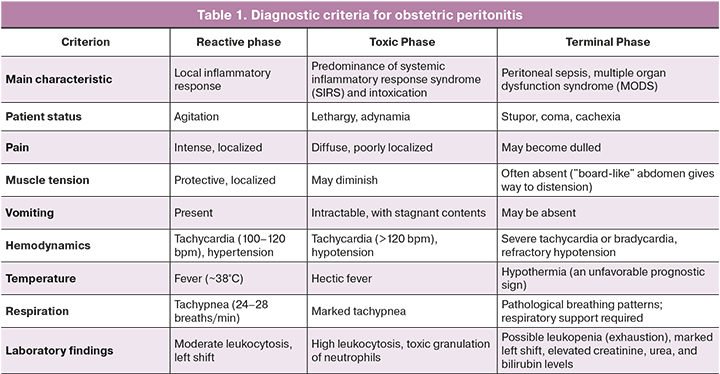
One of the most widely recognized and clinically established classifications of peritonitis, which remains relevant in contemporary surgery, was proposed by K. S. Simonyan in 1971 [22–27]. This classification distinguishes three successive phases of peritonitis: reactive (up to 24 hours after disease onset), toxic (24–72 hours), and terminal (more than 72 hours) (Table 1). This classification has been widely adopted in obstetric practice because it enables clinicians not only to assess the patient's condition but also to predict the risk of uterine suture dehiscence, spread of the purulent-inflammatory process to the parametrial tissues, and development of multiple organ dysfunction [28–31].
Several unresolved issues remain in the surgical management of obstetric peritonitis, one of the most controversial being the determination of the optimal extent of surgery [11–14]. The goal of preserving the uterus in young women of reproductive age often conflicts with the need for radical removal of the primary infectious focus in cases of complete uterine suture dehiscence, purulent destruction of the myometrial tissue, and widespread purulent-inflammatory involvement of the abdominal cavity [12–15]. Conversely, the conventional radical approach involving hysterectomy deprives women of the opportunity to have children in the future and is often associated with severe psychological consequences [16–19].
In contemporary surgery for generalized peritonitis, damage control surgery and the open abdomen technique are increasingly used [17–21]. These approaches involve delayed abdominal closure following initial source control, allowing repeated surgical revisions and removal of infected fluid, reduction of intra-abdominal pressure, and preparation of the tissues for definitive closure. One of the most effective methods of implementing this staged approach is vacuum-assisted laparostomy, which enables continuous active evacuation of wound exudate, facilitates the local application of antiseptics, and attenuates the systemic inflammatory response [22–24].
In gynecological practice, negative pressure wound therapy (NPWT) has traditionally been used to treat surgical site infections. In recent years, however, reports have emerged describing its successful application in generalized peritonitis of various etiologies. Studies specifically investigating this technology in obstetric peritonitis remain scarce and are insufficient to establish standardized management approaches for this patient population. This gap provided the rationale for summarizing our extensive experience in the treatment of these challenging patients [25–28].
This study aimed to improve the outcomes of surgical treatment in patients with generalized obstetric peritonitis by describing the clinical experience of using a staged surgical approach with vacuum-assisted laparostomy in a consecutive series of patients following cesarean delivery.
Materials and methods
Study design. This retrospective consecutive case series included 58 female patients with advanced obstetric peritonitis treated between January 1, 2018, and December 31, 2025. The study was conducted in accordance with the PROCESS 2023 (Preferred Reporting Of CasE Series in Surgery) guidelines [32], ensuring a standardized approach to describing and analyzing clinical observations. This single-center clinical study was based on a cohort of patients with a verified diagnosis of obstetric peritonitis. Participants were selected consecutively upon admission to the clinic, without preliminary randomization or selection.
Ethical approval and informed consent. The study protocol was approved by the Local Ethics Committee of the Professor V.F. Voino-Yasenetsky Krasnoyarsk State Medical University of the Ministry of Health of the Russian Federation (Protocol No. 15/04-2025 dated April 15, 2025). Written informed consent for the use of medical data for scientific purposes, including anonymous publication in peer-reviewed journals, was obtained from all patients included in the study. Consent was documented in writing and stored in medical records. All data were de-identified prior to statistical analysis.
Setting. The study was conducted at the gynecological department and purulent-septic center of the Regional Clinical Hospital in Krasnoyarsk. This institution is a large tertiary multi-profile hospital providing specialized, high-tech medical care to patients with purulent-septic complications in obstetrics and gynecology. These departments serve as clinical bases for the Department of Operative Gynecology of the Institute of Postgraduate Education and the Department of Hospital Surgery named after Prof. A.M. Dykhno, which includes a postgraduate education course at Krasnoyarsk State Medical University. The hospital is the primary regional center for managing patients with severe purulent-septic postpartum complications, leading to a concentration of highly complex clinical cases and the accumulation of significant clinical experience.
Study period. Patients treated in these departments between January 1, 2018, and December 31, 2025, were included. This time interval was selected for two main reasons. First, 2018 marked the inception of the systematic application of vacuum-assisted laparostomy in the department's clinical practice, using a newly developed technique. Second, including observations through the end of 2025 allowed for the evaluation of both immediate and long-term treatment outcomes, including cases of subsequent pregnancies. The total follow-up period was 8 years.
Inclusion and exclusion criteria. Patients were included if they met all of the following criteria: female sex, age older than 18 years (the lower age limit was determined by the onset of the reproductive period; no upper limit was established), abdominal delivery via cesarean section at a gestational age of 22 weeks or more, a verified diagnosis of obstetric peritonitis established based on a combination of clinical, laboratory, and intraoperative criteria, and provision of informed consent for the use of medical data for research purposes.
Patients were excluded if they met any of the following criteria: peritonitis of non-obstetric origin, including appendicular peritonitis, intestinal peritonitis (ulcer perforation, diverticulitis, intestinal obstruction), postoperative peritonitis following gynecological surgeries unrelated to pregnancy and childbirth, pancreatogenic peritonitis, or traumatic peritonitis. Additionally, patients who refused staged surgical treatment using vacuum-assisted laparostomy (or for whom its application was not technically feasible due to objective reasons), lacked informed consent for the use of medical data, or had incomplete medical documentation precluding a retrospective assessment of key study parameters were excluded.
Diagnostic criteria for obstetric peritonitis. The diagnosis of obstetric peritonitis was established based on a comprehensive assessment of clinical, laboratory, and instrumental data, identifying three phases of the pathological process (reactive, toxic, and terminal) in accordance with the classification of K.S. Simonyan (1971) [29–31, 33] (Table 1). This classification ensured a standardized approach to selecting patients for organ-sparing surgeries utilizing vacuum-assisted laparostomy.
Surgical technique
Vacuum-assisted laparostomy. Vacuum-assisted laparostomy was performed according to an original technique protected by Russian Federation Patent No. 2816058. The procedure comprised the following sequential steps:
- The "Reperen" polypropylene endoprosthesis was fixed to the aponeurosis.
- A polyurethane foam sponge was positioned within the wound to isolate the uterine suture zone.
- A catheter was inserted for fractional instillation of 300–500 mL of a 1% dioxidine solution at 6-hour intervals.
- A vacuum system with a negative pressure of 100–120 mmHg was connected, ensuring continuous evacuation of wound discharge and the antiseptic solution (except for 30-minute pauses following each instillation).
- Relaparotomy was performed after 48 hours, which was either intermediate (if continued sanitation was required) or definitive (upon achieving peritoneal cavity sanitation).
Organ-sparing surgery. Organ-sparing interventions adhered strictly to the following principles:
- Necroectomy was performed by excising non-viable tissues within healthy myometrium. The criterion for tissue viability was the appearance of capillary bleeding from the wound edges following excision. This procedure was feasible when the necrotic area did not exceed 70% of the myometrial volume.
- Metroplasty was performed by placing secondary sutures on the uterus.
- Uterine cavity drainage was established through the cervical canal to prevent hematometra and pyometra.
- Drainage of the abdominal cavity and lesser pelvis was performed by placing silicone drainage tubes.
- The final step involved the application of vacuum-assisted laparostomy according to the technique described above.
Radical surgery. Hysterectomy with salpingectomy was performed when any of the following criteria were present: diffuse purulent or fibrinous-purulent peritonitis, massive necrosis of the uterine walls (necrotic changes involving more than 70% of the myometrium), sepsis or septic shock, or ineffectiveness of vacuum-assisted laparostomy after 2 to 3 stages.
Laboratory methods
Venous blood samples for laboratory analysis were collected in the morning on an empty stomach (after 8–12 hours of fasting) from the cubital vein into VACUETTE vacuum tubes (Greiner Bio-One, Austria) containing a clot activator for serum acquisition and K3-EDTA for whole blood collection.
Complete blood counts were performed using a Sysmex XN-1000 hematology analyzer (Sysmex Corporation, Japan) with the manufacturer's standard reagents: Cellpack (diluent), Stromatolyser-4DS (erythrocyte lysing reagent), Fluorocell (leukocyte fluorescent dye), and Sulfolyser (hemoglobin lysing reagent). The following parameters were determined: white blood cell count (×10⁹/L), red blood cell count (×10¹²/L), hemoglobin (g/L), hematocrit (%), platelet count (×10⁹/L), and differential leukocyte count with automated 5-part cell population differentiation.
Biochemical assays (total protein, albumin, creatinine, urea, bilirubin and its fractions, alanine aminotransferase, aspartate aminotransferase, glucose) were performed on a Cobas c 311 automated biochemistry analyzer (Roche Diagnostics, Switzerland). Roche Diagnostics reagents were used: CREP2 (enzymatic creatinine), UREAL (urea by UV method), BILT3 (total bilirubin), ALB2 (albumin by bromcresol green), ALTL (ALT with pyridoxal phosphate), ASTL (AST with pyridoxal phosphate), and GLUC3 (glucose by hexokinase method).
C-reactive protein (CRP) was determined by immunoturbidimetry on a Cobas c 311 analyzer (Roche Diagnostics, Switzerland) using the CRPHS reagent (Roche Diagnostics) with a measurement range of 0.5–350 mg/L. Reference values for adults are <5 mg/L; an increase up to 10–20 mg/L is considered permissible for pregnant women in the second and third trimesters.
Procalcitonin (PCT) was measured by chemiluminescent immunoassay on a VIDAS analyzer (bioMérieux, France) using VIDAS PCT test kits (bioMérieux, France). The measurement range was 0.05–200 ng/mL The reference value for healthy individuals is <0.1 ng/mL Results were interpreted according to the manufacturer's recommendations: <0.1 ng/mL indicates a low probability of bacterial infection; 0.1–0.25 ng/mL indicates localized infection; 0.25–2.0 ng/mL indicates systemic infection (sepsis); and >2.0 ng/mL indicates severe sepsis or septic shock.
Coagulometry was performed on an STA-R Evolution automated coagulometer (Diagnostica Stago, France) using the following reagents: STA-PT (prothrombin time), STA-APTT (activated partial thromboplastin time), STA-Fibrinogen (Clauss fibrinogen), and STA-DDimer (D-dimer by immunoturbidimetry). Reference values were: fibrinogen 2.0–4.5 g/L (physiologically elevated up to 6.0 g/L during pregnancy) and D-dimer <0.5 mg/L (normal values in pregnant women may reach 1.0–1.5 mg/L depending on gestational age).
Blood gas composition and lactate levels were determined using an ABL800 FLEX analyzer (Radiometer Medical ApS, Denmark) with SafePICO disposable cartridges (Radiometer). Lactate was measured by an enzymatic amperometric method. The reference value for lactate is <2.0 mmol/L, and levels >2.0 mmol/L were considered a marker of tissue hypoperfusion and a criterion for septic shock.
Microbiological examination
Abdominal exudate sampling for microbiological analysis was performed intraoperatively immediately after opening the peritoneal cavity and prior to antiseptic administration. Material was collected using a sterile 10 mL syringe (BD Plastipak, Becton Dickinson, Ireland) or a sterile swab applicator with Amies transport medium (Copan Diagnostics, Italy) from the depth of the purulent focus and from various abdominal compartments (right and left subdiaphragmatic spaces, interloop spaces, and the lesser pelvis). Specimens were delivered to the laboratory within 2 hours at room temperature.
Inoculation onto nutrient media was performed in a class II laminar flow cabinet (Lamsystems, Russia) using standard nutrient media manufactured by FSUE NPO Microgen (Russia) and bioMérieux (France):
- Blood agar (5% sheep blood) for the isolation of aerobic and facultatively anaerobic microorganisms (incubation for 24–48 hours at 37 degrees Celsius with 5% carbon dioxide)
- Supplemented chocolate agar (bioMérieux, France) for the isolation of capnophiles and Haemophilus species (incubation for 24–48 hours at 37 degrees Celsius with 5–10% carbon dioxide)
- Sabouraud agar (FSUE NPO Microgen, Russia) for the isolation of Candida species (incubation up to 7 days at 30 degrees Celsius)
- Schaedler agar with 5% sheep blood (bioMérieux, France) for the isolation of anaerobes (incubation in an anaerobic jar with AnaeroGen gas-generating packets, Oxoid, United Kingdom, for up to 5 days at 37 degrees Celsius).
Pathogen identification was performed using two methods. Initial (genus-level) identification was based on morphological and staining properties (Gram staining using reagents: crystal violet, Lugol's solution, acetone-alcohol, and basic fuchsin, manufactured by Eco-Service, Russia). Final species-level identification was performed via matrix-assisted laser desorption/ionization time-of-flight mass spectrometry on a Vitek MS mass spectrometer (bioMérieux, France) using MALDI-TOF chips (bioMérieux) and matrix alpha-cyano-4-hydroxycinnamic acid (HCCA, bioMérieux). Identification results were evaluated with a confidence level >99.9% against the SARAMIS reference database (version 4.12, bioMérieux).
Antimicrobial susceptibility testing was performed by the disk diffusion method on Mueller-Hinton agar (FSUE NPO Microgen, Russia) using standard antibiotic disks (bioMérieux, France). Results were interpreted in accordance with the clinical guidelines "Determination of Antimicrobial Susceptibility of Microorganisms, Version 2025-01" and the standards of the European Committee on Antimicrobial Susceptibility Testing (EUCAST). The following antibiotics were tested: penicillins (amoxicillin/clavulanate), cephalosporins (ceftriaxone, cefotaxime, ceftazidime), carbapenems (imipenem, meropenem), aminoglycosides (gentamicin, amikacin), fluoroquinolones (ciprofloxacin, levofloxacin), glycopeptides (vancomycin), and oxazolidinones (linezolid). When methicillin-resistant staphylococci (methicillin-resistant Staphylococcus aureus, methicillin-resistant Staphylococcus epidermidis) were detected, susceptibility to fusidic acid, rifampicin, daptomycin, and tigecycline was additionally determined. When vancomycin-resistant enterococci were detected, linezolid was additionally tested.
Extended-spectrum beta-lactamase production in gram-negative bacteria (Escherichia coli, Klebsiella pneumoniae, Proteus species) was determined using the double-disk synergy test with ceftazidime, ceftazidime plus clavulanic acid, cefotaxime, and cefotaxime plus clavulanic acid disks (bioMérieux, France). An increase in the growth inhibition zone of >5 mm in the presence of clavulanic acid was regarded as a positive result.
Statistical analysis
Statistical data processing was performed using SPSS Statistics 26.0 software (IBM Corporation, Armonk, NY, USA). Due to the descriptive nature of the study, all applied methods were descriptive. Continuous variables were described using the median and quartiles (Me [Q1; Q3]). Categorical variables were described as count (n) and percentages (%). In view of the descriptive design and the absence of a control group, statistical hypothesis testing was not performed.
Results
Clinical and anamnestic characteristics of patients
Between 2018 and 2025, 58 patients with obstetric peritonitis following abdominal delivery were treated at the Department of Gynecology and the Purulent-Septic Center of the Regional Clinical Hospital (Krasnoyarsk). The main clinical and anamnestic data are presented in Table 2.
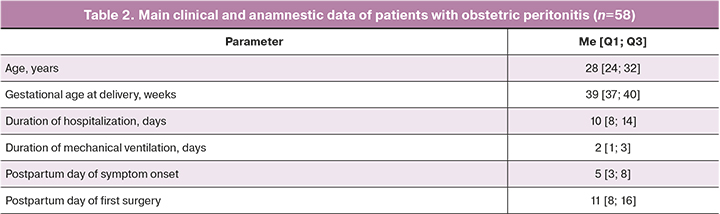
The vast majority of women (37/58, 63.8%) underwent emergency cesarean delivery. Premature rupture of membranes (19/58, 32.8%), labor dystocia (9/58, 15.5%), and a history of uterine scar from a previous cesarean section (8/58, 13.8%) were the leading indications for abdominal delivery.
Purulent-septic complications predominated in the study group. Localized fibrinopurulent peritonitis was diagnosed in 49/58 patients (84.5%), diffuse purulent peritonitis in 5/58 (8.6%), pelvic abscess in 2/58 (3.4%), and abdominal sepsis in 2/58 patients (3.4%).
Causes of obstetric peritonitis
Etiological analysis of obstetric peritonitis in this observational series revealed four main pathogenetic mechanisms that most frequently triggered the spread of purulent inflammation beyond the uterus. Notably, a combination of two or more causes occurred in the vast majority of patients (94.8%), reflecting the polyetiological and multifactorial nature of obstetric peritonitis development.
- Uterine dehiscence
The most frequent cause of obstetric peritonitis in our study was post-cesarean uterine suture breakdown/sequestration, recorded in 51 patients (87.9%). This complication typically developed against the background of preexisting postpartum endometritis, where inflammatory tissue infiltration compromised normal myometrial regeneration. In 24/51 cases (47.1%), the process was complicated by complete tissue sequestration: necrotic areas of the myometrium and ligatures sloughed off, forming a uterine wall defect through which infected contents (pus, ichorous exudate) entered the peritoneal cavity. Clinically, uterine dehiscence presented on postoperative days 4–7, manifesting with increasing lower abdominal pain, hectic fever, peritoneal signs, and, in some cases, purulent wound drainage. Ultrasound verification visualized a defect in the uterine scar area, free fluid in the peritoneal cavity, and soft tissue gas inclusions (a pathognomonic sign of anaerobic infection).
- Purulent-necrotizing endometritis extending to the myometrium
The second most common cause of obstetric peritonitis was purulent-necrotizing endometritis with myometrial involvement (endomyometritis), diagnosed in 46 patients (79.3%). Unlike isolated postpartum endometritis, in which the inflammatory process is limited to the decidua and superficial endometrial layers, endomyometritis was characterized by the extension of purulent inflammation into the uterine muscular layer, leading to purulent liquefaction necrosis and necrotic zone formation. In 18/46 patients (39.1%), myometrial necrosis was subtotal, involving over 70% of the wall thickness; this precluded uterine-sparing surgery and necessitated hysterectomy. Microbiologically, aerobic and anaerobic polymicrobial associations (Bacteroides fragilis, Peptostreptococcus spp., Escherichia coli) predominated in this group, explaining the aggressive clinical course and high propensity for tissue necrosis.
- Parametritis
Parametritis was identified in 12 patients (20.7%) and was combined with uterine dehiscence or endomyometritis in most cases (10/12, 83.3%). Parametritis developed via lymphatic and venous spread of infection from the uterine cavity or surgical wound into the parametrial cellular tissue. Clinically, this presented as unilateral lower abdominal pain radiating to the lower back and sacrum, effacement of the lateral vaginal fornices on bimanual examination, and the formation of a dense, tender mass. In 5/12 patients (41.7%), the process was complicated by parametrial abscess formation, requiring colpotomy or laparotomy drainage of the purulent focus. Isolated parametritis without involvement of other pelvic structures was rare (2/12, 16.7%); parametritis was typically a component of widespread pelvic inflammatory disease, reinforcing the need for comprehensive assessment of all potential infectious dissemination pathways.
- Pelvioperitonitis as a stage of infection propagation
Pelvic peritoneal inflammation (pelvioperitonitis) was recorded in 49 patients (84.5%). In the vast majority of cases, it was not an independent disease entity, but rather a progression stage of infection from the primary focus (uterus, parametrium, fallopian tubes) to the pelvic peritoneum. In 44/49 patients (89.8%), pelvioperitonitis co-occurred with uterine suture sequestration, confirming a direct route of peritoneal infection through the uterine wall defect. In the remaining 5/49 patients (10.2%), pelvioperitonitis developed via lymphogenous spread while uterine suture integrity was preserved. Clinically, pelvioperitonitis manifested with lower abdominal peritoneal signs (Blumberg sign, abdominal wall rigidity), paralytic ileus (abdominal distension, constipation, obstipation), and pelvic free fluid on ultrasound examination. In 5/49 patients (10.2%), inflammation was not confined to the pelvis and progressed to diffuse peritonitis involving all abdominal compartments, necessitating extensive surgical interventions and vacuum-assisted laparostomy placement.
Combination of causes and clinical implications
Crucially, a combination of two or more of these causes occurred in 55/58 patients (94.8%). The combination of uterine dehiscence, purulent-necrotizing endomyometritis, and pelvioperitonitis was the most typical pattern, identified in 44/55 patients (75.9%). This indicates that obstetric peritonitis rarely occurs in isolation; it is usually the result of generalization of an established purulent inflammatory process within the pelvis. This finding justifies the necessity for comprehensive intraoperative inspection not only of the uterus, but also of the parametrium, fallopian tubes, ovaries, and pelvic peritoneum to achieve complete debridement and lavage of all potential infectious foci.
Laboratory data
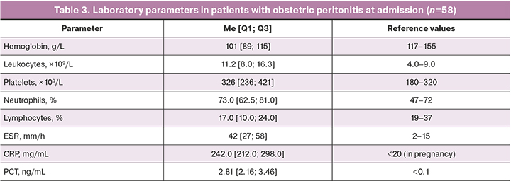
Laboratory parameters reflecting the activity of the systemic inflammatory response and the severity of endogenous intoxication in patients with obstetric peritonitis at admission are presented in Table 3. The evaluated women exhibited pronounced anemia, leukocytosis with a left shift, and significant elevations in CRP and PCT levels, consistent with the clinical severity of obstetric peritonitis.
Intraoperative findings
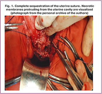
Intraoperative evaluation during surgical interventions revealed uterine suture dehiscence in 51/58 patients (87.9%), with complete tissue sequestration complicating the process in 24 cases (41.4%), reflecting extreme severity of purulent-destructive myometrial damage. An example of complete uterine suture sequestration, showing necrotic membranes protruding from the uterine cavity, is presented in Figure 1.
Microbiological characteristics of pathogens
Etiological analysis of pathogens isolated from intraoperative peritoneal exudate sampling demonstrated that Gram-positive cocci were most frequently identified. Staphylococcus epidermidis was isolated in 13 patients (22.4%) and Enterococcus faecalis in 12 patients (20.7%). Among Gram-negative bacteria, Escherichia coli predominated, identified in 6 patients (10.3%). Anaerobic microorganisms, Klebsiella spp., Proteus spp., Pseudomonas aeruginosa, and Candida species were isolated in single cases.
Surgical interventions performed
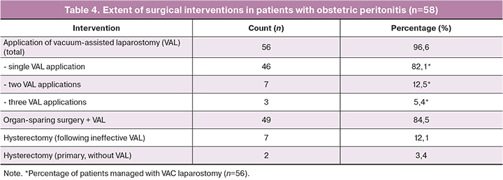
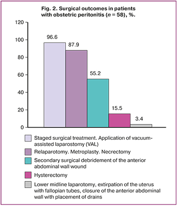
Analysis of surgical management demonstrated that a staged surgical strategy utilizing vacuum-assisted closure (VAC) laparostomy was employed in the vast majority of cases (56/58, 96.6%). A single application of VAC laparostomy was performed in 46/56 patients (82.1%), which was sufficient to resolve the purulent-inflammatory process. Repeat VAC application twice was required in 7 patients (12.5%), and three times in 3 patients (5.4%).
Uterine-sparing operations were successfully performed in 49/58 women (84.5%). Total abdominal hysterectomy was performed in 9/58 patients (15.5%). Among these, hysterectomy was performed in 7 patients (12.1%) due to VAC laparostomy failure (progressive sepsis, lack of laboratory improvement, and total myometrial necrosis). In 2 patients (3.4%), clinical conditions precluded uterine preservation and VAC application, necessitating primary total hysterectomy with bilateral salpingectomy. Data regarding the nature and extent of surgical interventions are summarized in Table 4.
Repeat application of VAC laparostomy was performed upon meeting one or more of the following criteria, evaluated intraoperatively (during planned relaparotomy at 48 hours) and dynamically (clinical and laboratory data):
- Persistent purulent or fibrinopurulent peritoneal exudate at relaparotomy
The most frequent indication (90% of repeat interventions). Criteria: exudate volume >300–500 mL, thick fibrin deposits on the peritoneum, purulent or turbid exudate, presence of gas bubbles (indicative of anaerobic infection), and lack of exudate clearance compared to the previous operation.
- Lack of favorable laboratory response (CRP, PCT) despite 48 hours of intensive care
Criteria: CRP reduction of less than 30% from baseline; persistent PCT >2.0 ng/mL; persistent bandemia >20–25% and toxic neutrophil granulation. A "rebound phenomenon" – a secondary surge in inflammatory markers following initial reduction – was observed in a subset of patients, indicating new purulent foci formation.
- Progression of organ dysfunction (SOFA Score)
Criteria: increase in Sequential Organ Failure Assessment (SOFA) score by ≥2 points at 48–72 hours post-primary surgery. Most common manifestations: respiratory deterioration (PaO₂/FiO₂<200 mm Hg), progressive renal failure (1.5- to 2-fold increase in serum creatinine, urine output <0.5 mL/kg/h), thrombocytopenia (<100×10⁹/L), and prolongation of prothrombin time by ≥50%.
- Persistent foci of myometrial necrosis requiring staged debridement
The most specific indication for obstetric peritonitis (70% of cases). Necrotic areas were excised in stages due to the risk of massive hemorrhage or extensive healthy tissue damage associated with single-stage necrosectomy. Staging allowed controlled tissue excision and dynamic assessment of myometrial viability.
Results of criteria application
In this series, two VAC laparostomy applications were required in 7 patients (12.5%), and three applications were required in 3 patients (5.4%). All patients requiring repeat interventions presented with a combination of two or more indications; the combination of "persistent exudate + lack of laboratory improvement" was the most frequent (8/10, 80%). Patients who underwent three VAC applications exhibited additional progression of organ dysfunction and persistent areas of myometrial necrosis.
Treatment outcomes
Implementation of this approach resulted in the discharge of 49/58 postpartum patients (84.5%) with clinical improvement and uterine preservation. No maternal mortality occurred in the study cohort (0%). Follow-up monitoring of patients with preserved uteri revealed subsequent successful pregnancy and full-term delivery in 2 women. This confirms the feasibility of preserving reproductive function in select patients undergoing organ-sparing treatment for obstetric peritonitis.
Adverse events
No intraoperative injuries to adjacent organs (bowel, ureters, urinary bladder, major vessels) occurred during surgical interventions. In 7 patients with VAC laparostomy failure, hysterectomy was performed due to progressive sepsis, lack of laboratory improvement (primarily CRP and PCT) despite intensive care, and development of total myometrial necrosis (>70% wall thickness involvement) (Fig. 2).
Discussion
The findings of this study, including uterine preservation in 84.5% of patients and no maternal deaths, are consistent with those reported by other authors who used a staged surgical approach with vacuum-assisted laparostomy to treat obstetric peritonitis.
In a systematic review by Maskin S.S. et al. (2023), the use of vacuum-assisted laparostomy for generalized peritonitis of various etiologies reduced mortality from 35.7% to 14.3% [25]. In a pilot study by Cherdantsev D.V. et al. (2023), the uterine preservation rate among patients with obstetric peritonitis treated with vacuum-assisted laparostomy reached 82.1%, which is comparable to our findings (84.5%) [13]. According to Dabuzov A.Sh. (2023), the rate of hysterectomy with conventional radical surgical management, without vacuum-assisted laparostomy, was 68–75%, substantially higher than that observed in our series (15.5%) [14].
Thus, our findings are consistent with the published literature supporting the effectiveness of staged surgical management with vacuum-assisted laparostomy for generalized obstetric peritonitis. As noted by Maskin S.S. et al., randomized clinical trials are needed to obtain evidence of higher certainty [25].
The laboratory findings in this study reflect a pronounced systemic inflammatory response and the severity of endogenous intoxication. Procalcitonin (PCT) levels above 2.0 ng/mL in most patients indicated a high likelihood of sepsis and provided a basis for aggressive antimicrobial therapy and surgical intervention. Changes in this biomarker over time, which were not fully analyzed in the present study, may have prognostic value.
The microbiological profile in this study was characterized by the predominance of Staphylococcus epidermidis (22.4%), Enterococcus faecalis (20.7%), and Escherichia coli (10.3%), consistent with published evidence regarding the polymicrobial etiology of obstetric peritonitis. The relatively low frequency of anaerobe detection (less than 5% in routine cultures) can be attributed to methodological difficulties in culturing these organisms and does not reflect the true etiological distribution.
The key finding of this study is that uterine preservation was feasible in the vast majority of patients with obstetric peritonitis managed using a staged surgical approach with vacuum-assisted laparostomy. These findings are consistent with contemporary damage control surgery principles, which involve performing the minimum necessary intervention during the initial stage, followed by definitive correction at a later stage. Vacuum-assisted technologies facilitate effective etiopathogenetic treatment and create optimal conditions for tissue healing.
It is important to emphasize that organ-preserving management of obstetric peritonitis is feasible only when several conditions are strictly met. These include early diagnosis and a limited defect, radical surgical debridement of the infectious focus through excision of nonviable tissue within the boundaries of healthy myometrium, effective drainage and source control, adequate antimicrobial and detoxification therapy, and dynamic monitoring with readiness to revise the treatment strategy promptly in favor of hysterectomy if signs of progression of the purulent-inflammatory process worsen.
The occurrence of two confirmed subsequent pregnancies after organ-preserving treatment is a clinically important observation demonstrating that preservation of reproductive function is possible in this patient population [33–39]. However, the small number of such cases precludes statistically robust conclusions regarding the likelihood of fertility recovery.
Study limitations. This study has several limitations that should be considered when interpreting the findings. First, the retrospective design precludes the complete exclusion of selection bias because patient enrollment was not standardized prospectively. Second, the absence of a control group comprising patients treated according to conventional management without vacuum-assisted laparostomy limits comparisons between the different approaches and precludes conclusions regarding the superiority of one method over another. Third, the single-center design may limit the generalizability of the findings to other medical institutions with different levels of equipment and staff expertise. Fourth, the follow-up period, which extended to 7 years for the earliest patients, was insufficient to assess long-term reproductive outcomes in all women whose uterus was preserved.
Conclusion
In this series of 58 consecutive patients with obstetric peritonitis, staged surgical management with vacuum-assisted laparostomy was associated with uterine preservation in 84.5% of patients and no deaths. These findings demonstrate the potential for preserving reproductive function in some patients who experience this complication.
This case series meets the PROCESS 2023 reporting requirements and may serve as a basis for planning prospective multicenter studies with control groups, which could provide higher-certainty evidence regarding the role of vacuum-assisted laparostomy in the treatment of obstetric peritonitis. Uterine preservation in women of reproductive age has not only medical but also important social and psychological implications, preserving the opportunity for future childbearing and improving quality of life.
References
- Бурьяк Д.В. Современные особенности антибактериальной терапии в акушерстве и гинекологии. Медицинские новости. 2022; 2: 76-8. [Buryak D.V. Current aspects of antibacterial therapy in obstetrics and gynecology. Meditsinskie novosti. 2022; (2): 76-8. (in Russian)].
- Ribeiro-do-Valle C.C., Bonet M., Brizuela V., Abalos E., Baguiya A., Bellissimo-Rodrigues F. et al.; WHO GLOSS research group. Aetiology and use of antibiotics in pregnancy-related infections: results of the WHO Global Maternal Sepsis Study (GLOSS), 1-week inception cohort. Ann. Clin. Microbiol. Antimicrob. 2024; 23(1): 21. https://dx.doi.org/10.1186/s12941-024-00681-8
- GBD 2017 Causes of Death Collaborators. Global, regional, and national age-sex-specific mortality for 282 causes of death in 195 countries and territories, 1980-2017: a systematic analysis for the Global Burden of Disease Study 2017. Lancet. 2018; 392(10159): 1736-88. https://dx.doi.org/10.1016/S0140-6736(18)32203-7
- Woodd S.L., Montoya A., Barreix M., Pi L., Calvert C., Rehman A.M. et al. Incidence of maternal peripartum infection: A systematic review and meta-analysis. PLoS Med. 2019; 16(12): e1002984. https://dx.doi.org/10.1371/journal.pmed.1002984
- Bonet M., Brizuela V., Abalos E., Cuesta C., Baguiya A., Chamillard M. et al. Frequency and management of maternal infection in health facilities in 52 countries (GLOSS): A 1-week inception cohort study. Lancet Global Health. 2020; 8: e661-e671. https://dx.doi.org/10.1016/S2214-109X(20)30109-1
- Лазарева И.В., Агеевец В.А., Сидоренко С.В. Антибиотикорезистентность: роль карбапенемаз. Медицина экстремальных ситуаций. 2018; 20(3): 320-8. [Lazareva I.V., Ageevets V.A., Sidorenko S.V. Antibiotic resistance: the role of carbapenemases. Medicine of Extreme Situations. 2018; 20(3): 320-8. (in Russian)].
- Министерство здравоохранения Российской Федерации. Клинические рекомендации. Сепсис (у взрослых). 2024. Доступно по: https://cr.minzdrav.gov.ru/preview-cr/898_1 [Ministry of Health of the Russian Federation. Clinical guidelines. Sepsis (in adults). 2024. Available at: https://cr.minzdrav.gov.ru/preview-cr/898_1 (in Russian)].
- Министерство здравоохранения Российской Федерации. Клинические рекомендации. Послеродовые инфекционные осложнения. 2024. Доступно по: https://cr.minzdrav.gov.ru/preview-cr/791_1 [Ministry of Health of the Russian Federation. Clinical guidelines. Postpartum infectious complications. 2024. Available at: https://cr.minzdrav.gov.ru/preview-cr/791_1 (in Russian)].
- Табеева Г.И., Думановская М.Р., Чернуха Г.Е. Современные представления о микробиоте в гинекологии. Акушерство и гинекология. 2020; 2: 38-44. https://dx.doi.org/10.18565/aig.2020.2.38-44 [Tabeeva G.I., Dumanovskaya M.R., Chernukha G.E., Priputnevich T.V. Current views on microbiota in gynecology. Obstetrics and Gynecology. 2020; (2): 38-44 (in Russian). https://dx.doi.org/10.18565/aig.2020.2.38-44].
- Полушин Ю.С. Сепсис и менеджмент: как улучшить исходы лечения больных сепсисом. Вестник хирургии имени И.И. Грекова. 2022; 181(5): 38-46. https://dx.doi.org/10.24884/0042-4625-2022-181-5-38-46 [Polushin Yu.S. Sepsis and management: how to improve the outcomes of treatment of patients with sepsis. Grekov’s Bulletin of Surgery. 2022; 181(5): 38-46 (in Russian). https://dx.doi.org/10.24884/0042-4625-2022-181-5-38-46].
- Абдураимов Т.Ф. Материнский сепсис: значимость научно обоснованных и доказательных подходов к профилактике критических ситуаций, обусловленных материнским сепсисом. В кн.: Тезисы IV Общероссийской научно-практической конференции акушеров-гинекологов «Оттовские чтения». М.; 2022: 3. [Abdurakhimov T.F. Maternal sepsis: the importance of evidence-based approaches to the prevention of critical situations caused by maternal sepsis. In: Abstracts of the IV All-Russian Scientific and Practical Conference of Obstetricians and Gynecologists «Ottovskie chteniya». Moscow, 2022: 3. (in Russian)].
- Омаров Н.С.М., Дабузов А.Ш., Меджидова Д.Р. Эфферентные методы в комплексном лечении акушерского перитонита. Охрана материнства и детства. 2022; 2(40): 36-9. [Omarov N.S.M., Dabuzov A.Sh., Medzhidova D.R. Efferent methods in complex treatment of obstetric peritonitis. Maternal and Child Health. 2022; 2(40): 36-39. (in Russian)].
- Черданцев Д.В., Дударовская Е.В., Галкина Д.Е., Макаренко Т.А., Базина М.И., Бохина Ж.В., Дятлов В.Ю., Лысенко О.А., Колесникова Н.А., Бикулова Т.В. Невозможное возможно: органосохраняющее лечение в условиях акушерского перитонита. Первая краевая. 2023; 4(90): 48-51. [Cherdantsev D.V., Dudarovskaya E.V., Galkina D.E., Makarenko T.A., Bazina M.I., Bokhina Zh.V., Dyatlov V.Yu., Lysenko O.A., Kolesnikova N.A., Bikulova T.V. The impossible is possible: organ-preserving treatment in conditions of obstetric peritonitis. Pervaya kraevaya. 2023; 4(90): 48-51. (in Russian)].
- Дабузов А.Ш. Оценка степени риска прогрессирования гнойно-воспалительных осложнений у родильниц с акушерским перитонитом и возможности органосохраняющих операций. Архив акушерства и гинекологии им. В.Ф. Снегирева. 2023; 10(1): 49-54. https://dx.doi.org/10.17816/2313-8726-2023-10-1-49-54 [Dabuzov A.Sh. Assessment of the risk of progression of purulent-inflammatory complications and the possibility of organ-preserving surgeries in puerperas with obstetric peritonitis. V.F. Snegirev Archives of Obstetrics and Gynecology. 2023; 10(1): 49-54 (in Russian). https://dx.doi.org/10.17816/2313-8726-2023-10-1-49-54].
- Галкина Д.Е., Макаренко Т.А., Ульянова И.О. Септические осложнения после абдоминального родоразрешения (обзор литературы). Проблемы репродукции. 2024; 30(6): 108-17. https://dx.doi.org/10.17116/repro202430061108 [Galkina D.E., Makarenko T.A., Ulyanova I.O. Septic complications after abdominal delivery (literature review). Russian Journal of Human Reproduction. 2024; 30(6): 108-17 (in Russian). https://dx.doi.org/10.17116/repro202430061108].
- Апресян С.В., Димитрова В.И., Слюсарева О.А. Диагностика и лечение послеродовых гнойно-септических заболеваний. Доктор.Ру. 2018; 6(150): 17-24. [Apresyan S.V., Dimitrova V.I., Slyusareva O.A. Diagnosis and treatment of postpartum purulent septic diseases. Doctor.Ru. 2018; 6(150): 17-24 (in Russian)].
- Knowles R., Sharland M., Hsia Y., Magrini N., Moja L., Siyam A. et al. Measuring antibiotic availability and use in 20 low- and middle-income countries. Bull. World Health Organ. 2020; 98(3): 177-187C. https://dx.doi.org/10.2471/BLT.19.241349
- Bonet M., Souza J.P., Abalos E., Fawole B., Knight M., Kouanda S. et al. The global maternal sepsis study and awareness campaign (GLOSS): study protocol. Reprod. Health. 2018; 15(1): 16. https://dx.doi.org/10.1186/s12978-017-0437-8
- Brizuela V., Cuesta C., Bartolelli G., Abdosh A.A., Abou Malham S., Assarag B. et al.; WHO GLOSS Research Group. Availability of facility resources and services and infection-related maternal outcomes in the WHO Global Maternal Sepsis Study: a cross-sectional study. Lancet Glob. Health. 2021; 9(9): e1252-e1261. https://dx.doi.org/10.1016/S2214-109X(21)00248-5
- Галкина Д.Е., Макаренко Т.А. Гнойно-септические осложнения послеродового периода: особенности клиники, диагностики и лечения. В кн.: Тезисы XVII Общероссийского семинара «Репродуктивный потенциал России: версии и контраверсии» и XII Общероссийской конференции «FLORES VITAE. Контраверсии в неонатальной медицине и педиатрии». М.; 2023: 13-4. [Galkina D.E., Makarenko T.A. Purulent-septic complications of the postpartum period: features of clinical presentation, diagnosis and treatment. In: Abstracts of the XVII All-Russian Seminar «Reproductive Potential of Russia: Versions and Controversies» and the XII All-Russian Conference «FLORES VITAE. Controversies in Neonatal Medicine and Pediatrics». Moscow; 2023: 13-4. (in Russian)].
- Орозалиева Б.К., Акматов Т.А., Сыдыгалиев К.С. Гнойно-септические осложнения в акушерстве. Наука, новые технологии и инновации Кыргызстана. 2020; 1: 59-62. [Orozalieva B.K., Akmatov T.A., Sydygaliyev K.S. Purulent-septic complications in obstetrics. Science, New Technologies, and Innovations of Kyrgyzstan. 2020; 1: 59-62. (in Russian)].
- Смирнова С.С., Егоров И.А., Голубкова А.А. Гнойно-септические инфекции у родильниц. Часть 1. Распространенность, факторы риска, эпидемиологический надзор (обзор литературы). Журнал микробиологии, эпидемиологии и иммунобиологии. 2022; 99(1): 109-25. https://dx.doi.org/10.36233/0372-9311-226 [Smirnova S.S., Egorov I.A., Golubkova A.A. Purulent-septic infections in puerperas. Part 1: prevalence, risk factors, epidemiological surveillance (literature review). Journal of microbiology, epidemiology and immunobiology. 2022; 99(1): 109-25 (in Russian). https://dx.doi.org/10.36233/0372-9311-226].
- Тарасова В.Г., Шувалова В.С. Акушерский сепсис в послеродовом периоде. Морфологические аспекты. В сб.: Современная патология: опыт, проблемы, перспективы. II Всероссийская научно-практическая конференция с международным участием. Самара; 2021: 171-4. [Tarasova V.G., Shuvalova V.S. Obstetric sepsis in the postpartum period. Morphological aspects. In: Modern pathology: experience, problems, prospects. II All-Russian Scientific and Practical Conference with international participation. Samara; 2021: 171-4. (in Russian)].
- Смирнова С.С., Егоров И.А., Голубкова А.А. Гнойно-септические инфекции у родильниц. Часть 2. Клинико-патогенетическая характеристика нозологических форм, этиология и антибиотикорезистентность (обзор литературы). Журнал микробиологии, эпидемиологии и иммунобиологии. 2022; 99(2): 244-59. https://dx.doi.org/10.36233/0372-9311-227 [Smirnova S.S., Egorov I.A., Golubkova A.A. Purulent-septic infections in puerperas. Part 2. Clinical and pathogenetic characteristics of nosological forms, etiology and antibiotic resistance (literature review). Journal of microbiology, epidemiology and immunobiology. 2022; 99(2): 244-59 (in Russian). https://dx.doi.org/10.36233/0372-9311-227].
- Маскин С.С., Александров В.В., Матюхин В.В., Дербенцева Т.В., Рашид А., Сигаев С.М., Бирюлев Д.С. Вакуум-ассистированная лапаростомия при тяжелой травме живота и ургентной абдоминальной патологии с компартмент-синдромом, перитонитом и сепсисом: сравнительные аспекты с другими вариантами многоэтапного хирургического лечения (систематический обзор и метаанализ). Acta Biomedica Scientifica. 2023; 8(1): 170-203. https://dx.doi.org/10.29413/ABS.2023-8.1.19 [Maskin S.S., Aleksandrov V.V., Matyukhin V.V., Derbentseva T.V., Rachid A., Sigaev S.M., Biriulev D.S. Vacuum-assisted laparostomy in severe abdominal trauma and urgent abdominal pathology with compartment syndrome, peritonitis and sepsis: comparison with other options for multistage surgical treatment (systematic review and meta-analysis). Acta Biomedica Scientifica. 2023; 8(1): 170-203 (in Russian). https://dx.doi.org/10.29413/ABS.2023-8.1.19].
- Kiponza R., Balandya B., Majigo M.V., Matee M. Laboratory confirmed puerperal sepsis in a national referral hospital in Tanzania: etiological agents and their susceptibility to commonly prescribed antibiotics. BMC Infect. Dis. 2019; 19(1): 690. https://dx.doi.org/10.1186/s12879-019-4324-5
- Amin R., Cruz J., Lee J. An unusual case of abdominal pain in the postpartum period: A case report of Vernix caseosa peritonitis. AIM Clinical Cases. 2025; 4: e250024. https://dx.doi.org/10.7326/aimcc.2025.0024
- Peng L., Chen X., Wang Z., Yi L., Jin Z. Maternal puerperal infection caused by Parabacteroides goldsteinii: a case report. Front. Med. (Lausanne). 2024; 11: 1450931. https://dx.doi.org/ 10.3389/fmed.2024.1450931.
- Yadav S., Mangla D., Anita. Pneumoperitoneum with peritonitis: an unseen, rare complication of postpartum caesarean uterine wound dehiscence. Int. J. Reprod. Contracept. Obstet. Gynecol. 2025; 14(8): 2772-5. https://dx.doi.org/10.18203/2320-1770.ijrcog20252359
- Kvalvik S.A., Zakariassen S.B., Overrein S., Rasmussen S., Skrede S., Baghestan E. Obstetric infections and clinical characteristics of maternal sepsis: a hospital-based retrospective cohort study. Sci. Rep. 2024; 14(1): 6067. https://dx.doi.org/10.1038/s41598-024-56486-4
- Zhang H., Ji G., Zhang Y. Ruptured ovarian tumor in a pregnant woman with chemical peritonitis. Asian Journal of Surgery. 2025; 48(8): 5021-2. https://dx.doi.org/10.1016/j.asjsur.2025.01.127
- Mathew G., Sohrabi C., Franchi T., Nicola M., Kerwan A., Agha R.; PROCESS Group. Preferred Reporting Of Case Series in Surgery (PROCESS) 2023 guidelines. Int. J. Surg. 2023; 109(12): 3760-9. https://dx.doi.org/10.1097/JS9.0000000000000940
- Рузибойзода К.Р., Саидзода П.А., Курбонов Ш.М., Али-Заде С.Г. Мониторинг лабораторных показателей и внутрибрюшного давления в ранней диагностике послеоперационных осложнений у пациенток с гинекологическим перитонитом. Вестник НМХЦ им. Н.И. Пирогова. 2025; 20(3): 69-73.https://dx.doi.org/10.25881/20728255_2025_20_3_69 [Ruziboyzoda K.R., Saidzoda P.A., Kurbonov Sh.M., Ali-Zade S.G. Monitoring of laboratory parameters and intra-abdominal pressure in early diagnosis of postoperative complications in patients with gynecological peritonitis. Bulletin of Pirogov National Medical & Surgical Center. 2025; 20(3): 69-73 (in Russian). https://dx.doi.org/10.25881/20728255_2025_20_3_69].
- Чурина Д.Е., Макаренко Т.А., Черданцев Д.В., Носков И.Г. Возможности органосохраняющего лечения акушерского перитонита: систематический обзор и метаанализ. Вестник РАМН. 2025; 80(5): 325-34. https://dx.doi.org/10.15690/vramn18108 [Churina D.E., Makarenko T.A., Cherdantsev D.V., Noskov I.G. Organ-preserving treatment options in obstetric peritonitis: systematic review and meta-analysis. Annals of the Russian Academy of Medical Sciences. 2025; 80(5): 325-34 (in Russian). https://dx.doi.org/10.15690/vramn18108].
- Самчук П.М., Ищенко А.И., Розалиева Ю.Ю. Органосохраняющие технологии при гнойных воспалительных осложнениях кесарева сечения. Вопросы гинекологии, акушерства и перинатологии. 2020; 19(2): 96-103. https://dx.doi.org/10.20953/1726-1678-2020-2-96-103 [Samchuk P.M., Ishchenko A.I., Rozalieva Yu.Yu. Organ-sparing surgery for suppurative inflammatory complications of caesarean section. Gynecology, Obstetrics and Perinatology. 2020; 19(2): 96-103 (in Russian). https://dx.doi.org/10.20953/1726-1678-2020-2-96-103].
- Matsumoto R., Kuramoto S., Muronoi T., Oka K., Shimojyo Y., Kidani A. et al. Damage control surgery for spontaneous perforation of pyometra with septic shock: a case report. Acute Med. Surg. 2021; 8(1): e657. https://dx.doi.org/10.1002/ams2.657
- Черданцев Д.В., Дударовская Е.В., Галкина Д.Е., Макаренко Т.А., Базина М.И., Бохина Ж.В., Дятлов В.Ю., Лысенко О.А. Способ органосохраняющего лечения акушерского перитонита после абдоминальных родов с использованием вакуум-ассистированной лапаростомии. Патент на изобретение RU 2816058 C1, 26.03.2024. Заявка № 2023121832 от 22.08.2023. [Cherdantsev D.V., Dudarovskaya E.V., Galkina D.E., Makarenko T.A., Bazina M.I., Bokhina Zh.V., Dyatlov V.Yu., Lysenko O.A. Method of organ-preserving treatment of obstetric peritonitis after abdominal delivery using vacuum-assisted laparostomy. Patent for invention RU 2816058 C1, 26.03.2024. Application No. 2023121832 dated 22.08.2023. (in Russian)].
- Arul G.S., Singh M., Ali A.M., Gee O.J. Damage control surgery in neonates: Lessons learned from the battlefield. J. Pediatr. Surg. 2019; 54(10): 2069-74. https://dx.doi.org/10.1016/j.jpedsurg.2019.04.001
- Черданцев Д.В., Первова О.В., Трофимович Ю.Г., Попов А.Е., Шапкина В.А., Тахтобин Е.Г., Наркевич А.Н. Возможности повышения эффективности периоперационной санации брюшной полости при перитоните. Сибирское медицинское обозрение. 2018; (1): 20-6. https://dx.doi.org/10.20333/2500136-2018-1-20-26 [Cherdantsev D.V., Pervova O.V., Trophimovich Yu.G., Popov A.E., Shapkina V.A., Takhtobin E.G., Narkevich A.N. Possibilities of improving the efficiency of perioperative sanitation of abdominal cavity at peritonitis. Siberian Medical Review. 2018; (1): 20-6 (in Russian). https://dx.doi.org/10.20333/2500136-2018-1-20-26].
Received 20.04.2026
Accepted 02.10.2026
About the Authors
Darya E. Churina, PhD, Associate Professor at the Department of Operative Gynecology, Institute of Postgraduate Education, Prof. V.F. Voino-Yasenetsky Krasnoyarsk State Medical University, Ministry of Health of the Russian Federation, 660022, Russia, Krasnoyarsk Territory, Krasnoyarsk, Partizan Zheleznyak str., 1, +7(923)376-94-33, dashsemch@mail.ru, https://orcid.org/0000-0001-7516-5203Tatiana A. Makarenko, Dr. Med. Sci., Professor, Head of the Department of Operative Gynecology, Institute of Postgraduate Education, Prof. V.F. Voino-Yasenetsky Krasnoyarsk State Medical University, Ministry of Health of the Russian Federation, 660022, Krasnoyarsk Territory, Krasnoyarsk, Partizan Zheleznyak str., 1,
+7(904)895-47-99, makarenko7777@yandex.ru, https://orcid.org/0000-0002-2899-8103
Dmitry V. Cherdantsev, Dr. Med. Sci., Professor, Head of the Department of Hospital Surgery named after Professor A.M. Dykhno with a Postgraduate Education Course, Prof. V.F. Voino-Yasenetsky Krasnoyarsk State Medical University, Ministry of Health of the Russian Federation, 660022, Krasnoyarsk Territory, Krasnoyarsk,
Partizan Zheleznyak str., 1, +7(902)-940-78-52, gs7@mail.ru, https://orcid.org/0000-0002-4743-4565
Corresponding author: Darya E. Churina, dashsemch@mail.ru
Similar Articles