
Unexplained antenatal fetal death in full-term pregnancy
Kurtser M.A., Kalashnikov S.A., Voskoboeva K.Yu.
Objective. To identify the features of anamnestic data, the course of pregnancy and the results of pathological examination in patients with antenatal death of full-term fetuses depending on the presence of meconium in the amniotic fluid.
Materials and methods. A retrospective analysis of individual pregnancy records, birth histories, and autopsy reports for 83 cases of stillbirth from 2021 to 2023 was conducted. The patients were divided into 2 groups: Group I included the patients with meconium-stained amniotic fluid (MSAF) (n=55); Group II included the patients with clear amniotic fluid (n=28).
Results. The patients in Group I most often had a history of induced abortion (p=0,048), abnormal placental attachment, and oligohydramnios, that can be a marker of MSAF (p=0,041). At the time of fetal death, the length of pregnancy in Group I was longer than in Group II (p=0,003). The frequency of fatty involution of thymus was comparable in stillbirths in both groups. The signs of chronic placental insufficiency and inflammatory-immune disorders were observed in most placental samples collected from both groups.
Conclusion. No direct association was found between fetal distress that led to antenatal death and MSAF. The probability of MSAF increases with increasing gestational age and oligohydramnios. More intensive fetal monitoring is essential in the second half of the third trimester of pregnancy in high-risk patients.
Authors' contributions. Kurtser M.A., Kalashmicov S.A. – the study concept and design, manuscript editing; Voskovoeva K.Yu. – material collection and processing, statistical data processing; Kalashnikov S.A., Voskoboeva K.Yu. – manuscript writing.
Conflicts of interest. The authors confirm that they have no conflict of interest to declare.
Funding. The authors received no funding for this study.
Ethical Approval. The study was approved by the local Ethics Committee of Pirogov Russian National Research Medical University, Ministry of Health of Russia (Pirogov University) (protocol No. №235 of December 18, 2023).
Generative Artificial Intelligence. No generative AI was used in preparing this article.
Patient Consent for Publication. All patients whose data are included in the study have signed informed voluntary consent that the results of examination and treatment will be used for scientific purposes and their data will be published.
Authors' Data Sharing Statement. The data supporting the findings of this study are available on request from the corresponding author after approval from the principal investigator.
For citation: Kurtser M.A., Kalashnikov S.A., Voskoboeva K.Yu. Unexplained antenatal fetal death in full-term pregnancy
Akusherstvo i Ginekologiya/Obstetrics and Gynecology. 2026; (6): 101-108 (in Russian)
https://dx.doi.org/10.18565/aig.2026.30
Keywords
antenatal fetal death
antenatal care
autopsy
meconium-stained amniotic fluid
stillbirth
placental morphology
sudden fetal death syndrome
Antenatal fetal death (AFD) is one of the most important medical and social issues in modern obstetric practice. According to the estimates of the World Health Organization about 2.6 million stillbirths occur globally every year [1]. As stated by some authors, intrauterine asphyxia occupies the leading position among the causes of stillbirth, and according to some estimates accounts for 80.5–88.0% [2, 3]. As reported in a number of studies, from 15% to 50% of all unexplained stillbirths in the world are classified as fetal death of unspecified cause (ICD code P95) [4, 5]. Due to this, Academician Kurtser M.A. et al. offered to use the term “sudden fetal death syndrome“ (SFDS) for antenatal deaths without a clear cause (not diagnosed during pregnancy and delivery) [6]. The issue of correlation between fetal hypoxia and meconium-stained amniotic fluid (MSAF) remains debatable in the scientific community. The presence of meconium in amniotic fluid is a common occurrence, and according to the estimates by different authors it occurs in 10–25% of all deliveries, primarily in full-term pregnancies, and increases up to 52% in post-term pregnancies [7, 8]. Some authors suggest that there is an association between fetal distress and MSAF [9, 10]. At the same time, other studies do not confirm this relationship, that casts doubt on the significance of this feature as a marker for adverse perinatal outcomes [8, 11]. Since there is no scientific consensus on this issue, the clinical interpretation of MSAF is still being actively explored and discussed in the context of predicting perinatal pregnancy outcomes.
The objective of this study was to identify the features of anamnestic data, the course of pregnancy and the results of pathological examination in patients with antenatal fetal death at full-term pregnancy depending on the presence of meconium in the amniotic fluid.
Materials and methods
A retrospective analysis of 83 birth histories (Form No, 096u), maternity records (Form No. 111/u-20), and protocols of fetal autopsy in stillbirth (Form No. 013u) was conducted from 2021 to 2023 in 14 perinatal centers in Moscow (Perinatal Center “Kommunarka”; Perinatal Center, L.A. Vorokhobov City Clinical Hospital No. 67; Perinatal Center, M.P. Konchalovsky City Clinical Hospital; Perinatal Center, G.M. Savelyeva Clinical Hospital No. 31; Maternity Hospital No. 8, O.M. Filatov City Clinical Hospital; Maternity Hospital No. 15, O.M. Filatov City Clinical Hospital; Maternity Hospital, F.I. Inozemtsev City Clinical Hospital; Maternity Hospital, A.K. Eramishantsev City Clinical Hospital; Maternity Hospital, City clinical hospital No. 52; Maternity Hospital No. 3, L.A. Vorokhobov City Clinical Hospital No. 67; Maternity Hospital, V.V. Veresaev City Clinical Hospital; Perinatal Center, S.S. Yudin City Clinical Hospital; Maternity Hospital, S.S. Yudin City Clinical Hospital, Center for family planning and reproduction”, group of companies “Mother and Child”. Inclusion criteria in the study were the following: singleton pregnancy, antenatal fetal death in full-term pregnancy, dispensary follow-up at women’s clinic from the first trimester of pregnancy in accordance with clinical guidelines, fetal death of unspecified cause during pregnancy, Exclusion criteria were preterm birth, placenta previa with hemorrhage, premature detachment of the normally located placenta, umbilical cord abnormalities (true umbilical cord knot, tight nuchal cord loop, umbilical cord thrombosis), uterine rupture, preeclampsia, fetal growth restriction, hemolytic disease, congenital fetal anomalies, acute infections during pregnancy, types 1 and type 2 diabetes mellitus, gestational diabetes mellitus, coronavirus infection (COVID-19), and influenza.
The patients were divided into two groups. Group I comprised the patients with MSAF (n=55). Group II comprised the patients with clear amniotic fluid (CAF) (n=28).
Comparison analysis of clinical and anamnestic data of patients in both groups included the following parameters: age, external genital pathology, obstetric and gynecological anamnesis, the features of pregnancy course.
Analysis included fetal autopsy reports, height, weight, calculation of the Ponderal index (PI) (calculated as body weight (kg)/length (m)3), as well as gestational age and gender using the INTERGROW-21st growth charts [12, 13].
Morphological examination of the placenta was conducted, including macroscopic and microscopic examination of the material. Placental weight was assessed using the percentile scale that has been developed at Moscow Regional Research Institute of Obstetrics and Gynecology [14]. Also, the weight of stillborn babies in relation to placental weight was calculated [15].
Statistical analysis
Statistical analysis was performed using IBM SPSS Statistics 26.0 (IBM, USA) and StatTech v. 3.1.8 (LLL “Stattech, Russia) software programs. The normality test (for <50 patients) or the Kolmogorov–Smirtnov test (for >50 patients). The normal distribution of the quantitative parameters was described as the arithmetic mean (M) and standard deviation (SD). Non-normal distribution was described as the median (Me) and the lower and upper quartiles [Q1; Q3]. The categorical data were presented as absolute values the percentage (%). The quantitative data with normal distribution were compared in both groups using Welch's t-test. In non-normal distribution, the groups were compared using the Mann–Whitney U test. The data in fourfold contingency tables were analyzed using Pearson’s chi-squared test. The obtained results were considered statistically significant at p<0.05.
Results
The initial clinical and anamnestic characteristics in the studied groups were comparable, with the exception of the frequency of the disorders of lipid metabolism, which were noted almost twice more often in patients in group II compared with patients in Group I, although the differences were not statistically significant (Table 1).
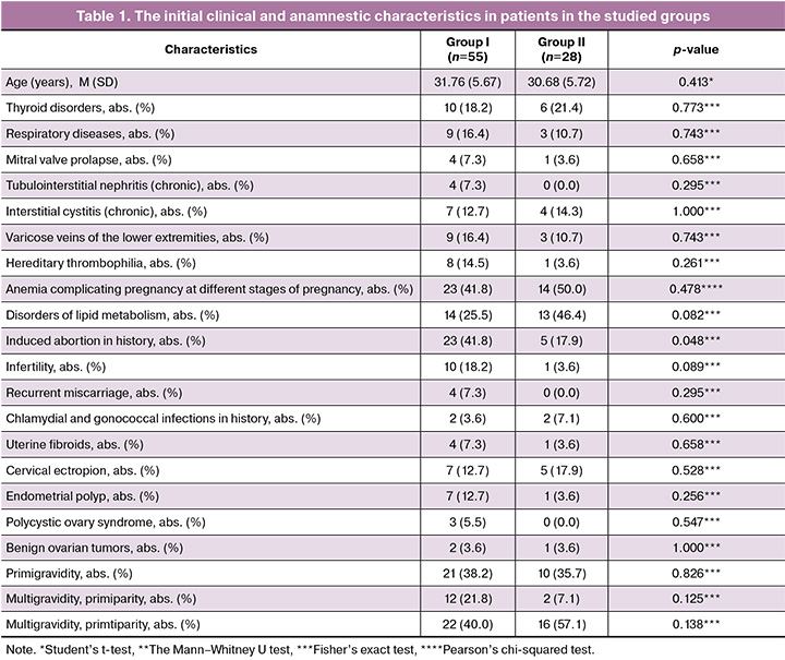
No differences were found between the groups in parity and gravidity. However, analysis of the reproductive anamnesis showed that induced abortion was most often in patients in group I (23/55 (41.8%) and 5/28 (17.9%), respectively; р=0.048; OR=3.306; 95% CI 1.094–9.988).
Also, no differences were found between the groups in the time of registering for pregnancy care, predicted risks of fetal growth restriction, preeclampsia and chromosomal abnormalities based on the results of the first prenatal screening, as well as in the timing of cardiotocography (CTG) and Doppler ultrasound test during dispensary follow-up at women’s clinic (р>0.05). During antenatal follow-up no signs of intrauterine fetal hypoxia (decreased variability, tachycardia and bradycardia, recurrent decelerations, prolonged decelerations) were found according to CTG results and Doppler ultrasound assessment of the mother-placenta-fetus system in both patient groups. Time interval from the first visit to women’s clinic to detection of antenatal fetal death (AFD) was 9.50 [5.25; 14.00] days in group I and 9.50 [3.75; 14.00] days in group II (р=0.917).
Similar frequency of pregnancy complications – threatened abortion, acute respiratory viral infections, inflammatory diseases of the vulva and vagina (Candida albicans, Enterococcus faecalis, Escherichia coli, Klebsiella, Corynebacterium, Staphylococcus aureus, Staphylococcus haemoliticus, Staphylococcus epidermidis at a concentration of 105 CFU/mL) were in patients in both groups (Table 2). Asymptomatic bacteriuria was observed significantly more often in group I (the growth of Enterococcus faecalis, Escherihia coli, Staphylococcus aureus, Streptococcus agalactiae, Streptococcus epidermidis at a concentration of 105 CFU/mL), though it did not reach statistical significance. Based on the results of bacterial culture and antibiotic sensitivity tests, therapy was provided for all patients in both groups with inflammatory diseases of the vulva and vagina, and asymptomatic bacteriuria. Before cervical ripening, the results of cervical swab culture were available only in 8/55 (14.5%) patients in group I and in 7/28 (25.0%) patients in group II. The growth of bacterial or opportunistic pathogens was observed in 6/8 (75.0%) patients in group I and in 4/7 (57.1%) in group II (Candida albicans, Enterococcus faecalis, Escherihia coli, Klebsiella, Corynebacterium, Staphylococcus aureus, Staphylococcus haemoliticus, at a concentration of higher than 105 CFU/mL). The results of histological examination of placentas showed changes specific for deciduitis in more than half of patients in both groups, who exhibited pathogenic or opportunistic bacterial growth in the cervical canal microbiota.
Reduced fetal movements were registered in medical documentation in 43/55 (78.1%) patients in group I and in 19/28 (67.9%) patients in group II. At admission to hospital, 30/43 (69.8%) patients in group I and 17/19 (89.5%) in group II reported the absence of fetal movements during 24 hours (р=0.118), 14/43 (32.6%) and 2/19 (10,5%) women during 48 hours, respectively (р=0.114).
Despite the fact that previous ultrasound assessments showed normal amniotic fluid volume in patients in both groups, oligohydramnios was more often detected in patients with MSAF at hospital admission – 21/55 (38.2%) and 4/28 (14.3%), respectively; р=0.041; OR =3.706; 95% CI 1.127–12.183).
Gestational age at the time of fetal death confirmation was significantly higher in group I compared with group II. In group II, the patients predominantly gave birth at 37.0–38.6 weeks – 22/55 (40.0%) and 20/28 (71.4%), respectively; р=0.007; OR=0.267; 95% CI 0.100–0.712), whereas the patients with MSAF gave birth at 41 weeks.
The tendency toward higher frequency of cesarean section was noted in group II (Table 2), due to uterine scars after previous CSs (100%). Vaginal deliveries in group I were complicated by partially or completely adherent placenta and placental abnormalities, whereas hypotonic bleeding was observed predominantly in Group II – 1/55 (1.8%) and 6/28(21.4%), respectively; р=0.005; OR=0.068; 95% CI 0.008–0.597). The patients in both groups had no postpartum complications.
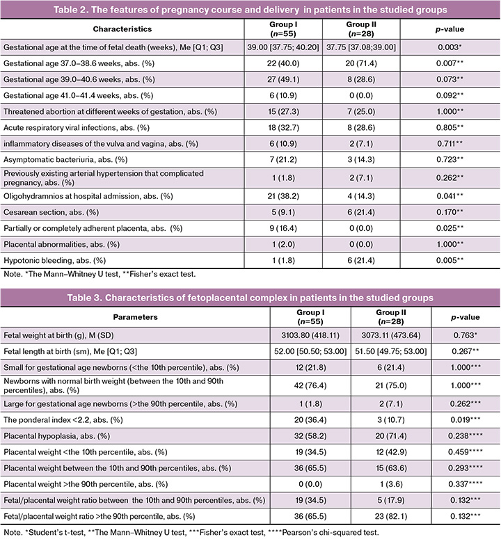
In both groups, every fifth newborn was underweight (Table 3).
The results of pathological examination of stillborn babies in both groups showed no differences, including premature thymic fatty involution. The frequency of occurrence of placental hypoplasia was similar both in the groups with MSAF and CAF, as evidenced by mismatch between the fetal/placental weight ratio (FPR) and the gestational age, as well as reduction in placental weight below the 10th percentile (Table 3). FPR was 0.13 [0.11; 0.14] in group I, and 0.12 [0.10; 0.13] in group II (р=0.183). Histological examination of placenta samples in both groups showed chronic placental insufficiency including vascular malperfusion, premature maturation of the villous tree, increased number of syncytial knots, villous immaturity, villous infarctions, and the presence of non-functional zones. In addition, lymphoid-plasmacytic infiltration of chorion structures, involutional-dystrophic changes were identified (Table 4).
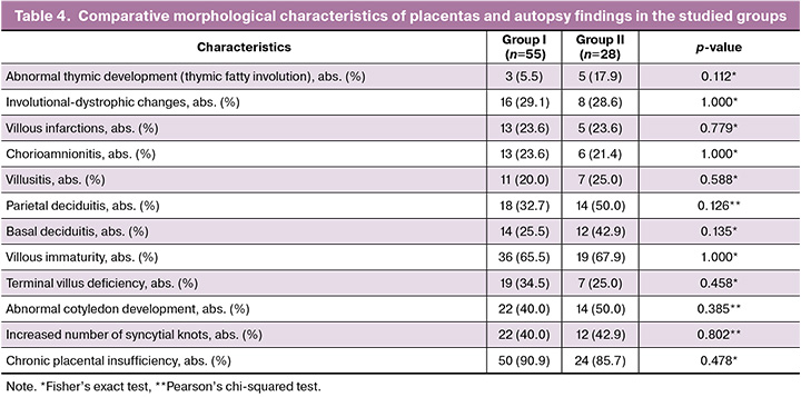
Acute inflammatory and immune pathologies, such as deciduitis, chorioamnionitis and villusitis were detected in more than half of placental samples from both groups. However, the clinical diagnosis of chorioamnionitis was present in none of patients in the studied groups during delivery and in the postpartum period (Table 4). Chronic placental inflammation (basal deciduitis) was observed in one fourth of patients in group I and in more than one third of patients in group II.
Discussion
According to the established concepts, one of the key clinical signs indicating intrauterine fetal hypoxia is the presence of meconium in the amniotic fluid [9, 10]. It is believed that meconium is released in response to fetal distress: Vagus nerve stimulation in different conditions accompanied by hypoxia can cause increased peristalsis, anal sphincter relaxation, leading to meconium release [16]. However, according to some researchers, MSAF can be also a physiological process [8. 11].
Comparative analysis of the clinical and anamnestic characteristics found the relationship between gestational age and MSAF: 73.1% of patients with CAF delivered at 37.0–38.6 weeks of gestation, while most patients with MSAF delivered at ≥39 weeks. There were no cases of leaking clear amniotic fluid among 7 patients who gave birth after 41 weeks. Similar data regarding the relationship between AFD and gestational age at AFD were obtained in the study by Avagliano L. et al. (36.47±1.80 in the group with CAF and 38.13±1.93 in the group with MSAF; р=0.0006), and in the study by Jacques S.M. et al. Their study reported that 13 (72%) patients with MSAF delivered at 39.0–40.6 weeks (full- term pregnancy) and at 41.0–41.6 weeks of gestation (late-term pregnancy), while 16 (94%) patients with CAF delivered at 37.0–38.6 weeks (early-term pregnancy) (р=0.0001) [8, 11]. MSAF can also be a consequence of some other physiological or pathological processes at 39–42 weeks of gestation, possibly related to a certain stage of fetal gestrointestinal tract development, but absent in most fetuses at 37–39 weeks of gestation. Determination of these processes requires further research.
Disorders of lipid metabolism were observed almost twice more often in patients in group II compared with patients in Group I. According to Åmark H. et al., оbesity is recognized as a state of low-grade chronic inflammation and is characterized by increased systemic inflammatory markers, that leads to the release of adipokines, which are ultimately responsible for the dysmetabolic syndrome and can lead to AFD [17].
Vaginal deliveries in every fifth patient in group I were complicated with partially or completely adherent placenta and placental abnormalities, whereas these complications did not occur in group II, that can be associated with a high frequency of medical abortions in history compared with the group with CAF. It was noted that almost half of patients (5/11) with abnormal placental attachment had a history of medical abortions, that indicates the possibility of developing chronic placental insufficiency associated with impaired implantation due to previous intrauterine interventions and chronic endometriris, which became decompensated and led to fetal distress accompanied by intrauterine meconium release.
Our study also found that the patients in group I had oligohydramnios significantly more often, that could be a consequence of chronic placental insufficiency associated with increased risk of MSAF, adverse pregnancy outcome, and a consequence of intrauterine hypoxia, that can result in AFD. This does not contradict the data reported by Sawant A.A. et al., that oligohydramnios correlates with MSAF (14.9%), meconium aspiration syndrome (1,2%), fetal distress (25.3%), stillbirth (1.59%) [18]. According to Bynarowicz T. et al., to minimize the risk of AFD due to oligohydramnios, it is prudent to schedule delivery at 36–37 weeks of gestation [19].
Currently, pathological examination of stillborn babies is the most informative method for establishing the causes and pathogenetic mechanisms of AFD [20]. In our study, the frequency of acute thymic involution was comparable in both groups and was not associated with MSAF. Jacques S.M. et al. suggested that acute thymic involution is a histopathological confirmation of long-term fetal distress resulting in AFD, that is probably caused by chronic placental insufficiency, ultimately leading to thymic injury [11]. The authors reported that severe thymic involution was observed in 11 stillborn babies in the group with MSAF compared with 4 stillborn babies in the group with CAF (p=0.04), that indicate a longer time interval between initiation of distress and fetal death in the group with MSAF [11]. The opposite data were obtained by Avagliano L. et al., who stratified the degree of changes in the thymus depending on the degree of amniotic fluid staining, and found that the degree of changes in the thymus did not correlate with the intensity of meconium stained amniotic fluid [8].
One of the causes of AFD in late pregnancy can be reduced functional capacity and insufficient compensatory abilities of the placenta due to hypoplasia, high frequency of which was identified in both groups, that could be a cause of low birth weight in every fifth stillborn baby. According to Hayward C.E. et al., fetal/placental weight ratio is an indicator of adaptation of the placental size and function to fetal needs to ensure fetal growth [15]. In our study, increased FPR was in 70% of patients in both groups due to reduced placental weight. This could have resulted in imbalance between fetal needs and functional capacity of the placenta, that led to AFD.
Histological examination of the placentas showed the signs of chronic placental insufficiency in placental samples from both groups without significant differences and were not associated with the presence of meconium in the amniotic fluid, that does not contradict the data obtained by Avagliano L. et al. [8].
One of possible causes of AFD can be inflammatory changes in the placenta. Despite treatment of infectious complications in the first trimester of pregnancy, the data on follow-up bacteriological testing of urine and cervical discharge in the third trimester were absent in patients’ medical records. At hospital admission, cervical specimens were collected for cervical swab culture only from 9/58 (15.5%) patients in group I and 7/30 (23.3%) patients in group II, while inflammatory immune pathology was found in one third of tissue specimens from patients in both group. This confirms the importance of cervical swab culture or PCR test to investigate urogenital tract microbiota in all patients with AFD for a more thorough analysis of the causes of stillbirth.
Contrary to сommon belief that meconium release is a consequence of fetal distress, the results of our study found no direct association between the presence of meconium in the amniotic fluid and fetal distress in a number of observations.
Limitations of the study. This is a retrospective study with a small sample size.
Conclusion
Thus, the results of our study found no direct relationship between fetal distress, that led to AFD, and MSAF. The probability of MSAF increases with increasing gestational age. Olygohydramnios is associated with the increased risk of MSAF and can be a marker for adverse perinatal outcomes. It should be noted that high frequency of abnormal placental attachment was in the group of patients with MSAF, where medical abortions in history were observed also more often.
Detection of inflammatory changes during histological examination of the placenta without clinical manifestations in patients necessitates bacteriological examination of the discharge from the cervical canal both in the third trimester of pregnancy and after delivery in patients with antenatal fetal death.
Our study showed the need for more intensive follow-up of fetal condition in the second half of the third trimester. It is supposed that remote CTG examination will provide timely detection of the risk of fetal distress and improve perinatal outcomes.
References
- Thornton H.V., Cornish R.P., Lawlor D.A. Non-linear associations of maternal pre-pregnancy body mass index with risk of stillbirth, infant, and neonatal mortality in over 28 million births in the USA: a retrospective cohort study. EClinicalMedicine. 2023; 66: 102351. https://dx.doi.org/10.1016/j.eclinm
- Щеголев А.И., Туманова У.Н., Чаусов А.А., Шувалова М.П. Сравнительный анализ причин мертворождения в Российской Федерации в 2019 и 2020 годах. Акушерство и гинекология. 2022; 2: 80-90. https://dx.doi.org/10.18565/aig.2022.2.80-90 [Shchegolev A.I., Tumanova U.N., Chausov A.A., Shuvalova M.P. Comparative analysis of stillbirth in the Russian Federation in 2019 and 2020. Obstetrics and Gynecology. 2022; (2): 80-90 (in Russian). https://dx.doi.org/10.18565/aig.2022.2.80-90].
- Гусак Ю.К., Чикин В.Г., Хованов А.В., Новикова А.В., Гусак Н.Ю. Антенатальная гибель плода: клинико-биохимические параллели и особенности родоразрешения. Главный врач Юга России. 2020; 5(75): 18-23 [Gusak Yu.K., Chikin V.G., Khovanov A.V., Novikova A.V., Gusak N.Yu. Antenatal fetal death: clinical and biochemical parallels and features of delivery. Glavnyy Vrach Yuga Rossii. 2020; 5(75): 18-23 (in Russian)].
- Merc M.D., Peterlin B., Lovrecic L. The genetic approach to stillbirth: a «systematic review». Prenat. Diagn. 2023; 43(9): 1220-8. https://dx.doi.org/10.1002/pd.6354
- Lavezzi A.M., Piscioli F., Pusiol T., Jorizzo G., Ferrero S. Sudden intrauterine unexplained death: time to adopt uniform postmortem investigative guidelines? BMC Pregnancy Childbirth. 2019; 19(1): 526. https://dx.doi.org/10.1186/s12884-019-2603-1
- Курцер М.А., Кутакова Ю.Ю., Сонголова Е.Н., Белоусова А.В., Каск Л.Н., Чемезов А.С. Синдром внезапной смерти плода. Акушерство и гинекология. 2011; 7(1): 79-83. [Kurtser M.A., Kutakova Yu.Yu., Songolova E.N., Belousova A.V., Kask L.N., Chemezov A.S. Sudden fetal death syndrome. Obstetrics and Gynecology. 2011; 7(1): 79-83 (in Russian)].
- Мочалова М.Н., Мудров В.А., Мудров А.А. Роль состава околоплодных вод в структуре перинатальной патологии. Журнал акушерства и женских болезней. 2019. 68(2): 95-108. https://dx.doi.org/10.17816/ JOWD68295-108 [Mochalova M.N., Mudrov V.A., Mudrov A.A. Amniotic fluid composition and its role in perinatal pathology. Journal of Obstetrics and Women's Diseases. 2019; 68(2): 95-108 (in Russian). https://dx.doi.org/10.17816/ JOWD68295-108].
- Avagliano L., Massa V., Bulfamante G. Meconium-stained amniotic fluid and histologic signs of fetal distress in stillbirths. Eur. J. Obstet. Gynecol. Reprod. Biol. 2021; 266: 55-62. https://dx.doi.org/10.1016/j.ejogrb.2021.09.016
- Karpe N.S., Tagad M.B., Holkar R.R., More V.S. Evaluation and association of meconium-stained amniotic fluid with fetal distress. Cureus. 2025; 17(5): e83295. https://dx.doi.org/10.7759/cureus.83295
- Gallo D.M., Romero R., Bosco M., Gotsch F., Jaiman S., Jung E. et al. Meconium-stained amniotic fluid. Am. J. Obstet. Gynecol. 2023; 228(5S): S1158-78. https://dx.doi.org/10.1016/j.ajog.2022.11.1283
- Jacques S.M., Qureshi F. Does in utero meconium passage in term stillbirth correlate with autopsy and placental findings of hypoxia or inflammation? J. Matern. Fetal. Neonatal. Med. 2022; 35(10): 1853-9. https://dx.doi.org/10.1080/14767058.2020.1770217
- Zong X.N., Li H., Zhang Y.Q., Wu H.H. Reference values and growth curves of weight/length, body mass index, and ponderal index of Chinese newborns of different gestational ages. Chin. J. Pediatr. 2021; 59(3): 181-8. https://dx.doi.org/10.3760/cma.j.cn112140-20201130-01063
- The International Fetal and Newborn Growth Consortium for the 21st Century. Available at: https://www.intergrowth21.org.uk
- Баринова И.В., Никольская И.Г., Котов Ю.Б., Прокопенко Е.И., Кондриков Н.И. Плацента при хронической болезни почек у беременных. Акушерство и гинекология. 2020; 1: 169-77. https://dx.doi.org/10.18565/aig.2020.1.169-177 [Barinova I.V., Nikolskaya I.G., Kotov Yu.B., Prokopenko E.I., Kondrikov N.I. Placenta in pregnant women with chronic kidney disease. Obstetrics and Gynecology. 2020; (1): 169-77 (in Russian). https://dx.doi.org/10.18565/aig.2020.1.169-177].
- Hayward C.E., Lean S., Sibley C.P., Jones R.L., Wareing M., Greenwood S.L. et al. Placental adaptation: what can we learn from Birthweight:Placental weight ratio? Front. Physiol. 2016; 5: 7-28. https://dx.doi.org/10.3389/fphys.2016.00028
- Пестрикова Т.Ю., Юрасова Е.А., Ткаченко В.А. Мекониальное окрашивание амниотической жидкости и его влияние на материнские и перинатальные исходы. Российский вестник акушера-гинеколога. 2024; 24(3): 42-7. https://dx.doi.org/10.17116/rosakush20242403142 [Pestrikova T.Yu., Yurasova E.A., Tkachenko V.A. Meconial staining of amniotic fluid and its influence on maternal and perinatal outcomes. Russian Bulletin of Obstetrician-Gynecologist. 2024; 24(3): 42-7 (in Russian). https://dx.doi.org/10.17116/rosakush20242403142].
- Åmark H., Westgren M., Sirotkina M., Hulthén Varli I., Persson M., Papadogiannakis N. Maternal obesity and stillbirth at term; placental pathology – a case control study. PLoS One. 2021; 16(4): e0250983. https://dx.doi.org/10.1371/journal.pone.0250983
- Sawant A.A., Wankhede S., Thakare S., Narayan G.N., Saha I., Vernekar A. et al. Maternal and perinatal outcomes in oligohydramnios: a cross-sectional analysis of pregnancies between 28 to 42 weeks of gestation. Cureus. 2025; 17(2): e79232. https://dx.doi.org/10.7759/cureus.79232
- Bynarowicz T., Jenkins S.M., Shanks A.L. Oligohydramnios. [Updated 2025 May 4]. In: StatPearls [Internet]. Treasure Island (FL): StatPearls Publishing; 2026 Jan-. Available from: https://www.ncbi.nlm.nih.gov/books/NBK562326/
- Blythe C., Vazquez Rez., Cabrera M.S., Zekic Tomas S., Oc Anumba D., Cohen M.C. Results of full postmortem examination in a cohort of clinically unexplained stillbirths: undetected fetal growth restriction and placental insufficiency are prevalent findings. J. Perinatol. 2019; 39(9): 1196-203. https://dx.doi.org/10.1038/s41372-019-0412-z
Received 02.02.2026
Accepted 09.06.2026
About the Authors
Mark A. Kurtser, Academician of the RAS, Dr. Med. Sci., Professor, Head of the Academician G.M. Savelyeva Department of Obstetrics and Gynecology, Institute of Motherhood and Childhood, Pirogov Russian National Research Medical University, Ministry of Health of Russia, 119421, Russia, Moscow, Novatorov str., 3,+7(495)719-78-96, kurtser_ma@rsmu.ru, https://orcid.org/0000-0003-0175-1968
Sergey A. Kalashnikov, Dr. Med. Sci., Professor, Academician G.M. Savelyeva Department of Obstetrics and Gynecology, Institute of Motherhood and Childhood,
Pirogov Russian National Research Medical University, Ministry of Health of Russia, 119421, Russia, Moscow, Novatorov str., 3, +7(495)719-78-96, kalashnikov_sa@rsmu.ru, https://orcid.org/0000-0003-2658-5417
Ksenia Yu. Voskoboeva, PhD student, Academician G.M. Savelyeva Department of Obstetrics and Gynecology, Institute of Motherhood and Childhood, Pirogov Russian National Research Medical University, Ministry of Health of Russia, 119421, Russia, Moscow, Novatorov str., 3, +7(495)719-78-96, voskoboeva96@bk.ru,
https://orcid.org/0000-0003-4484-2229
Corresponding author: Ksenia Yu. Voskoboeva, voskoboeva96@bk.ru
Similar Articles

