В настоящее время в связи с совершенствованием лапароскопических операций в гинекологии значительно сократилось количество операций абдоминальным доступом. С целью сохранения репродуктивного потенциала предпочтения отдаются малоинвазивным реконструктивно-пластическим органосохраняющим операциям. Миомэктомия относится к распространенным операциям, которая может быть проведена гистероскопическим, лапароскопическим и робот-ассистированным доступами. Насколько безопасно выполнять данную операцию, не зная морфологической характеристики опухоли? Этот важный вопрос волнует отечественных и зарубежных клиницистов не первый год.
В этой связи нашей целью явился анализ данных, имеющихся в научной медицинской литературе, посвященных современным аспектам предоперационной дифференциальной диагностики доброкачественных и злокачественных гладкомышечных опухолей матки.
В 2018 г. было выявлено 26 948 случаев заболевания злокачественными новообразованиями тела матки. За 20 лет этот показатель вырос на 87,8%, средний темп прироста составил около 3% в год. В возрастных группах 50–54 и 55–59 лет злокачественные новообразования тела матки занимают 2-е ранговое место в структуре онкологической заболеваемости, в возрасте 45–49, 60–64 и 65–69 лет – 3-е место [1].
Саркомы матки (СМ) – это достаточно редкие, но крайне злокачественные неэпителиальные опухоли, которые составляют приблизительно 3% всех злокачественных заболеваний матки. К СМ относятся мезенхимальные, а также смешанные эпителиальные и мезенхимальные опухоли. Группу злокачественных мезенхимальных опухолей составляют лейомиосаркома (ЛМС) (наиболее часто встречающаяся опухоль), эндометриальные стромальные и родственные опухоли. К смешанным эпителиальным и мезенхимальным опухолям относятся аденосаркома и карциносаркома. Генетические и молекулярные исследования продемонстрировали сходство молекулярных профилей карциносаркомы матки и низкодифференцированного рака тела матки, что свидетельствует об эпителиальном происхождении карциносаркомы [1].
ЛМС отличается крайне агрессивным течением и плохим прогнозом. Поскольку ЛМС – быстрорастущая опухоль, заболевание на начальных стадиях протекает бессимптомно, и диагноз устанавливается только при морфологическом исследовании операционного материала. Крайне редко (около 13%) эта опухоль диагностируется при исследовании материала, полученного при раздельном диагностическом выскабливании полости матки [2].
Макроскопически ЛМС представлена одиночными или множественными интерстициальными узлами, в среднем более 8,0 см в диаметре, мягкими, с плохо определяемыми границами. Поверхность разреза обычно серо-желтая или розовая с зонами кровоизлияний и некрозов. ЛМС является клеточной опухолью, состоящей из пучков веретеновидных клеток с обильной эозинофильной цитоплазмой. Основным критерием для установления гистологического диагноза ЛМС является наличие коагуляционного некроза опухолевых клеток. При этом диагноз ЛМС должен устанавливаться с большой осторожностью у женщин моложе 30 лет и только после исключения причин развития некроза, идентичного коагуляционному некрозу в опухоли. Выделяют эпителиоидно-клеточный и миксоидный варианты ЛМС [3].
Эпителиоидные ЛМС сочетают «эпителиоидный» фенотип с обычными чертами злокачественности, т.е. большой клеточностью, цитологической атипией, некрозом опухолевых клеток и высокой митотической активностью. Миксоидная ЛМС является крупной студенистой опухолью, которая часто выглядит ограниченной при макроскопическом исследовании. Гладкомышечные клетки широко разделены миксоидным материалом. Эти опухоли часто инвазируют миометрий, в редких случаях – кровеносные сосуды [3, 4].
Наиболее распространенной доброкачественной гладкомышечной опухолью женской репродуктивной системы является лейомиома. Частота заболевания среди женщин репродуктивного возраста достигает 70%. Средний возраст выявления миомы матки (ММ) – 32–34 года, а пик заболеваемости приходится на начало менопаузы. В настоящее время отмечается рост частоты встречаемости ММ у молодых женщин до 30 лет, не реализовавших репродуктивную функцию [5].
В настоящее время миомэктомия с применением миниинвазивного доступа является ведущим методом хирургического лечения при ММ у молодых пациенток. Вместе с тем при лапароскопической или робот-ассистированной миомэктомии извлечение препаратов из брюшной полости производится с использованием морцелляторов, с помощью которых интракорпорально фрагментируют узлы, в результате чего возможен контакт опухоли с органами и стенкой брюшной полости.
В 2014 г. Американская ассоциация Food and Drug Administration (FDA) опубликовала заявление, обусловленное увеличением количества случаев поздней диагностики ЛМС при выполнении плановых операций по поводу ММ [5]. Ключевое заключение подразумевало полный отказ в США от проведения морцелляции при удалении матки или миоматозных узлов вне зависимости от характера опухоли. Несмотря на это, европейские хирурги не стали принимать столь жесткие меры и отказываться от такого способа удаления препарата полностью. Одним из направлений развития безопасной морцелляции стало производство медицинскими компаниями специальных мешков (endobag) для обеспечения абластичности процедуры [6]. В российских клинических рекомендациях по лечению ММ нет указаний на ограничение использования морцелляции при гистерэктомии [4].
Следует отметить, что до операции по поводу ММ хирург не имеет морфологической верификации образования, да и возможностью проведения срочного интраоперационного гистологического исследования располагают далеко не все клиники. Подобные ограничения иногда приводят к повторным операциям после получения результатов планового морфологического исследования или к раннему рецидивированию опухоли, характер которой не был установлен своевременно и адекватно.
В этой связи, безусловно, не теряет актуальности предоперационная диагностика специфических изменений в миоматозных узлах, позволяющих заподозрить пролиферативные, дистрофические изменения или СМ.
Задачи, стоящие перед патологами и клиницистами, усложняются еще и тем, что в настоящее время выделена отдельная гетерогенная группа гладкомышечных опухолей с неопределенным потенциалом злокачественности – лейомиоматозы – группа опухолей, которые нельзя надежно диагностировать как доброкачественные или злокачественные на основании обычно используемых критериев.
Для нозологий данной категории характерно формирование гладкомышечных опухолей с доброкачественными морфологическими характеристиками, но с нетипичной клинической картиной: внутрисосудистым распространением, формированием отдаленных метастазов, диссеминацией по брюшине, массивным вовлечением мягких тканей. Заболевания описаны преимущественно у женщин детородного возраста и являются гормонозависимыми. Следует подчеркнуть, что дифференциальная диагностика гладкомышечных опухолей матки, особенно редко встречающихся ЛМС и различных вариантов лейомиоматозов, по плечу только грамотному патоморфологу, имеющему достаточный опыт в конкретной области.
Лучевая диагностика (ультразвуковое исследование (УЗИ), магнитно-резонансная томография (МРТ), компьютерная томография (КТ), ПЭТ/КТ) гладкомышечных опухолей на предоперационном этапе имеет большое значение, однако в полной мере зависит от опыта специалиста. Ультразвуковая диагностика как в нашей стране, так и за рубежом имеет более длительную историю в сравнении с другими лучевыми методами. В свое время ряд специалистов имели возможность изучить особенности диагностики и лечения СМ различной морфологической принадлежности в результате анализа более чем 200 наблюдений [7, 8]. Это дало возможность выделить ряд ультразвуковых признаков, наиболее характерных для каждого морфологического варианта.
ЛМС характеризуется агрессивным течением; при этом новообразования небольших размеров не проявляют себя какой-либо симптоматикой, поэтому выявление ЛМС до 5 см в диаметре является случайной находкой [8].
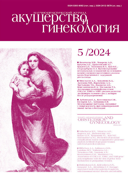
В большинстве наблюдений уже при первичном обследовании определяется значительно увеличенная матка, соответствующая 10 и более неделям беременности, с неровными, бугристыми контурами, нередко может достигать гигантских размеров и занимать всю брюшную полость [8]. В таких случаях достаточно сложно визуализировать неизмененную структуру миометрия, что нередко соответствует тотальному поражению матки. На рисунке 1 приведен пример ультразвуковой картины ЛМС матки пациентки, проходившей обследование и лечение в МКНЦ им. А.С. Логинова.
Для ультразвуковой семиотики ЛМС свойственны многообразие и неоднозначность эхографических признаков.
Условно можно выделить 3 варианта ультразвукового изображения ЛМС:
1) многоузловое образование (интерстициальное и/или субсерозное расположение) (50%);
2) солидный единичный узел, расположенный субсерозно, чаще по ребру нижней трети тела матки (39%);
3) единичные, расположенные преимущественно в полости матки опухолевые узлы, «растягивают» полость и спускаются в цервикальный канал (11%).
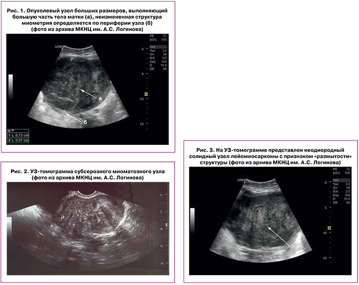
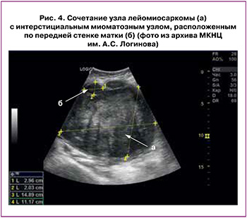
Если для первого и второго вариантов характерно наличие узлов размером более 8 см в диаметре, то при третьем варианте опухолевые узлы матки не превышают 8 см в продольном измерении. Приблизительно в половине наблюдений ультразвуковая картина характеризуется наличием дольчатости, при этом сами дольки-узлы имеют гипоэхогенную структуру, а между ними визуализируются гиперэхогенные перетяжки. Структура долек узлов может быть представлена мелкозернистой диффузной или мелкозернистой гипоэхогенной структурой; появляется такой признак, как «размытость». Плотность опухоли, как правило, значительно ниже, чем при типичной лейомиоме (рис. 2), отсутствуют дистальные акустические тени. Контуры опухолевых образований неровные. Границы опухоли с миометрием нечеткие (рис. 3).
На рисунках 2, 3 представлены ультразвуковые изображения опухоли матки у пациенток. Послеоперационный морфологический диагноз соответствовал данным ультразвукового исследования.
Помимо указанных признаков, в первом варианте ультразвукового изображения ЛМС в редких наблюдениях (при условии небольших размеров опухоли) при цветовом допплеровском картировании могут визуализироваться сосуды радиальной направленности, что нельзя отнести к специфическим признакам. При больших размерах опухоли сосудистые локусы не всегда выражены из-за обширных полей некрозов. Использование современных ультразвуковых технологий, позволяющих фиксировать микроваскуляризацию и хаотично расположенные мелкие опухолевые сосуды, безусловно, могло бы быть полезно для раннего выявления ЛМС при наличии двух условий: с одной стороны, использование ультразвуковых систем экспертного класса широкой врачебной аудиторией, с другой – возможность ранней диагностики бессимптомно развивающейся редкой опухоли. На наш взгляд, соблюдение этих условий в настоящее время проблематично. Серьезных данных, основанных на внушительном количестве исследований, об использовании показателей сосудистого сопротивления для раннего выявления ЛМС или дифференциальной диагностики лейомиомы и ЛМС матки в нашей стране не представлено, а зарубежные публикации неоднозначны.
В структуре СМ могут определяться крупные анэхогенные участки, внутри которых не регистрируются сосудистые локусы, доступные для измерения показателей сосудистого сопротивления. Более того, эти показатели имеют идентичные значения при дегенеративных изменениях в миоматозном узле. Таким образом, значения RI неоптимальны для дифференциации доброкачественных и злокачественных опухолей [8].
В ретроспективной серии из 179 наблюдений Ludovisi M. et al. [9] показали, что СМ представляют собой гетерогенные образования неправильной формы, неоднородной структуры за счет кистозных участков; при этом в их структуре отсутствовали акустические тени, характерные для лейомиоматозных узлов. Около трех четвертей СМ были умеренно васкуляризованы.
В ретроспективном когортном исследовании, опубликованном в 2023 г., авторы оценили СМ с 1997 по 2019 гг. и лейомиомы матки с 2016 по 2019 гг. пациенток, которые лечились в специализированном центре. Ультразвуковые изображения были повторно оценены независимо двумя специалистами с использованием терминов и определений MUSA. Полученные данные статистически обработаны [10]. В исследование были включены 107 пациенток, у 16 из которых была СМ и у 91 – лейомиома матки. ЛМС была наиболее частым гистологическим типом (6/16, 38%). Группой экспертов сделаны выводы о том, что пациентка в постменопаузе с аномальным маточным кровотечением и вновь образованным или резко увеличивающимся мезенхимальным образованием, с неровными границами опухоли и неоднородной эхогенностью при УЗИ, умеренной или обильной центральной или периферической васкуляризацией, наличием кистозных участков и отсутствием кальцификатов имеет более высокий риск в отношении вероятности СМ. Согласие между наблюдателями по большинству терминов и определений MUSA является умеренным. Будущие исследования направлены на проспективный анализ мезенхимальных опухолей матки [10].
Применение современных технологий (эластография, контрастно усиленное УЗИ с применением Соновью) не продвинуло нас вперед по пути усовершенствования критериев дифференциальной диагностики лейомиомы матки и ЛМС матки; однако подтверждает выделенные ранее характеристики, свидетельствующие о наличии массивных участков некроза в структуре опухолевых узлов.
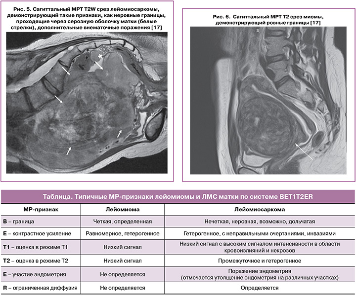
Менее половины наблюдений (41,7%) ЛМС тела матки сочетается с лейомиомой матки (рис. 4).
Макроскопически, а следовательно, и при УЗИ очень сложно отличить ЛМС от нетипичной лейомиомы матки (клеточная лейомиома, внутрисосудистый (интравенозный) лейомиоматоз и т.д.) [11]. По мнению авторов [12], такие же сложности возникают при дифференциальной диагностике между дистрофическими изменениями в миоматозном узле и СМ.
Учитывая все вышесказанное, дифференциальная диагностика между доброкачественными и злокачественными мезенхимальными опухолями при помощи УЗИ малоэффективна.
По сравнению с УЗИ, МРТ имеет ряд преимуществ в диагностике объемных образований (хорошее разрешение и визуализация с учетом большого количества параметров), что, в свою очередь, оказывает решающую роль в сложных диагностических задачах [13].
Для гиалиновой дегенерации лейомиом при МРТ характерен типичный сигнал от стромы узла на Т2-взвешенных изображениях (Т2-ВИ), более низкий, чем для миометрия, и изоинтенсивный для поперечнополосатых мышц. При УЗИ участки гиалиноза обычно эхонегативны и могут напоминать мелкодисперсную несмещаемую взвесь. Контуры таких лейомиом четкие, но часто неровные, фиброзные изменения стромы узла сопровождаются повышением его эхогенности. Кальциноз лейомиом сопровождается появлением в их строме гипоинтенсивных включений во всех типах взвешенности при МРТ; при УЗИ кальцинаты представлены характерным ультразвуковым изображением – различной формы включений, гиперэхогенных с эффектом дистального затенения [14].
Кистозная форма дегенерации лейомиом при УЗИ проявляется наличием в строме узла гипо- или анэхогенных полостей, лишенных кровотока; при МРТ кистозные включения характеризуются гиперинтенсивным сигналом на Т2-ВИ и гипоинтенсивным – на Т1-ВИ с высокими показателями измеряемого коэффициента диффузии. При геморрагической дегенерации строма лейомиомы на МР-изображениях характеризуется повышенным сигналом в Т1-взвешенности [15].
Наиболее сложными по структуре, МР- и эхо-характеристикам являются лейомиомы с миксоматозной дегенерацией. При эхографии лейомиомы такого типа характеризуются анэхогенными включениями вне клиники нарушения питания узла, появлением зон сниженной эхогенности в узле без признаков акустического усиления, а также участками повышенной эхогенности в узле. При МРТ лейомиомы данного типа демонстрируют негомогенную структуру, вариабельную интенсивность сигнала на Т1-ВИ, при этом они гипер- или изоинтенсивны миометрию на Т2-ВИ [16].
Такое разнообразие семиотики определяет сложности дифференциальной диагностики дегенеративно измененных лейомиом с ЛМС.
В связи с этим ряд авторов рекомендуют при дифференциальной диагностике СМ и ММ использовать МРТ, включая диффузионно-взвешенные изображения (ДВИ) и ИКД-карты (индекс коэффициента диффузии). ДВИ позволяет количественно измерять значения ИКД на соответствующих производных картах. Эти значения варьируют в зависимости от плотности клеток и соотношения объема/размера ядра к цитоплазме [17]. Обычно лейомиомы демонстрируют низкий МР-сигнал на ДВИ, что соответствует низкому МР-сигналу на Т2-ВИ (так называемый «эффект затемнения на Т2-ВИ») из-за присутствия гиалинизированного коллагена [17], в то время как СМ демонстрируют высокую интенсивность МР-сигнала на ДВИ (с высоким значением b фактора), что говорит о злокачественной природе опухоли.
Sato K. et al. (2014) показали результаты, где лейомиомы характеризовались низким МР-сигналом на ДВИ, а ЛМС, в свою очередь, имели промежуточный и высокий МР-сигнал на ДВИ. Кроме этого, в данной работе был произведен анализ значений ИКД, где показатели ИКД более 1,1×10⁻³ мм2/с относились к лейомиомам, а показатели менее 1,1×10⁻³ мм2/с отмечались в ЛМС или атипичных лейомиомах [18]. Другими исследователями были получены схожие результаты. Среднее значение ИКД составило 0,91×10⁻³ мм2/с (диапазон 0,70–1,44×10⁻³ мм2/с) для ЛМС, 1,14×10⁻³ мм2/с (диапазон 0,61–2,00×10⁻³ мм2/с) – для лейомиом [19].
В 2015 г. Lin et al. обнаружили, что, хотя МРТ с контрастным усилением приводит к более высокой точности диагностики СМ, чем ДВИ (значение b=1000 с/мм2), комбинация значений ДВИ и ИКД-карты (<1,08×10⁻³ мм2/с) смогла установить диагноз саркомы с точностью, эквивалентной точности МРТ с контрастным усилением [20]. Однако последовательность ДВИ и количественное картирование ИКД не считаются отдельными параметрами для дифференциальной диагностики СМ и лейомиом, поэтому данные ДВИ/ИКД следует сопоставлять с МР-характеристиками Т1/Т2-ВИ. Эти результаты подчеркивают необходимость разъяснений, которые могут возникнуть в результате объединенного анализа.
В связи с этим, Smith J. et al. (2021) в своей работе опубликовали алгоритм для дифференциальной диагностики СМ от ММ по результатам полученных МР-признаков [21].
Система BET1T2ER включает оценку объемного образования по нескольким МР-признакам: В (Borders) – оценка границ объемного образования, E (Enhancement) – контрастное усиление, T1/T2 – оценка сигналов в соответствующих режимах, E (Endometrial involvement) – вовлечение эндометрия, R (Restricted diffusion) – ограниченная диффузия (таблица).
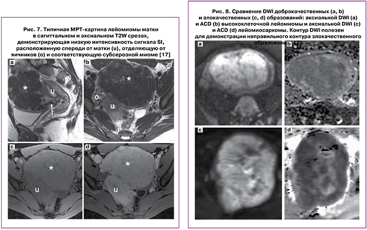
Неровность контуров, неправильная или плохо визуализирующаяся граница новообразования является подозрительным признаком, который гораздо чаще выявляли при ЛМС в сравнении с лейомиомой (чувствительность 74–84% и специфичность 86–91%) (рис 5, 6) [17].
Использование контрастного усиления повышает точность диагностики ЛМС матки [21]. СМ демонстрируют различные характеристики усиления контрастного вещества (КВ) по сравнению с лейомиомами; но на примере наблюдений показано, что структура опухолевых узлов чаще характеризуется гетерогенным накоплением КВ, с отсутствием накопления КВ в центральных отделах в соответствии с наличием некроза [21]. Этот признак наблюдается в большинстве случаев сарком (80%). Lakhman Y. et al. сообщили, что на МРТ зоны некроза с отсутствием накопления КВ в ЛМС имеют чувствительность 95–100% и специфичность 68–73% [22]. Кроме того, саркомы демонстрируют раннее усиление КВ (III тип кривой) при анализе данных динамических кривых накопления КВ по сравнению с лейомиомами [22].
Лейомиомы с признаками дегенерации также имеют участки отсутствия накопления КВ. Поэтому очень важно, чтобы интенсивность МР-сигнала в этих областях сравнивалась с Т1/Т2-ВИ, чтобы охарактеризовать область гиалиновой и кистозной дегенерации, а также наличие геморрагических изменений, которые имеют типичную карту на Т1/Т2-ВИ [22].
Следует отметить, что и те, и другие признаки не являются надежными критериями злокачественности (рис. 7) [17].
При оценке Т1-ВИ ЛМС дают более высокий сигнал, чем ММ; вместе с тем стоит учитывать, что увеличение Т1-ВИ характерно также и для лейомиом с кровоизлиянием. Высокий Т1-сигнал в ЛМС характеризуется большей неоднородностью, плохо очерченными границами, меньшей интенсивностью, чем высокий Т1-сигнал в структуре лейомиомы матки. По сравнению с миомами ЛМС матки имеют значительно более высокий сигнал интенсивности в Т2; однако, если лейомиома имеет различные виды дегенераций, такие как кистозную, муцинозную и т.д. или различные клеточные гистологические подтипы, уровень сигнала интенсивности в Т2 также возрастает, что, в свою очередь, затрудняет точность дифференциальной диагностики. По мнению авторов [22], при проведении дифференциальной диагностики между ЛМС и лейомиомой матки важными параметрами МР-картины являются характер контура эндометрия и серозной оболочки матки. Безусловно, очевидно, что указанные критерии субъективны и дискутабельны, и для серьезных выводов необходимо большее количество наблюдений.
При оценке доброкачественных образований Abdel Wahab C. et al. (2020) в своем исследовании указали, что в случаях, когда интенсивность сигнала на ДВИ была меньше, чем у миометрия, или больше, чем у миометрия, но меньше, чем у эндометрия и лимфатических узлов, можно утверждать, что данное образование является доброкачественным, и наоборот, при интенсивности сигнала на ДВИ больше, чем у эндометрия, а ИКД меньше или равном 0,91×10⁻³ мм2/с, с большей вероятностью следует предполагать злокачественный процесс (рис. 8) [17].
КТ играет ограниченную роль в диагностике опухолей миометрия. В основном КТ рекомендовано для проведения стадирования, оценки распространенности процесса, с целью исключения отдаленных метастазов и рецидивов после первоначального лечения, учитывая преобладающий путь метастазирования ЛМС – гематогенный (рис. 9).
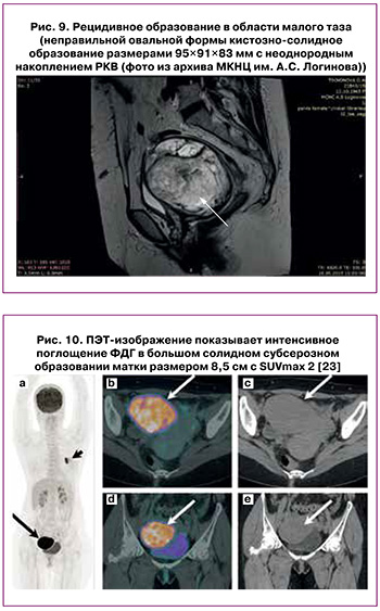
Использование высокотехнологичного метода радионуклидной диагностики – ПЭТ – позволило качественно расширить возможности лучевой диагностики в стадировании многих злокачественных процессов у человека. При этом исследовании возможно визуализировать очаг накопления радиофармпрепарата (РФП). Широко используемым в клинических целях РФП для ПЭТ-диагностики является 18F-фтордезоксиглюкоза (18F-ФДГ), более чем 95% всех ПЭТ-исследований проводятся именно с ним.
Принцип применения 18F-ФДГ в онкологии основывается на разнице дифференциальных скоростей метаболизма глюкозы в доброкачественных и злокачественных тканях (рис. 10) [23]. Возможности предоперационной ПЭТ с ФДГ оценили в своем исследовании Ho K.S. et al. при дифференциальной диагностике ЛМС и мезенхимальных опухолей неясного злокачественного потенциала [24].
Данное исследование основано на наблюдениях с использованием специального параметра ПЭТ-визуализации, называемого «соотношение метаболическая опухоль/некроз», который авторы разработали для количественной оценки коагуляционного некроза в ЛМС и опухолях неясного злокачественного потенциала (STUMP). Его определяли как соотношение между поверхностным метаболизмом опухоли и метаболизмом некротического ядра. Значения указанного параметра при ЛМС/STUMP (диапазон: 3,7–11,8) были значительно выше, чем наблюдаемые при лейомиоме матки (диапазон: 2,0–9,4; P=0,003). Все ЛМС/STUMP выявили характерный паттерн поглощения ФДГ, определяя специфический признак «полого шара» (соответствующий участкам коагуляционного некроза опухоли). Напротив, этот признак неизменно отсутствовал у пациентов с лейомиомой. Вместе с тем достоверных результатов исследователи не получили, в связи с чем клиническая значимость полученных результатов пока сомнительна [24].
Несмотря на относительную редкость, ЛМС могут быть диагностированы и у молодых пациенток; при этом характеризуются агрессивным течением и неблагоприятным прогнозом. По мнению ряда отечественных авторов, успешная предоперационная диагностика имеет решающее значение для планирования лечения и прогноза заболевания [25].
Заключение
К настоящему времени не определены надежные методы, позволяющие улучшить раннюю диагностику новообразований с неопределенным или явным злокачественным потенциалом. К сожалению, в большинстве наблюдений диагноз ЛМС по-прежнему устанавливается по результатам послеоперационного гистологического исследования.
Вместе с тем комплексное использование лучевых методов, развитие современных технологий позволяют получить дополнительную информацию, необходимую хирургу для принятия правильного решения на предоперационном и интраоперационном этапах.