Беременность является нормальным физиологичным состоянием женщины, однако это и большая нагрузка на ее организм. Одна из целей врачей – повысить качество жизни, по возможности уменьшить количество дискомфортных состояний и максимально снизить риски, связанные с беременностью. Избыточным воздействиям подвержена вся сердечно-сосудистая система, но особенно выраженные изменения происходят в венозном русле.
Сосуды венозной системы относят к емкостным; их строение таково, что они способны существенно увеличивать свою емкость и кумулировать большое количество крови. Стенка вен тонкая, с большим количеством коллагеновых волокон, в структуре венозной стенки слабо развита внутренняя эластическая мембрана, преобладает продольный мышечный слой, а циркулярный мышечный слой развит слабо. Наличие клапанов внутри вен обеспечивает одностороннее движение крови. При движении вены сдавливаются скелетными мышцами и опорожняются. Сочетанная работа «мышечной помпы» и клапанного аппарата обеспечивает движение крови в сторону сердца.
Венозная система приспособлена к определенному типу работы: вены легко переносят сдавление, скорость кровотока в венах в норме довольно низкая, венозные сосуды способны растягиваться и депонировать значительные объемы крови. Существуют значительные отличия в работе венозной системы в зависимости от положения тела. Если в положении лежа давление в венозной системе ног составляет 8–20 мм рт. ст., то нагрузка на венозную систему в вертикальном положении значительно возрастает и давление в венозной системе ног повышается до 85–100 мм рт. ст. В таких условиях вероятность появления дополнительных патологических признаков изменений в венах возрастает [1].
Во время беременности в венозной системе происходят как функциональные, так и структурные изменения. Основные неблагоприятные факторы, влияющие на венозную систему во время беременности: увеличение объема циркулирующей крови, увеличение матки, набор веса (особенно избыточный), снижение физической активности, изменения гормонального статуса. Увеличение уровня прогестерона и эстрогена способствует расслаблению гладкой мускулатуры стенки венозных сосудов и ослаблению коллагеновых волокон, что может приводить к расширению вен и возникновению клапанной недостаточности. Связь между уровнем гормонов и эффективностью работы мышечной помпы еще не до конца изучена. Однако известно, что прогестерон снижает сократительную способность гладких мышц венозной стенки, а эстрогены вызывают снижение синтеза и ухудшение связи между нитями коллагена, что может приводить к возникновению сосудистых звездочек (телеангиэктазий), даже в случаях отсутствия венозной гипертензии [2].
Сочетанное влияние перечисленных факторов приводит к возникновению отеков, телеангиэктазий, варикозной болезни или ее прогрессированию у большинства беременных женщин. Растущий плод, плацента и увеличивающаяся матка оказывают давление на вены полости малого таза, нижнюю полую и подвздошные вены, что вызывает увеличение внутрисосудистого давления и дополнительно ухудшает венозный отток.

Симптомы варикозного расширения вен, связанные с венозной недостаточностью, крайне разнообразны и неоднократно описаны в литературе. В исследовании H. Hall и соавт., включающем 1835 беременных женщин, было отмечено, что не только визуальная картина расширения подкожной сети вен, но и выраженная клиническая симптоматика, такая как вечерние отеки нижней части голени, ночные боли, тяжесть и дискомфорт в икроножных мышцах, судороги, болезненность, зуд в области кожи над измененной веной, являются частыми проявлениями патологии венозной системы [3, 4].
Варикозная болезнь вен нижних конечностей – самое частое заболевание периферических сосудов, встречающееся у 35–55% взрослого населения, в любом возрасте и неуклонно прогрессирующее. Варикозная болезнь значительно чаще встречается у женщин [5]. Ее обнаруживают у 25–60% женщин и 15–40% мужчин трудоспособного возраста [6]. Основным предрасполагающим фактором, увеличивающим частоту варикозной болезни вен у женщин, считается беременность, особенно повторная.
Это полиэтиологическое заболевание, связанное с совокупным воздействием разных факторов, среди которых наибольшее значение имеют возраст, наследственность, пол, многократные беременности, интервал между беременностями, характер трудовой деятельности и малоподвижный образ жизни. Ключевой механизм развития варикозной болезни – недостаточность венозных клапанов. Обратный ток венозной крови приводит к ее задержке и дальнейшему расширению вен, поскольку мышечно-венозная помпа голени оказывается не способной обеспечить отток крови. Перераспределение кровотоков со временем приводит к изменениям в трофике поверхностных тканей голени [2, 6].
Разброс в оценке встречаемости варикозной болезни обусловлен наличием двух принципиально разных подходов в ее выявлении: по клиническим признакам и методом ультразвуковой диагностики. В первом случае оценивают клинические проявления заболевания, прежде всего степень выраженности подкожных отеков. Данный подход позволяет легко оценить эффективность проводимой терапии. Однако, согласно рекомендациям «Национального руководства» 2014 года, только после ультразвукового исследования возможно подтверждение клинического диагноза, установление диагноза варикозной болезни с использованием классификации CEAP, выбор консервативной терапии или хирургического лечения [6, 7].
 Для оценки венозной системы используют ультразвуковое дуплексное ангиосканирование (УЗДАС) (другое название – цветовое дуплексное сканирование (ЦДС)). Эта методика позволяет получить изображение глубоких и подкожных вен, а также рядом расположенных артерий, оценить ток крови в реальном режиме времени и записать спектрограмму кровотока в любом участке сосуда. Важным достоинством метода считают возможность не инвазивно оценить венозное сосудистое русло практически на всем протяжении. Данный метод позволяет объективно оценить анатомические особенности венозных сосудов, выявить наличие ретроградных токов крови по венам, то есть признаки клапанной недостаточности с детализацией локализации указанных изменений. Несомненным достоинством методики является возможность диагностики венозного тромбоза независимо от места расположения и протяженности, а также оценка риска развития тромбоэмболии легочной артерии [7].
Широко используемая ранее методика ультразвуковой допплерографии (УЗДГ) для исключения патологии венозной системы в настоящее время не применяется, поскольку этот «слепой» метод исследования не позволяет достоверно установить даже наличие тромбоза, как в глубоких, так и в подкожных венах, тем более определить степень его выраженности и протяженности [7].
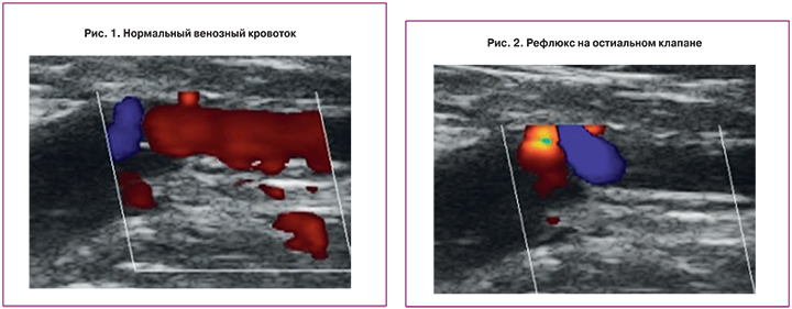
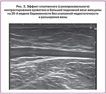
При ультразвуковом сканировании венозную систему исследуют на нескольких уровнях. Наиболее важной задачей является исключение тромбоза глубоких и подкожных вен даже при отсутствии симптоматики. Следует отметить, что только ЦДС позволяет абсолютно точно подтвердить или опровергнуть подозрение на тромбоз. Другим направлением исследования вен является оценка работы клапанного аппарата глубоких и подкожных вен на разных уровнях. Режим допплеровского картирования кровотока позволяет выявить наличие и выраженность ретроградного тока крови на клапанах (рис. 1, рис. 2) при проведении функциональных тестов в горизонтальном и вертикальном положениях. Современное оборудование позволяет выявить так называемое «позднее закрытие клапанов», которое не является проявлением клапанной недостаточности, но может быть свидетельством особенностей их строения и первым сигналом их недостаточной работы при нагрузках. Кроме функциональных изменений обязательно оценивают и некоторые анатомические характеристики: расширение основных стволов подкожных вен, расширение их притоков, варикозное расширение стволов и притоков, расширение перфорантных вен [8, 9].
Данная последовательность исследования позволяет определить локализацию и степень выраженности патологических изменений в венах для выбора тактики лечения, прогнозировать возможность развития варикоза, разработать адекватную стратегию профилактических мер, прогнозировать состояние венозной системы в послеродовом периоде у беременных. Одной из особенностей варикозной болезни у беременных является выраженность симптоматики, в том числе отеков, на фоне равномерного расширения стволов больших и малых подкожных вен, преобладание относительной клапанной недостаточности. Такие изменения в венозной системе, как правило, подвержены значительному регрессу в послеродовом периоде. В случае значительного неравномерного расширения притоков и особенно стволов подкожных вен прогноз в послеродовом периоде не может быть благоприятным. Пациентка должна быть ориентирована на необходимость повторного исследования венозной системы после родов и консультацию сосудистого хирурга с целью выработки оптимальной тактики лечения.
Необходимо отметить, что почти у всех беременных, особенно в третьем триместре, при ультразвуковом исследовании в серошкальном изображении отмечается эффект спонтанного (самопроизвольного) контрастирования потоков крови в больших подкожных венах (рис. 3). Это гемореологический феномен, особенно хорошо фиксируемый на аппаратах экспертного класса; становится заметным ток крови из-за сладж-феномена на фоне замедления тока крови [1]. Данный эффект является нормальной эхографической находкой и не является предвестником тромбозов [10].
Еще одной особенностью варикозной болезни во время беременности является расширение притока с передней поверхности брюшной стенки, половых губ, варикозное расширение вен таза и прямой кишки.
Ведение пациентов с варикозной болезнью вен нижних конечностей состоит из хирургического, медикаментозного и немедикаментозного лечения. Согласно данным Кокрановского (Cochrane) обзора 2015 г., при ведении беременных с варикозом предпочтительны немедикаментозные методы лечения, направленные на уменьшение симптоматики и замедление прогрессирования расширения вен. Оперативное лечение беременным пациенткам проводят только в случае такого тяжелого осложнения, как восходящий тромбофлебит подкожных вен [3, 7, 11].
Основным методом консервативной терапии является эластическая компрессия. При этом предпочтение необходимо оказывать медицинскому компрессионному трикотажу, так как правильное компрессионное бандажирование с помощью бинтов для беременной женщины затруднительно.
Согласно данным того же Кокрановского (Cochrane) обзора 2015 г., широко использующиеся флеботропные препараты у беременных не показаны, так как их эффективность и безопасность не были должным образом подтверждены [3]. Однако некоторые из них имеют широкий опыт клинического применения, в том числе и у беременных женщин. В «Национальных рекомендациях» 2014 года особо отмечаются препараты группы флавоноидов (микронизированные фракции), во многих исследованиях показавшие выраженное противоотечное действие, без выраженных побочных эффектов [7]. Поэтому при выраженной отечности и трудностях с ношением компрессионного трикотажа данные препараты могут использоваться во время беременности. Из методов профилактики отеков у беременных наибольшим клиническим эффектом обладают отдых и возвышенное положение ног [3].
В настоящее время считается, что при должном обследовании и предоставлении беременным информации о заболевании в большинстве случаев удается снизить риск возникновения и прогрессирования варикозной болезни [12].
Для коррекции или отмены терапии проводят ультразвуковое исследование венозной системы ног через 3 месяца после родов. К этому времени у основной массы пациенток анатомические и функциональные изменения вен, присутствовавшие на фоне беременности, возвращаются к исходному уровню [13].
Заключение
Таким образом, помня об анатомических и функциональных изменениях в венозной системе нижних конечностей, всем женщинам во время беременности, особенно при повторной беременности, при наличии жалоб, при наличии факторов риска варикозной болезни, необходимо рекомендовать ультразвуковое исследование вен (УЗДАС, ЦДС). Данное исследование может быть рекомендовано уже в первом триместре беременности, для оценки риска развития или прогрессирования варикозной болезни, перед и после родоразрешения, в том числе для исключения тромбоза. Оценку послеродовых изменений венозной системы проводят не ранее чем через 3 месяца. Всем пациенткам рекомендовано ношение компрессионного трикотажа, который подбирается флебологом на основании данных ультразвукового исследования, а также отдых лежа с возвышенным положением ног.


