Медицинская и социальная значимость проблемы глубоко недоношенных детей и сегодня сохраняет свою крайнюю актуальность, в первую очередь в связи с тем, что кажущаяся на первый взгляд невысокой в общей когорте новорожденных частота рождения младенцев с очень низкой (0,66%) и экстремально низкой (0,35%) массой тела формирует высокую долю структуры ранней неонатальной смертности, достигающей почти 49% (по данным формы № 32 Росстата, 2015).
Глубоко недоношенные дети, особенно с очень низкой (1500 г и менее) и экстремально низкой (1000 г и менее) массой тела рождаются, как правило, в тяжелом состоянии, требующем проведения реанимационных мероприятий и интенсивной терапии. Организация высокотехнологичной системы выхаживания в условиях перинатальных центров страны обеспечивает выживание глубоко недоношенных детей – одной из самых тяжелых категорий пациентов. Однако глубокая незрелость, функциональная полигландулярная и органная недостаточность при рождении определяют сложный, мало предсказуемый процесс дальнейшего формирования организма ребенка и повышенный риск развития инвалидизирующей патологии [1–3].
Наиболее часто отсроченные неблагоприятные исходы у глубоко недоношенных детей обусловлены психоневрологической патологией: детским церебральным параличом (ДЦП), гидроцефалией, нарушениями двигательного, речевого, когнитивного и психоэмоционального развития, а также расстройствами зрения и слуха [4–6]. Так, среди выживших ДЦП встречается у 8–15%, что в 40–100 раз чаще по сравнению с его частотой у доношенных детей; обусловленные тяжелой ретинопатией глубокие нарушения зрения, нередко приводящие к слепоте, регистрируются у 1–6%, нарушения слуха – у 2,3% [5, 7]. При этом когнитивные, моторные и поведенческие расстройства наблюдаются даже при отсутствии таких перинатальных поражений мозга, как внутрижелудочковые кровоизлияния и перивентрикулярная лейкомаляция [5–9]. Отдаленные расстройства в виде трудностей школьного обучения, освоения чтения и письма, необходимости в специальном обучении отмечаются почти у половины глубоко недоношенных детей [4, 10, 11]. При этом по мере уменьшения гестационного возраста риск неблагоприятных психоневрологических исходов прогрессивно увеличивается [2–11].
В условиях, когда критический период активного развития коры, увеличения массы и площади ее поверхности, появления борозд и извилин головного мозга, тонкой дифференцировки его структур протекает не во внутри-, а во внеутробных условиях, безусловно, неблагоприятное воздействие на незрелую нервную систему оказывают световые, звуковые, тактильные и болевые раздражители. Созревание головного мозга могут ухудшать также заболевания неонатального периода, нутритивные проблемы, нарушение оксигенации и гемодинамики, характерные для таких детей [12–14]. Все вышеизложенное определяет неослабевающую заинтересованность в изучении с помощью современных методов нейровизуализации закономерностей постнатального развития головного мозга у глубоко недоношенных детей с целью поиска ранних предикторов неблагоприятных неврологических исходов.

Магнитно-резонансная томография (МРТ) рассматривается в качестве наиболее информативного метода оценки головного мозга у недоношенных детей. Однако продолжительность исследования, невозможность его многократного повторения, необходимость транспортировки с нарушением щадящего режима выхаживания и анестезиологического пособия (седации) резко ограничивают использование метода для рутинного или серийных обследований этой категории пациентов. В то же время лишенное указанных недостатков эхографическое сканирование представляется перспективным как для первичной диагностики, так и для динамического наблюдения за развитием структур головного мозга у глубоко недоношенных детей. Результаты МРТ-исследований продемонстрировали, что задержка развития коры головного мозга, замедление гирификации и недостаточная площадь ее поверхности могут рассматриваться в качестве неблагоприятных прогностических признаков для последующего неврологического развития [9, 15]. Вместе с тем возможности ультразвуковых методов визуализации, в том числе трехмерной (3D) нейросонографии, для оценки созревания коры головного мозга у глубоко недоношенных детей отражены лишь в единичных научных публикациях [16, 17].
Цель нашего исследования – определить с помощью 3D-нейросонографии этапы формирования коры головного мозга и сроки первичной эхографической визуализации его борозд у глубоко недоношенных детей до достижения постконцептуального возраста 40 недель.

Материал и методы исследования
В исследование включены 39 детей (22 мальчика и 17 девочек), родившихся в сроке от 25 до 31 недели беременности с массой тела от 640 до 1490 г (1102±218 г М±SD) в ФГБУ НЦАГиП им. В.И. Кулакова МЗ РФ. Из их числа 25 детей родились от многоплодной, а 14 – от одноплодной беременности. «Гестационный возраст» устанавливался традиционно, с учетом первого дня последней менструации и показателя копчико-теменного размера плода антенатального ультразвукового исследования 1-го триместра беременности.
Оценка физического развития (масса тела, длина, окружность головки) недоношенных детей осуществлялась по центильным графикам Фентона 2013 года. В исследование были включены новорожденные, степень развития которых превышала 10-й центиль.
Эхографическое сканирование головного мозга проводилось около 2 минут через большой родничок с помощью педиатрического конвексного 3D-датчика частотой 5–9 МГц переносного прибора Voluson I (Дженерал Электрик, США) при непосредственном нахождении ребенка в инкубаторе. Начиная с 1–3-х суток жизни исследования повторялись еженедельно, до момента выписки младенца из стационара. Всего было проведено 273 ультразвуковых исследования.
При отборе исключались недоношенные дети с задержкой внутриутробного развития, генетическими заболеваниями, врожденными пороками развития центральной нервной системы, внутрижелудочковыми кровоизлияниями 2-й и 3-й степени, перивентрикулярной лейкомоляцией и наличием других тяжелых деструктивных изменений головного мозга. Сканирование производилось в двух проекциях: среднесагиттальной и коронарной.
После активации 3D-режима полученные при каждом заборе в трех ортогональных плоскостях (сагиттальная, коронарная, аксиальная) изображения головного мозга архивировались. Для анализа 3D-цифровых файлов применялись мультиплоскостной (рис. 1В), мультипланарный (рис. 1 С, D) и поверхностный режимы (рис. 1А). Поверхностный режим (Surface rendering) при исследовании поверхностных структур головного мозга предоставлял отчетливую картину извитости борозд и извилин (рис. 1А). Мультипланарный режим (Multiplanar view) – основа объемного ультразвукового исследования обеспечивал визуализацию изображения одновременно в трех или четырех плоскостях (рис. 1С, D). При перемещении «точки интереса» в каждую из плоскостей осуществлялся анализ множества срезов эхографического изображения головного мозга. Применение мультиплоскостного режима (Tomographic ultrasound imaging – TUI) позволяло выбрать толщину «среза» полученного объема головного мозга в одной из плоскостей и получить на экране в виде томограмм параллельные послойные изображения интересующих структур (рис. 1В).
Исследованию подвергались следующие борозды головного мозга: обонятельные, орбитальные, поясные, лобные, верхние височные, теменно-затылочная и шпорная. Также изучалась островковая доля мозга (островок): сроки его закрытия, появление длинных и коротких борозд. При этом для анализа обонятельных и орбитальных борозд использовалась коронарная проекция, для остальных – и сагиттальная, и коронарная проекции, преимущественно в мультиплоскостном режиме. Характеризуя структуры борозд, оценивали следующие признаки: гиперэхогенность в проекции будущей борозды, четкость эхо-контура борозды, фрагментация борозды, появление изгибов борозды, наличие вторичных и третичных борозд.
Результаты исследования
Большинство глубоко недоношенных детей, включенных в исследование, родились путем операции кесарева сечения (38/97%). Среди доминирующих соматических заболеваний и патологии беременности были: артериальная гипертензия (5/13%), угроза прерывания беременности (29/74%), преэклампсия (5/13%), гестационный сахарный диабет (4/10%), холестаз (3/8%). Показаниями к досрочному оперативному родоразрешению служили: начало родовой деятельности (9/23%), преждевременное излитие околоплодных вод (8/21%), в том числе при высоком боковом надрыве плодного пузыря (5/13%), нарастание тяжести преэклампсии (5/13%), отслойка нормально расположенной плаценты (5/13%), при хронической плацентарной недостаточности – резкая отрицательная динамика показателей фетоплацентраного кровотока (4/10%), не купируемое нарастание холестаза (3/8%).
Все глубоко недоношенные дети для стабилизации дыхательной функции уже в родильном зале требовали проведения респираторной поддержки. В длительной респираторной поддержке при сформированной бронхо-легочной дисплазии нуждались 7/18% детей. Большинство детей (35/90%) в постконцептуальном возрасте 36–45 недель были выписаны из стационара в удовлетворительном состоянии с неврологическим статусом, соответствующим указанному сроку. Четыре младенца по достижении 1–1,5 месяцев в стабильном состоянии были переведены в другие стационары.
Для изучения возможного влияния «многоплодности» на формирование борозд головного мозга у 25 глубоко недоношенных новорожденных от многоплодной и у 14 – от одноплодной беременности сравнительный анализ сроков ультразвукового появления борозд проведен в зависимости от паритета.
В ходе проведенной работы установлено, что наиболее рано выявляемыми бороздами головного мозга при эхографическом исследовании являлись шпорная и теменно-затылочная, визуализация изображения которых на медиальной поверхности затылочной доли была возможна уже при первом сканировании у всех детей, в том числе рожденных в 25 недель гестации.
В большинстве случаев в этом сроке данные борозды представлялись фрагментированными структурами, прямолинейность и четкость контуров которых определялись к 28-й неделе, а при дальнейшем развитии обнаруживались характерные для них изгибы (рис. 2).
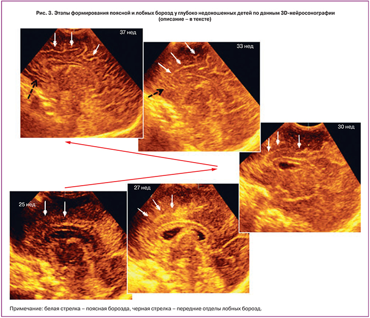
Эхографическое появление контура поясной борозды, как показал проведенный нами анализ, наблюдалось в возрасте 27 недель. При этом в парасагиттальной плоскости данная борозда отчетливо визуализировалась в виде линии, располагающейся над мозолистым телом. В дальнейшем к 29-й неделе формировались ее изгибы. Ранее, на сроках 25–26 недель гестации, в проекции поясной борозды представлялось возможным визуализировать зону гиперэхогенности и появляющиеся нечеткие фрагменты будущей борозды (рис. 3).
Формирование вторичных борозд поясной борозды у 14/36% детей приходилось на 31-ю неделю, а более чем у 2/3 (31/79%) – на 32-ю неделю, что совпадало с первичным эхографическим выявлением передних отделов лобных борозд. Так, передние отделы последних визуализировались в этом возрасте у 27/69% пациентов, в то время как в 33 недели – практически у всех обследуемых (38/97%). К постконцептуальному возрасту 34–35 недель на фоне преобладания в эхографической семиотике изображений первичных борозд наблюдалось дальнейшее увеличение длины и количества вторичных, а к 37–40 неделям – по всей видимой медиальной поверхности полушарий констатировалось развитие «сети» хорошо выраженных, с прогрессивным увеличением изогнутости борозд (рис. 3).
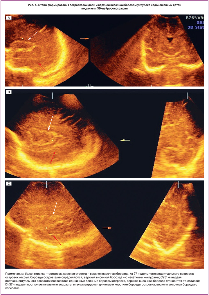
Закрытие островка мозга, по данным проведенного нами анализа, происходило в возрасте 31–32 недели; также в это же время начинали визуализироваться единичные длинные борозды островка, количество которых прогрессивно возрастало к 34-й неделе. Короткие борозды островка на сканограммах появлялись только после 34-й недели (рис. 4).
Верхняя височная борозда при исследовании в парасагиттальном срезе в сроке до 26–27 недель имела размытые прерывистые контуры, а с 28-й недели в большинстве случаев становилась отчетливой прямой линией, повторяющей задний контур островка. Формирование ее изгибов приходилось на возраст 34 недели (рис. 4).
При объемной нейросонографии обонятельные и орбитальные борозды исследовались в коронарной плоскости и представляли собой линейные структуры, располагающиеся по обе стороны от межполушарной щели на нижней поверхности лобной доли. Обонятельные борозды визуализировались в непосредственной близости от межполушарной щели и определялись у всех обследуемых с 27–28 недель. Вместе с тем латеральнее от обонятельных, выявление орбитальных борозд оказалось возможным гораздо позже, только на 32–34-й неделе.
В результате сравнительного анализа статистически значимых различий в сроках эхографической визуализации изучаемых борозд головного мозга у недоношенных новорожденных от многоплодной и одноплодной беременностей не выявлено.
Обсуждение
Диагностика формирования коры головного мозга с помощью современных методов визуализации для выявления ранних объективных критериев прогнозирования неврологических расстройств у глубоко недоношенных детей остается крайне актуальной [9, 15, 17]. Несмотря на возросшее количество публикаций, содержащих эхографические данные о поэтапном (последовательном) созревании коры головного мозга плодов в различные сроки беременности, до настоящего времени отсутствуют общепринятые критерии первичного ультразвукового выявления ее борозд. Кроме того, также недостаточно изучены диагностические возможности 3D-нейросонографии в процессе анте- и постнатального формирования коры головного мозга у глубоко недоношенных детей.
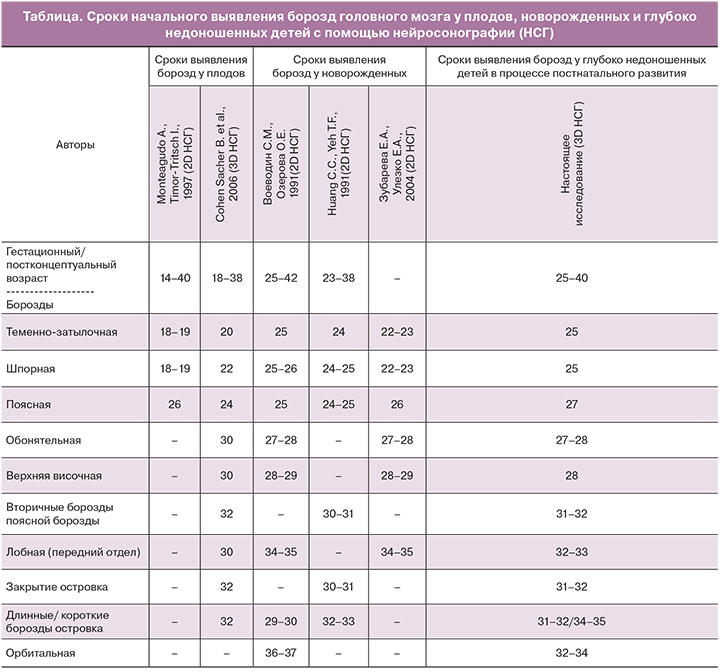
Проведенное нами исследование позволило представить основные закономерности эхографического выявления борозд головного мозга у рассматриваемой категории пациентов в процессе их постнатального развития. При сравнении результатов антенатальных ультразвуковых исследований сроки выявления ряда борозд (поясной, обонятельной, верхней височной, лобной) у плодов отличались от полученных нами при 3D-нейросонографии данных на 2–3 недели, как в большую, так и в меньшую сторону [18, 19] (таблица).
При сравнении представленной в современной научной литературе результатов 2D-эхографических исследований у новорожденных, родившихся в различные сроки беременности, были выявлены как сходства, так и отличия [14–16]. Так, всеми авторами отмечалось наличие теменно-затылочной и шпорной борозд при гестационном возрасте 24–25 недель. Также оказались сравнимы сроки первичной эхографической визуализации для обонятельных, верхних височных и вторичных борозд поясной борозды. В то же время на основании проведенного нами исследования поясная борозда выявлялась на 2 недели позже, а передние отделы лобной и орбитальные борозды – на 2–3 недели раньше, чем в работах других авторов (таблица).
Детальный анализ серийных срезов архивированных данных позволил нам констатировать определенную этапность формирования изображения борозд в процессе постнатального развития коры головного мозга: первоначально – в виде зоны гиперэхогенности, позднее – выявление размытых нечетких ее фрагментов и, наконец, дальнейшие формирование четкой линейной структуры (или четких линейных фрагментов, при условии сохранения фрагментарного строения борозды).
Таким образом, описанные выше несоответствия сроков эхографического выявления борозд можно объяснить как различиями в качестве визуализации, так и различной интерпретацией авторами полученных ультразвуковых изображений. В нашем исследовании сроком первичного выявления борозды считалась визуализация четкой эхо-структуры борозды или четких линейных фрагментов в ее проекции.
Согласно результатам отдельных авторов, на формирование борозд головного мозга может влиять «многоплодность». Так, у плодов от многоплодной беременности по сравнению с одноплодной отставание в развитии борозд достигало 3 недель [23, 24]. Однако нам не удалось выявить четкие различия в сроках эхографической первичной визуализации борозд головного мозга у глубоко недоношенных детей, родившихся как от одноплодной, так и от многоплодной беременностей.
Следует принять во внимание, что согласно протоколу, из нашего исследования исключались дети с задержкой внутриутробного развития, у которых, по данным ряда публикаций, имелись особенности формирования коры головного мозга, имеющие на МРТ характер дискордантного процесса с относительным уменьшением поверхности коры по отношению к «индексу сулькации» [23, 24].
Заключение
3D-нейросонография предоставляет возможность в течение короткого времени получить эхографическое изображение головного мозга в трех режимах (поверхностном, мультиплановом и мультиплоскостном), и с помощью последующего изучения архивированных данных провести детальный анализ полученных изображений. Благодаря своим преимуществам данный метод является приоритетным как при первичном обследовании структур коры головного мозга, так и при мониторинге их развития у глубоко недоношенных детей.
Результаты проведенного исследования, отражающие процесс формирования и сроки выявления борозд головного мозга, могут быть использованы для оценки постнатального структурного развития коры головного мозга у глубоко недоношенных детей с помощью ультразвуковых методов визуализации.
Необходимы дальнейшие исследования по изучению формирования коры головного мозга у различных групп новорожденных и недоношенных детей, с разработкой линейных и объемных характеристик, для использования их при прогнозировании неврологического, психического и когнитивного развития.