Традиционным методом определения вида, позиции и уровня положения головки плода во время родов является влагалищное исследование. Тем не менее данный метод является субъективным и недостаточно точным. Частота ошибки определения прохождения головкой плоскости узкой части полости малого таза может превышать 30–40% вне зависимости от опыта врача. Неточности в определении положения головки плода в полости таза негативно отражаются на тактике ведения родов и могут приводить к неблагоприятным перинатальным исходам [1, 2].
Объективизировать оценку положения головки плода в родах позволяет ультразвуковое исследование (УЗИ). Использование данного метода может служить дополнительным инструментом, определяющим тактику ведения родов и диагностику возможных осложнений [3–5]. Выделенный в качестве диагностически значимого ультразвуковой параметр оценки динамики течения родов должен удовлетворять следующим требованиям: простота измерения, воспроизводимость, связь с положением головки, продолжительностью и исходом родов. В настоящее время известно около десятка таких эхографических показателей, которые используют для прогнозирования течения родов. Тем не менее на сегодняшний день не существует единой точки зрения на показания, время проведения исследования, интерпретацию отдельных показателей и анализ полученных результатов [6].
В связи с вышеизложенным, целью данной работы явилось определение показателей УЗИ в родах, которые наиболее точно отражают динамику их течения. Выделение этих параметров позволит объективизировать оценку течения родов и будет способствовать ранней диагностике осложнений.
Материалы и методы
В исследование были включены 120 пациенток, родоразрешенных в родильном отделении ФГБУ «НМИЦ АГП им. В.И. Кулакова» Минздрава России с мая 2017 по сентябрь 2018 гг.
Критерии включения: доношенная одноплодная беременность, головное предлежание плода, информированное согласие пациентки на участие в исследовании.
Для ультразвукового обследования использовали аппарат Medison MySono-U6 с конвексным датчиком для трансабдоминального сканирования с частотой 3,5–7,0 МГц. Обследование пациентов проводили путем УЗИ в В-режиме. Определение положения головки плода проводили путем трансабдоминального исследования в положении пациентки на спине при пустом мочевом пузыре в соответствии с методикой, описанной Rayburn et al. [7]. При эхографии для определения продвижения головки плода датчик располагали в сагиттальной плоскости ниже лобкового симфиза между половыми губами в соответствии с методикой, описанной Barbera et al. [8].
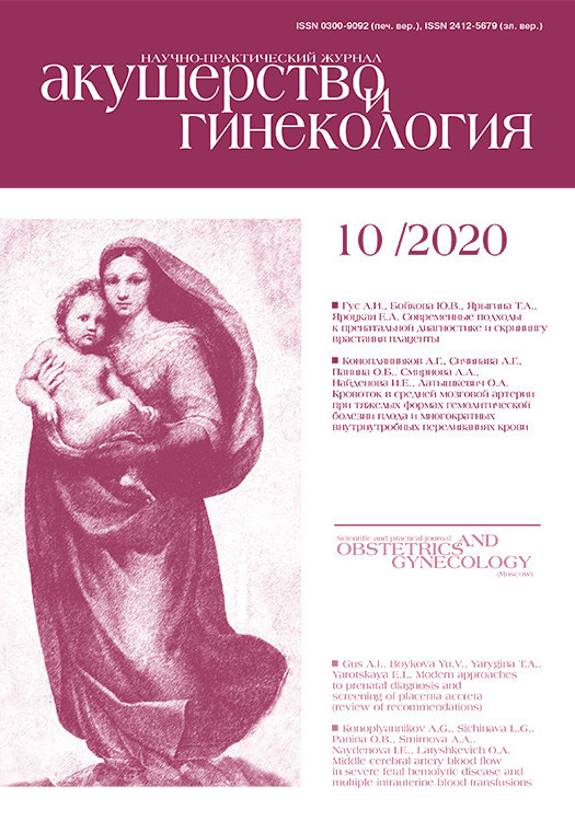
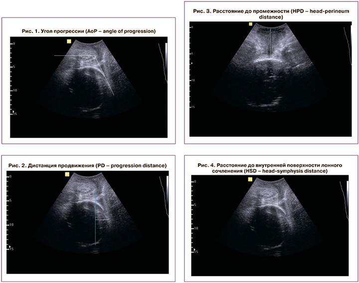
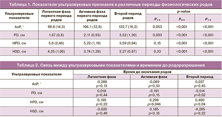
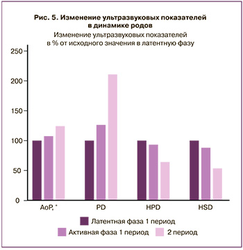
С помощью УЗИ проводили оценку 4 показателей, которые, по данным литературы, наиболее часто используются для характеристики течения родового акта [2, 9, 10]: угол прогрессии (AoP – angle of progression) измеряли при трансперинеальном продольном сканировании. Датчик устанавливали по средней линии лонного сочленения, чтобы его изображение располагалось горизонтально. Первый вектор строили от нижнего до верхнего края лонного сочленения, второй – от нижнего края лона по касательной к контуру головки плода (рис. 1) [8]; дистанцию продвижения (PD – progression distance) измеряли трансперинеально при продольном сканировании. Изображение лонного сочленения выводили горизонтально, как при измерении угла прогрессии. Первый вектор проводили вертикально вниз от нижнего края лонного сочленения (linia infrapubica). Измеряли расстояние от linia infrapubica до наиболее удаленной точки головки плода (рис. 2) [11]; расстояние до промежности (HPD – head-perineum distance) (наименьшая дистанция между промежностью и головкой плода) измеряли при трансперинеальном поперечном сканировании в горизонтальном направлении от задней спайки до контура головки (рис. 3) [12]; расстояние до внутренней поверхности лонного сочленения (HSD – head-symphysis distance) измеряли при трансперинеальном продольном сканировании от нижнего края середины внутренней поверхности лона до контура головки плода (рис. 4) [13]. Определение значений выбранных ультразвуковых показателей осуществляли в начале латентной, активной фазы первого периода и начале второго периода родов.
Статистический анализ
Статистическую обработку данных и построение графиков выполняли при помощи электронных таблиц Microsoft Excel и пакета программ GraphPad Prism 6 (GraphPad Software, USA). Для определения нормальности распределения использовали обобщенный тест Д’Агостино–Пирсона. Все данные были определены как параметрические, поэтому при их анализе рассчитывали среднее значение и среднеквадратичное отклонение; при множественном сравнении использовали одномерный дисперсионный анализ повторных наблюдений (Repeated measures ANOVA), в случае наличия различий проводили тест Turkey для попарного сравнения. Для выявления корреляции между признаками определяли коэффициент корреляции Пирсона. При значении коэффициента корреляции от 0 до 0,299 связь расценивали как слабую, от 0,3 до 0,699 – как среднюю, от 0,7 до 1 – как сильную. Различия считали значимыми при p<0,05.
Результаты
Все пациентки, которые вошли в исследование, имели одноплодную беременность доношенного срока (в среднем 39,4 (0,9) недели) с головным предлежанием плода. Во всех случаях роды завершились через естественные родовые пути без применения каких-либо оперативных методов родоразрешения. 82 (68,3%) пациентки были первородящими, 38 (31,7%) – повторнородящими. Средний возраст пациенток, включенных в исследование, составил 30,2 (4,4) года, индекс массы тела – 25,8 (3,8) кг/м2. Преиндукция родов была применена у 30 (25%) пациенток; эпидуральная аналгезия в родах – в 63 (52,5%) наблюдениях; инфузия окситоцина с целью коррекции родовой деятельности – у 38 (31,7%) пациенток. Средняя продолжительность первого периода родов составила 523,7 (169,6) минуты, второго периода родов – 77,7 (49,3) минуты, из них потужной период составил 20 (14,5) минут. Средняя величина послеродовой кровопотери составила 315 (76) мл.
В первом периоде родов передний вид затылочного предлежания имел место в 56 (43,7%), задний вид – в 64 (53,3%), 1-я позиция – в 72 (60%), 2-я позиция – в 48 (40%) наблюдений. При анализе положения после рождения в переднем виде оказалось 95 (79,2%), в заднем виде – 25 (20,8%), в 1-й позиции – 65 (54,2%), во 2-й позиции – 55 (45,8%) новорожденных.
Состояние всех детей по шкале Апгар оценено от 7 до 9 баллов. Вес новорожденных колебался от 2800 до 4280 (3452,0 (389,8)) г, длина тела – от 49 до 58 (52,0 (2,1)) см, окружность головки – от 33 до 39 (35,0 (1,4)) см.
Оценка результатов эхографического исследования в процессе неосложненных родов демонстрировала прогрессивное увеличение показателей AoP и PD в сочетании с уменьшением HPD и HSD (табл. 1).
 АоР в латентной фазе первого периода родов составлял в среднем 98,8˚. К началу активной фазы первого периода родов он увеличивался на 7,4%, а к началу второго периода родов – еще на 24,2% от исходного значения в латентную фазу (p<0,001) (рис. 5).
Динамика изменения PD была более выраженной. Так, по сравнению со значением в латентной фазе, к началу активной фазы первого периода родов показатель прироста составил 26,3%, а к началу второго периода – 110,8%.
Изменение HPD между латентной и активной фазами первого периода родов было незначительным и составляло в среднем 5,6 см (6,8%) (p=0,15), однако существенно (на 35,9%) сокращалось к началу второго периода родов (p<0,001).
HSD к активной фазе родов уменьшалось на 12% (p=0,33). К началу второго периода родов уменьшение было уже значительным и составляло в среднем 46,6% от исходного значения в латентную фазу (p<0,001).
На следующем этапе работы мы проанализировали связь между значениями ультразвуковых показателей и временем до родоразрешения (табл. 2). В латентной фазе первого периода родов значимой связи между ультразвуковыми показателями и продолжительностью родов выявлено не было. В активной фазе родов была выявлена положительная слабая корреляция между временем до окончания родов и HPD (r=0,298; p=0,046). Во втором периоде родов была выявлена средняя положительная связь времени до окончания родов с HPD (r=0,40; p=0,04) и средняя отрицательная – с PD (r=-0,514; p=0,02).
Обсуждение
УЗИ в родах позволяет более точно определить положение головки плода в полости таза матери. Как показали полученные нами результаты, положение головки плода затылком кзади в первой позиции является наиболее часто встречающимся вариантом в первом периоде родов. Однако рождение в большинстве наблюдений происходит затылком кпереди, что подтверждает поворот плода в полости таза во втором периоде родов.
Согласно практическим рекомендациям Международного общества ультразвука в акушерстве и гинекологии (ISUOG), основными ультразвуковыми показателями, отражающими механизм неосложненных родов, являются АоР и HPD, а дополнительными – PD и HSD. Однако в представленной литературе недостаточно сведений, позволяющих расценивать вышеуказанные показатели в качестве предикторов исхода родов.
Вместе с тем T. Chi et al. (2018) подтверждают, что определение AoP является точным и легко воспроизводимым методом определения положения головки плода в родах [6].
Согласно данным B. Tutschek et al. (2013), в течение родов AoP изменяется от 84° (положение головки «-3») до 170° (положение головки «+5») [14]. В нашем исследовании показатель AoP находился в диапазоне от 98,8° до 122,7°. Полученные отличия максимальных значений AoP, по-видимому, можно объяснить проведением измерений при уровне положения головки «+3».
PD отнесена ISUOG к дополнительным методам оценки положения головки плода, поскольку данный показатель реже используется в клинической и научной практике. В практических рекомендациях ISUOG вместо PD предлагается рутинно использовать AoР [6]. Согласно полученным данным, показатель PD демонстрирует динамичное и достоверное увеличение с течением родов, что позволяет рассматривать его в качестве одного из критериев продвижения головки плода.
HPD отражает расстояние, которое предстоит пройти головке плода по родовому каналу [15]. Следовательно, уменьшение его с течением родов является объективным признаком опускания головки плода. Несмотря на то что HSD отнесено к дополнительным методам, тем не менее, его значения, являясь простым и воспроизводимым ультразвуковым показателем, четко коррелируют с положением головки плода: чем меньше HSD, тем ниже располагается предлежащая часть [6, 16].
Проведенное нами исследование показало, что все четыре изученных показателя проявили значимую динамику при переходе от активной фазы первого периода родов ко второму периоду родов, что закономерно отражает механизм родов. Однако только AoР и PD достоверно менялись от латентной к активной фазе первого периода. При этом динамика изменения PD была наиболее значимой (более 100% от исходного значения).
Одним из наиболее сложных вопросов при определении тактики ведения родов является прогнозирование их течения, которое опирается на временные рамки. В широкой клинической практике обычно используют усредненный подход, основанный на средней скорости раскрытия шейки матки. Однако определение степени раскрытия шейки матки является недостаточно объективным показателем. Также на точность результата влияет степень сглаженности шейки матки, толщина ее стенок. В связи с этим представляет интерес поиск объективных критериев продвижения головки в связи с продолжительностью родов.
Заключение
Полученные нами результаты показали, что наиболее часто применяемые ультразвуковые параметры продвижения головки, оцениваемые в первом периоде родов, неинформативны или мало информативны для прогнозирования их продолжительности и закономерно отражают лишь механизм родов (период раскрытия шейки матки). Однако во втором периоде родов (период изгнания плода) показатели PD и HPD демонстрируют значимую связь с продолжительностью родов, что позволяет использовать их для оценки динамики родового процесса.
Результаты проведенного исследования свидетельствуют о необходимости дальнейшего изучения возможностей интранатальной ультразвуковой диагностики положения головки плода, что позволит объективизировать оценку течения родового акта и улучшит прогнозирование осложнений.