
Surgical treatment for multiple uterine leiomyoma with intravascular leiomyomatosis
Background. Cases of intravascular leiomyomatosis are extremely rare, and the treatment of patients with this pathology is of particular interest in terms of surgical tactics.Zavaruev A.V., Mazurenko A.A., Shumsky S.V., Solovyev V.V., Zavarueva I.G.
Description. The article describes a clinical case of successful surgical treatment for multiple uterine leiomyoma with intravenous and intracardiac leiomyomatosis. The patient was operated on urgently. Thrombus leiomyomectomy from the right atrium, right ventricle, all segments of the inferior vena cava and right iliac vein with hysterectomy without appendages was performed via total median sternolaparotomy in the presence of temporary occlusion of the vena cava and pulmonary artery. The postoperative period was uncomplicated. The patient was discharged from hospital in a satisfactory condition on the 14th day.
Conclusion. The treatment could achieve not only the main goal of the operation to prevent massive pulmonary thromboembolism, but could also follow the ablastic principles of treatment for uterine leiomyomas.
Keywords
leiomyomatosis
leiomyoma
uterus
inferior vena cava
Tumor thrombosis of the inferior vena cava and the right chambers of the heart is not common and is mainly due to the cancerous process in the organs of the urinary system [1, 2]. Leiomyomas of the uterine body in rare cases can cause widespread tumor invasion through the uterine, internal iliac and inferior vena cava and reach the right parts of the heart [3, 4]. Further progressive growth of the tumor thrombus increases the risk of death due to the narrowing of the tricuspid valve or pulmonary embolism [5]. Surgical treatment of intravenous leiomyomatosis in such cases is not an easy task, which requires the involvement of specialists of different profiles and even the use of cardiopulmonary bypass.
Clinical observation
In June 2017, a 41-year-old patient S. was admitted to the Emergency Department of the Amur Regional Clinical Hospital with complaints of pressing pain behind the sternum. The pain was not relieved by nitrates, the patient’s condition became worse within a month.
There were not any operations or injuries in the past history. She had pneumonia a few years ago. There were no infectious and other diseases. Her menstrual cycle was regular; she had two pregnancies and two deliveries.
On examination her condition was severe, consciousness was clear. She was of the normosthenic type. Body mass index was 29 kg/m2. Body temperature was normal. Skin and mucous membranes were of normal color, the patient exhibited acrocyanosis. Feet and lower legs were slightly swollen. Peripheral lymph nodes and thyroid gland were not enlarged. No abnormality detected in organs of the musculoskeletal system. Breathing was normal through the nose. The respiratory rate was 18 breaths per minute. Oxygen saturation was 98%. On auscultation the breath was vesicular, without any sounds. The pulse was rhythmic, its rate was 86 beats per minute. The heart sounds are muffled and rhythmic. Blood pressure measured in both arms was 120/80 mm Hg. The tongue was uncoated. The abdomen showed the correct form, without bloating, on palpation it was soft and not tender. Palpation revealed the growth of 10×15×20 cm size, which was painless and had tight and elastic consistency, bumpy. The liver did not extend beyond the edge of the costal arch. The spleen was not palpable. Stools and defecation were normal. There were no changes in the lumbar region. Murphy’s punch sign was negative. The kidneys were not palpable. The color of the urine was normal, urine output was adequate.
Blood tests were within normal limits. Troponin test was negative. Electrocardiogram did not reveal ischemia; there was sinus rhythm, tachycardia, and increased load on the right heart chambers. Sonographic examination detected floating thrombus in the right chambers of the heart extending the inferior vena cava, the dilation of the right atrium, no hypokinetic zones. Ejection fraction was 67%, pressure in the right ventricle was 25 mm Hg (mild pulmonary hypertension). Ultrasonography revealed diffuse changes in the pancreatic parenchyma, the induration of the pelvicalyceal system of both kidneys. There was a heterogeneous tumor in the pelvis, its size was 128×178×215 mm. Occlusive thrombosis of the internal iliac vein with non-occlusive outflow to the common iliac vein and inferior vena cava along all their length was detected. All the rest veins were passable.
Computer tomography angiography revealed thrombosis of the inferior vena cava, right atrium and right ventricle. The giant cystic solid multichamber tumor in the pelvic cavity up to 22 cm in the maximum vertical dimension was identified.
 The patient was urgently operated on. Complete median sterno-laparotomy was performed. There was a small amount of effusion of the light color in the abdomen. The uterus had a size of that up to 20 weeks of pregnancy, it had numerous nodes of different sizes and localization passing to the cervix. The ovaries and tubes were not changed. Lymph nodes were not enlarged. There was up to 200 ml of light effusion in the pericardium. The right chambers were dilated, and a blood clot was palpated. During the operation the superior vena cava and pulmonary trunk were blocked. The blood circulation was stopped. The right atrial appendage was opened. Tumor thrombus of 2.5×5 cm size was removed, it continued distally into the inferior vena cava, it was resected. The inferior vena cava was clamped. The clamp was applied on the atrial appendage, tourniquets from the pulmonary trunk and superior vena cava were removed. The blood circulation restored (the time of arrest was 60 sec). The atrial appendage was sutured. The inferior vena cava was mobilized along all its length. Tourniquets were applied on the renal, hepatic veins, and infrarenally. Longitudinal cavatomy was performed and tumor thrombus was removed along all its length. The blood flow in the vena cava restored. It was impossible to mobilize infrarenal and iliac sections due to the enlarged size of the uterus. Extirpation of the uterus without appendages was carried out. The iliac veins on the right and the vena cava were mobilized infrarenally. The tourniquets were applied, the longitudinal operation on the common iliac vein was performed. The rest tumor thrombus was removed from the vena cava, common and internal iliac veins. The blood flow restored. There were no signs of pulmonary thromboembolism. The veins were palpable. Hemostasis was stable. The sanation and drainage of the right pleural cavity, mediastinum, abdomen and pelvis were performed.
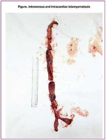
The total size of the removed tumor thrombus was 45 cm and is shown in Figure.
The operation lasted for 6 hours. The blood loss was 9000 ml (cell saver). The blood return was 2500.0 ml with cell saver. The patient was transfused 1500.0 ml of donor erythrocyte mass, and 4000.0 ml of blood plasma. Thus, the total volume of transfusion was 5500.0 ml, autotransfused blood composed 2500.0 ml. Taking into account the volume of surgery and the high postoperative risk of pulmonary thromboembolism, dynamic observation and treatment were carried out for 5 days in the intensive care unit. In order to prevent thromboembolic complications, continuous intravenous infusion of heparin with activated partial thromoplastine time of 60-80 sec. was given, the compression of the lower extremities was performed. The postoperative period was unremarkable.
The patient was discharged on the 14 day in a satisfactory condition. The histology results showed intravascular leiomyomatosis, the leiomyoma of the myometrium.
Conclusion
The relatively young age of the patient, the use of modern blood-saving technologies and the benign nature of the tumor, despite the volume of the operation performed without the use of a heart-lung machine, made it possible to avoid severe postoperative complications. During the treatment, the main goal of the operation was achieved since massive pulmonary thromboembolism was prevented; moreover, uterine extirpation, complete removal of leiomyomatous tissue, including intravascular tissue, were performed. The described clinical observation is unique for the Far Eastern region of Russia and demonstrates the possibility of cardiovascular surgeons together with gynecologists to perform the optimal volume of surgery for uterine leiomyoma with intravascular and intracardiac leiomyomatosis. The patient has been still under dynamic supervision, there are no data on relapse of the disease.
References
- Хубулава Г.Г., Тарасов В.А., Гаврилов Е.К., Ларин И.А. Опухолевая непроходимость нижней полой вены и ее притоков: диагностика, особенности оперативных вмешательств и их результаты. Часть I. Вестник хирургии им. И.И. Грекова. 2018; 177(2): 86-90. [Khubulava G.G., Tarasov V.A., Gavrilov E.K., Larin I.A. Tumor obstruction of the inferior vena cava and its tributaries: diagnosis, features of surgical interventions and their results. Part I. Bulletin of Surgery them. I.I. Grekov. 2018; 177 (2): 86-90. (in Russian)].
- Заваруев В.Н., Мазуренко А.А., Заваруев А.В., Шумский С.В., Ачкасов И.А. Хирургическое лечение рака почки с протяженной наддиафрагмальной опухолевой инвазией нижней полой вены. Вестник РОНЦ им. Н.Н. Блохина РАМН. 2016; 27(4-1): 115-7. [Zavaruev V.N., Mazurenko A.A., Zavaruev A.V., Shumsky S.V., Achkasov I.A. Surgical treatment of kidney cancer with extended supraphrenic tumor invasion of the inferior vena cava. Bulletin RCRC them. N.N. Blokhin RAMS. 2016; 27 (4-1): 115-7. (in Russian)].
- Давыдов М.И., Чарчян Э.Р., Герасимов С.С., Дземешкевич С.Л., Давыдов М.М., Груздев В.Е., Ломидзе С.В., Никуличев Л.А., Серебрянская М.В. Хирургическое лечение больных внутривенным лейомиоматозом с опухолевым тромбозом правых отделов сердца. Клиническая и экспериментальная хирургия. 2016; 4(4): 48-55. [Davydov M.I., Charchyan E.R., Gerasimov S.S., Dzemeshkevich S.L., Davydov M.M., Gruzdev V.E., Lomidze S.V., Nikulichev L.A., Serebryanskaya M. AT. Surgical treatment of patients with intravenous leiomyomatosis with tumor thrombosis of the right heart. Clinical and experimental surgery. 2016; 4 (4): 48-55. (in Russian)].
- Стилиди И.С., Бохян В.Ю., Паяниди Ю.Г., Жорданиа К.И. Внутривенный лейомиоматоз. Хирургия. Журнал им. Н.И. Пирогова. 2012; 5: 81-3. [Stilidi I.S., Bohyan V.Yu., Payanidi Yu.G., Jordania K.I. Intravenous leiomyomatosis. Surgery. Journal them. N.I. Pirogov. 2012; 5: 81-3. (in Russian)].
- Парамонова Т.И., Вдовкин А.В., Палькова В.А., Горностаева О.С. Комплексная лучевая диагностика внутривенного лейомиоматоза с экспансивным ростом в просвет нижней полой вены, полость правого предсердия и правого желудочка (клиническое наблюдение). Диагностическая и интервенционная радиология. 2012; 6(1): 105-12. [Paramonova T.I., Vdovkin A.V., Palkova V.A., Gornostaeva O.S. Comprehensive radiation diagnosis of intravenous leiomyomatosis with expansive growth into the lumen of the inferior vena cava, the cavity of the right atrium and the right ventricle (clinical observation). Diagnostic and Interventional Radiology. 2012; 6 (1): 105-12. (in Russian)].
Received 17.09.2018
Accepted 21.09.2018
About the Authors
Zavaruev, Artem V., assistant professor, Department of Surgery, University hospital, Amur State Medical Academy. 675000, Blagoveshchensk, Gorkogo str., 95;doctor-vascular surgeon, oncologist, Department of Vascular Surgery, Amur Regional Clinical Hospital.
675000, Blagoveshchensk, Voronkova str., 26. E-mail: zavdoc@mail.ru. Tel: +79246733816.
Mazurenko, Artem A., PhD, head, Department of Vascular Surgery, Amur Regional Clinical Hospital.
675000, Blagoveshchensk, Voronkova str., 26. E-mail: mazart@mail.ru. Tel: +79098107626.
Shumsky, Sergey V., head, Department of Anesthesiology and Resuscitation №1, Amur Regional Clinical Hospital.
675000, Blagoveshchensk, Voronkova str., 26. Tel: +79145535900.
Soloviev, Vitaly V., obstetrician-gynecologist, Department of Gynecology, Amur Regional Clinical Hospital. 675000, Blagoveshchensk, Voronkova str., 26. Tel: +79098134725.
Zavarueva, Irina G., obstetrician-gynecologist, women’s consultation №2, Blagoveshchensk Town Clinical Hospital.
675000, Blagoveshchensk, street bitter, d. 247. E-mail: 11ishenko@mail.ru . Tel: +79638091235.
For citation: Zavaruev A.V., Mazurenko A.A., Shumsky S.V., Solovyev V.V., Zavarueva I.G. Surgical treatment for multiple uterine
leiomyoma with intravascular leiomyomatosis. Akusherstvo i Ginekologiya/Obstetrics and Gynecology.2019; (4): 114-17. (in Russian)
https://dx.doi.org/10.18565/aig.2019.4.114-117
Similar Articles

