
Giant myoma of uterine rudiments in uterovaginal aplasia
Makiyan Z.N., Adamyan L.V., Yarygina N.K., Asaturova A.V.
Objective. To optimize surgical treatment for uterine aplasia. Subjects and methods. Sixty-three patients with uterovaginal aplasia were examined and operated on at the Department of Operative Gynecology, Academician V.I. Kulakov National Medical Research Center of Obstetrics, Gynecology, and Perinatology, in 2016 to 2020. Results. Multiple uterine myoma was detected in 8 patients, which was 12.7%, of whom 3 patients had giant uterine myoma. The authors had not found similar cases in the literature. Uterine rudiments in complete uterovaginal aplasia had a certain proliferative potential, which was confirmed by clinical cases of an increase and manifestations of functional activity, new formation of leiomyoma, including giant myoma of uterine rudiments. Conclusion. To optimize surgical treatment, it is advisable to verify clinical and anatomical options, to assess the likelihood of potential growth of pluripotent cells, and to expand indications for the preventive removal of uterine rudiments.
Keywords
uterovaginal aplasia
uterine rudiments
abnormalities of the uterus
Uterine and vaginal aplasia is an extreme form of uterine agenesis and considered to be a rare congenital anomaly, as it affects 1 out of 4000–5000 newborn girls [1–5].
The uterus and vagina develop from paired Mullerian ducts, therefore, in the case of complete or partial agenesis (aplasia), there are persistent rudimentary formations developed at the early stages of embryogenesis. Complete uterine agenesis in all cases has uterine rudiments which appear as muscle strands or rolls. Isolated cases of leiomyoma originating from uterine rudiments have been reported in the literature [5–14].
We have studied and presented unique clinical observations of multiple myoma originating from uterine rudiments, and a giant myoma of uterine rudiments was detected in three patients.
The objective of the study is to optimize surgical treatment for uterine and vaginal aplasia.
Materials and Methods
Sixty-three patients with uterovaginal aplasia were examined and operated on in the Department of Operative Gynecology, Academician V.I. Kulakov National Medical Research Center for Obstetrics, Gynecology and Perinatology, Moscow, Russia. The study was conducted from 2016 to 2020. A complete clinical and laboratory examination was performed, including ultrasound assessment of the pelvic organs and the urinary system. Operative treatment was performed by laparoscopy, according to the patients’ complaints, detected abnormalities, and findings of an instrumental examination.
The research was approved by the Local Ethics Committee of the Academician V.I. Kulakov National Medical Research Center for Obstetrics, Gynecology and Perinatology, Moscow, Russia (Protocol No. 9 dated November 22, 2018).
Results
Surgical treatment was performed for uterine and vaginal aplasia: artificial vagina was made from the pelvic peritoneum in 63 patients; uterine rudiments were removed simultaneously in 26 patients (non-functioning rudiments were removed in 13 patients, and functioning uterine rudiments were excised in 13 patients).
Paired uterine rudiments in all 63 patients with uterine and vaginal aplasia were macroscopically presented by oval-shaped muscle rollers or strands located in the true pelvis in the lateral areas, mesoperitoneally in the area where the round ligaments cross the fallopian tubes and the ovarian ligaments.
Multiple uterine myoma was detected in eight patients (12.7%). The materials were presented earlier in the article «Uterine rudiments: clinical and morphological variations and optimization of surgical management» [1].
During the clinical examination and surgical treatment, three patients were found to have a giant myoma of uterine rudiments, which was of particular interest for clinical consideration and surgical treatment.
Clinical observation
This is a unique clinical observation of a giant myoma of uterine rudiments in a 44-year-old patient T., who complained of epigastric pain and increased abdominal volume. Congenital aplasia of the uterus and vagina was diagnosed at the age of 22 years, karyotype 46, XX. An artificial vagina was made using the method of treatment with colpoelongation by Sherstnev.
The gynecological examination revealed that there was a normal development of the external genitals and vaginal vestibule. Neovagina was about 8 cm long, and stretchable up to 10 cm. In the pelvic and abdominal areas, there was a rounded formation of about 25.0x30.0 cm in size, dense consistency, painful on palpation, and difficult to dislocate. According to the ultrasound examination, it was a rounded formation of 25.0x30.0x35.0 cm in size, which occupied the total space of the true pelvis and abdominal cavity up to the epigastric area, apparently originating from the right adnexa. The patient was diagnosed with uterine and vaginal aplasia; she had a solid formation, probably a fibroma of the right ovary.
The patient underwent a standard procedure of laparoscopy in the Trendelenburg position. After applying pneumothorax and introducing the primary trocar, Storz laparoscope was introduced in the umbilical area. Additionally, two trocars were introduced, namely, in the right (diameter 5 mm) and left (diameter 12 mm) iliac areas.
Laparoscopy revealed aplasia of the uterus, paired uterine rudiments at the junction of the round ligaments with the fallopian tubes and the ovarian ligaments. Uterine rudiments appeared as elongated muscle rollers from 1.0 to 2.0 cm thick without signs of functional activity. In the abdominal cavity there was a rounded formation originating from the right uterine rudiment; it had a wide base and its diameter was about 30.0 cm. The upper pole of the formation reached the transverse colon. In the area of the left uterine rudiment, there was a myoma up to 5.0–6.0 cm in diameter. The ovaries had a normal size and structure, and the follicular apparatus was normal, too. The fallopian tubes did not show any visible pathology.
The right uterine rudiment was fixed with tenaculum forceps and moved to the opposite (contralateral) side of the pelvic wall. Coagulation and bilateral transection of round ligaments and ovarian ligaments were performed; the vesico-uterine peritoneum was opened. The uterine vessels were coagulated and transected on both sides. The right uterine rudiment with myomatous node was excised. Then the left uterine rudiment was fixed with tenaculum forceps, it was moved to the contralateral side, and the left uterine rudiment was removed. Uterine rudiments with myomatous nodes were morcellated and removed from the abdominal cavity.
The examination of the abdominal organs showed that the anterior surface of the liver was smooth and shiny, the edge of the liver was sharp; the loops of the small and large intestines, as well as the large omentum did not reveal any visible pathologies; the parietal and visceral peritoneum was smooth and shiny; there was no effusion in the abdominal cavity and pelvis.
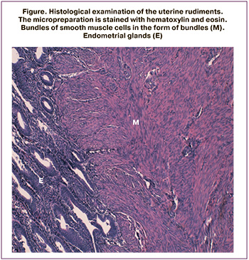 The pathomorphological study showed that the left uterine rudiment (Figure) was 5.0x6.0x3.0 cm in size and contained a myomatous node with a diameter of 4.0x5.0 cm, consisting of smooth muscle tissue (myometrium). Most of the myocytes were relatively large and spindle-shaped; they had oval nuclei with a predominance of euchromatin. Fewer smooth muscle cells demonstrated the signs of metabolic disorders (dystrophy), and had sharply elongated ribbon-like and hyperchromic nuclei. Myocytes were located chaotically and formed uneven bundles and trabeculae. The structure of the myometrium differed from normal muscle tissue as the myometrium had a high number of fibrocytes. Among the bundles of smooth muscle cells, there were endometrial glands that formed small endometrial cavities.
The right uterine rudiment consisted of bundles of myocytes without endometrial glands. A giant myomatous node was a benign tumor consisting of bundles of smooth muscle fibromyoma cells with a total volume of 3.5 liters.
Thus, uterine rudiments contained the smooth muscle tissue, which was not homogeneous in its histological characteristics. Deep dystrophic changes and impaired tissue architectonics were detected in the myocytes of both rudiments.
Conclusion
Thus it is worth taking into account anatomical characteristics for the verification of uterine rudiments, especially in the presence of tumor-like formations that make it difficult to identify the topographic anatomy. Uterine rudiments have some proliferative potential, which is confirmed by clinical cases of growth and functional activity of uterine rudiments, neoplasms of uterine myoma and endometriosis, including recurrent ones.
The obtained clinical findings indicate the need for timely diagnosis, verification of anatomical variations of uterine rudiments in uterine aplasia, assessment of the potential for growth and proliferation of uterine rudiments, and dynamic observation of asymptomatic variants.
It is reasonable to expand the indications for removal of uterine rudiments with functional activity identified by ultrasonography and Doppler ultrasound and in the presence of pain or gynecological comorbidity, as well as for the prevention of neoplasms originating from uterine rudiments.
A video presentation of the operation is provided for this clinical observation (available at: https://aig-journal.ru/archive).
References
- Макиян З.Н., Адамян Л.В., Асатурова А.В., Ярыгина Н.К. Маточные рудименты: клинико-морфологические варианты и оптимизация хирургического лечения. Акушерство и гинекология. 2019; 12: 126-32. [Uterine rudiments: clinic-morphological variants and optimization of surgical treatment. Akusherstvo i ginecologia/Obstetrics and gynecology. 2019; 12: 126-32. (in Russian)]. https://dx.doi.org/10.18565/aig.2019.12.126-132.
- Адамян Л.В., Курило Л.Ф., Глыбина Т.М., Окулов А.Б., Макиян З.Н. Аномалии развития органов женской репродуктивной системы: новый взгляд на морфогенез. Проблемы репродукции. 2009; 15(4): 10-9. [Adamyan L.V., Kurilo L.F., Glybina T.M., Okulov A.B., Makiyan Z.N. Female reproductive organs anomalies: new look on the embryogenesis. Problemi reproduktsii/Problems of reproduction. 2009;4:10-19. (in Russian)].
- Makiyan Z. New theory of uterovaginal embryogenesis. Organogenesis. 2016; 12(1): 33-41. https://dx.doi.org/10.1080/15476278.2016.1145317.
- Окулов А.Б., Магомедов М.П., Поддубный И.В., Богданова Е.А., Файзулин А.К., Макиян З.Н., Глыбина Т.М., Смирнов В.Ф., Володько Е.А., Мираков К.К., Бровин Д.Н. Синдром Майера-Рокитанского-Кюстера-Хаузера у девочек, его варианты. Органосохраняющая тактика лечения. Андрология и генитальная хирургия. 2007; 8(4): 45-52. [Okulov A.B., Magomedov M.P., Poddubniy I.V. et al. Mayer-Rockitansky-Kuster-Hauser syndrome in adolescents and its variations. Operative treatment. Andrology and genital surgery. 2007; 8(4): 45-52. (in Russian)].
- Fletcher H.M., Campbell-Simpson K., Walcott D., Harriott J. Müllerian remnant leiomyomas in women with Mayer-Rokitansky-Küster-Hauser syndrome. Obstet. Gynecol. 2012; 119(2, Pt 2): 483-5. https://dx.doi.org/10.1097/AOG.0b013e318242a9b5.
- Makiyan Z. Endometriosis origin from primordial germ cells. Organogenesis. 2017; 13(3): 95-102. https://dx.doi.org/10.1080/15476278.2017.1323162.
- Lamarca M., Navarro R., Ballesteros M.E., García-Aguirre S., Conte M.P., Duque J.A. Leiomyomas in both uterine remnants in a woman with the Mayer-Rokitansky-Küster-Hauser syndrome. Fertil. Steril. 2009; 91(3): 931. e13-5. https://dx.doi.org/10.1016/j.fertnstert.2008.08.132.
- Papa G., Andreotti M., Giannubilo S.R., Cesari R., Ceré I., Tranquilli A.L. Case report and surgical solution for a voluminous uterine leiomyoma in a woman with complicated Mayer-Rokitansky-Küster-Hauser syndrome. Fertil. Steril. 2008; 90(5): 2014. e5-6. https://dx.doi.org/10.1016/j.fertnstert.2008.04.06.
- Fukuda J., Kumazawa Y., Fujimoto T., Tanaka T. Mayer-Rokitansky-Kustner Hauser syndrome complicated by either uterine leiomyoma or ovarian tumor. J. Obstet. Gynaecol. Res. 2010; 36(1): 191-4. https://dx.doi.org/10.1111/j.1447-0756.2009.01116.x.
- Rawat K.S., Buxi T., Yadav A., Ghuman S.S., Dhawan S. Large leiomyoma in a woman with Mayer-Rokitansky-Kuster-Hauser syndrome. J. Radiol. Case Rep. 2013; 7(3): 39-46. https://dx.doi.org/10.3941/jrcr.v7i3.1267.
- Blontzos N., Iavazzo C., Vorgias G., Kalinoglou N. Leiomyoma development in Mayer-Rokitansky-Küster-Hauser syndrome: a case report and a narrative review of the literature. Obstet. Gynecol. Sci. 2019; 62(4): 294-7. https://dx.doi.org/10.5468/ogs.2019.62.4.294.
- Sharma R., Guleria K., Suneja A., Bhaskaran S., Tanveer N. Giant leiomyoma with extensive myxoid degeneration in Mayer-Rokitansky-Küster-Hauser syndrome. Int. J. Gynaecol. Obstet. 2017; 138(1): 125-7. https://dx.doi.org/10.1002/ijgo.12162.
- Salem Wehbe G., Bitar R., Zreik T., Samaha M., Walter C., Sleiman Z. Intra-peritoneal leiomyoma of the round ligament in a patient with Mayer-Rokitansky-Küster-Hauser (MRKH) syndrome. Facts Views Vis. Obgyn. 2016; 8(4): 233-5.
- Amaratunga T., Kirkpatrick I., Yan Y., Karlicki F. Ectopic pelvic fibroid in a woman with uterine agenesis and Mayer-Rokitansky-Küster-Hauser syndrome. Ultrasound Q. 2017; 33(3): 237-41. https://dx.doi.org/10.1097/RUQ.0000000000000284.
Received 10.03.2020
Accepted 24.04.2020
About the Authors
Zograb N. Makiyan, Doctor of medicine, Principal Investigator, National Medical Research Center for Obstetrics, Gynecology and Perinatology named after Academician V.I. Kulakov. Tel.: +7(910)437-62-21. E-mail: makiyan@mail.ru, z_makiyan@oparina4.ru. ORCID: 0000-0002-0463-1913. 4, Oparina str., Moscow, 117997, Russian Federation.Leyla V. Adamyan, Academician of RAS, professor, Doctor of medicine, the Head of Operative Gynecology Department, National Medical Research Center for Obstetrics, Gynecology and Perinatology named after Academician V.I. Kulakov. Tel.: +7(495)222-37-37. 4, Oparina str., Moscow, 117997, Russian Federation.
Nadezhda К. Yarygina, Researcher, National Medical Research Center for Obstetrics, Gynecology and Perinatology named after Academician V.I. Kulakov.
Tel.: +7(925)574-83-87. 4, Oparina str., Moscow, 117997, Russian Federation.
Aleksandra V. Asaturova, Doctor of medicine, Principal Investigator,
National Medical Research Center for Obstetrics, Gynecology and Perinatology named after Academician V.I. Kulakov. Tel.: +7(926)994-43-14.
4, Oparina str., Moscow, 117997, Russian Federation.
For citation: Makiyan Z.N., Adamyan L.V., Yarygina N.K., Asaturova A.V. Giant myoma of uterine rudiments in uterovaginal aplasia.
Akusherstvo i Ginekologiya/Obstetrics and Gynecology. 2020; 8: 149-152 (in Russian).
https://dx.doi.org/10.18565/aig.2020.8.149-152
Similar Articles

